Bio 301 - Ch. 3 Book Notes
Bacteria & Archaea
both classified as prokaryotes
bacteria have phospholipid bilayers similar to the ones eukaryotes have
thick, complex outer envelope that protects the cell from environmental stress
contains a compact genome which maximizes the production of cells from limited resources
tightly coordinated functions to form a highly coordinated mechanism
archaea have unique membrane and envelope structures
an example is ether membranes
archaea live in moderate environments
Eukaryotic cells
possess extensive membranous organelles
endoplasmic reticulum & Golgi complex
mitochondria and chloroplasts evolved by endosymbiosis with engulfed bacteria
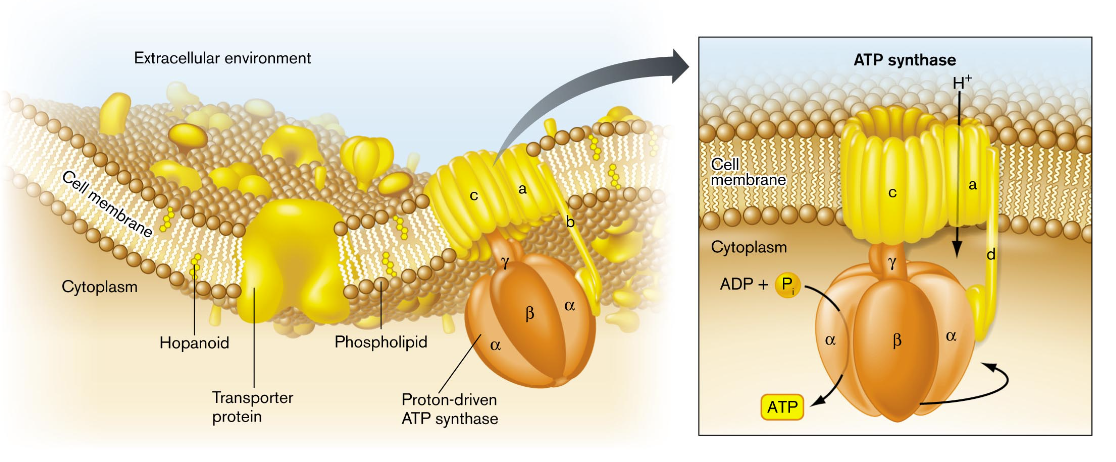
Model of a bacterial cell
a cell is composed of a cell membrane and cytoplasm which form the physical qualities of a cell which are reinforced by inner and outer membranes
the inner membrane is made up of phospholipids, transporter proteins, and other molecules; functions to prevent cytoplasmic proteins from escaping and maintains a gradient of ions & nutrients
the cell wall lies between the inner and outer membrane of the cell which is formed by sugar chains linked covalently by peptides
limits the expansion of the cytoplasm and keeps the cell membrane intact when water flows in (turgor pressure)
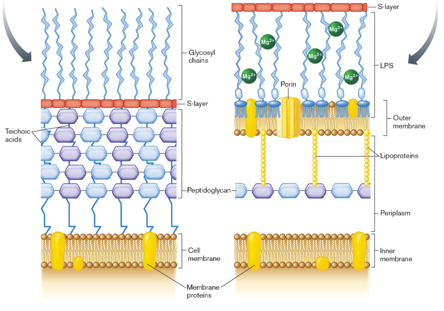
a gram-positive species would have the cell wall outside its one plasma membrane
a gram-negative species has a cell wall that lies within the periplasm and has phospholipids & lipopolysaccharides (LPS) outside the cell wall
the bacterial envelope includes cell-surface proteins that enable the bacterium to interact with specific host organisms
motile bacteria has an array of chemoreceptors the bind molecules from outside the cell or the periplasm and converts this binding intel into signals within the cytoplasm
the signaling molecules direct the rotation of flagella to propel the corresponding movement
the membrane is a 2D fluid of lipids and proteins
a phospholipid bilayer that has lipid-soluble proteins
the bilayer behaves as a 2D fluid which lipids and proteins can diffuse across
proteins embedded in the cell membrane often function as a complex
the subunits of a complex are usually adjacent and fit together like a puzzle (eg. ATP synthase)
the cell membrane and envelope provide an attachment point for one or more chromosomes
organized as a system of looped coils, nucleoids, which are not enclosed by a membrane
instead, the loops of DNA extend throughout the cytoplasm & can be transcribed by RNA polymerase to form mRNA, rRNA, and tRNA
recall during the transcription process, chaperones are present to aid in the proper DNA folding
biochemical composition of bacteria
all cells share common chemical components:
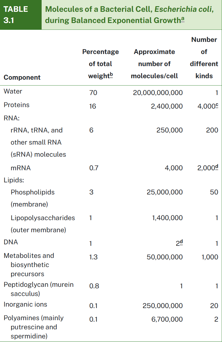
water - the fundamental solvent of life
essential ions - these include K+, Mg2+, and Cl-
small organic molecules - these include lipids & sugars which are incorporated in numerous cell structures and provide nutrition by catabolism
macromolecules - these include nucleic acids and proteins which contain information, catalyze reactions and mediate transport
small molecules & ions - these include phospholipids, enzyme cofactors, and charged organic molecules
the cell’s genomic DNA directs expression of its proteins
a cell uses different genes to make different proteins while factors such as temperature, nutrient levels, and entry to a host organism are accounted for
the proteins expressed by a cell under given conditions are known as a proteome
another important component of cells is the bacterial cell wall that consists of peptidoglycan
this component limits the volume of the enclosed cell meaning water rushing in will create high turgor pressure
cell fractionation
a procedure to separate cell components that often includes ultracentrifugation
this process also provides purified proteins that act as antigens for candidate vaccines
the process of ultracentrifugation was refined
the gravitational force separates molecules by weight and density
cell fractionation requires techniques that lyse the cell
there must be enough force to separate the membrane lipids but not enough to disintegrate complexes of protein and RNA
for gram-negative cells, the method requires more specificity to separate the compartments because it has inner & outer membranes, the cytoplasm and the periplasm
the membrane vesicles proteins are analyzed on gel electrophoresis
these proteins can be identified by bands on the gel by enzyme digestion and mass spectrometry
limitations to cell fractionation
provides little information about processes that require an intact cell (eg cell divison)
an alternation approach to studying a portion of the cell without breaking it open would be genetic analysis
this process includes mutating a strain so it alters a gene and then select the mutant strains for loss of a given function
the phenotype of the mutant can provide insight about the function of the altered part
steps for cell wall lysis and spheroplast formation
1: permeabilize the bacterial outer membrane by removing the Mg2+ & Ca2+
this allows sucrose to cross and fill the periplasm which maintains an osmotically stable solution
2: lysozyme cleaves peptidoglycan and breaks down the cell wall
without the cell wall, the cell forms into a spheroplast
these spheroplasts can be seen by TEM
3: to isolate the periplasmic contents, the spheroplasts are transferred to distilled water
water rushes in through the EDTA-weakened outer membrane
this causes osmotic shock of the periplasmic compartment, but the inner membrane remains intact
4: after osmotic shock, the spheroplasts undergo ultracentrifugation to separate the periplasmic contents from the other three type of cell compartments
5: the membranes are then broken open by a French press device
6: a second step of ultracentrifugation now pellets the inner and outer membrane vesicles while removing the cytoplasm in the supernatant
7: the inner and outer membranes are separated by density gradient ultracentrifugation
the gradient is created by a solute concentration
the lower-density fractions contain inner membrane vesicles
the higher-density fractions contain outer membrane vesicles
membrane lipids
the phospholipid bilayer creates membrane fluidity and gives the cell consistent thickness
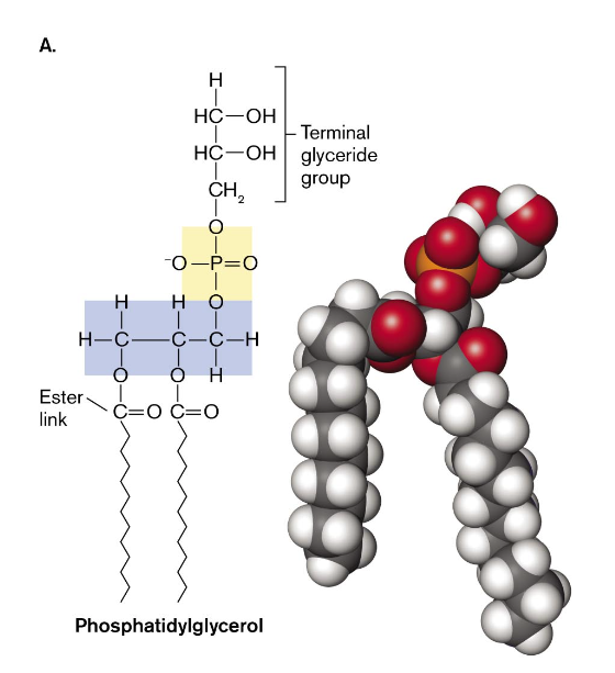
a phospholipid consists of glycerol with ester links to each of two fatty acids and a phosphoryl polar head group (phosphatide)
the negatively charged head group of a phosphatide can contain various organic groups or have a side chain with a positive charge (typically on amine group)
lipid biosynthesis is a key process that can make some cells vulnerable to antibiotics
factors that can effect the formation of membrane lipids to maintain structural integrity and function & uniform thickness
environmental stress
starvation stress increases bacterial production of lipids with an unnatural type of phosphoryl head group
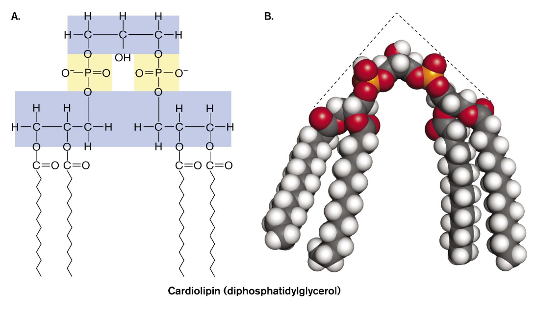
cardiolipin, a double phospholipid linked by a glycerol, concentration will increase in bacteria grown to starvation
cardiolipin helps define the polar structure of a bacterial cell and diffuses in concentration patches called “domains” near the cell poles
at the cell pole cardiolipin binds certain environmental stress proteins and a phospholipid can have specific functions associated with specific membrane proteins
the fatty acid components of phospholipids varies between being saturated and unsaturated (most are cis - forms a kink)
the enhanced fluidity of a kinked phospholipid improves the function of the membrane at low temperatures
cyclization of the part of the chain to form a stiff planar ring with decreased fluidity
the double bond of unsaturated fatty acids can generate a cyclopropane fatty acid
stiff planar molecules can reinforce the membrane and reduce the membrane fluidity
for eukaryotes reinforcing agents are sterols like cholesterol
in some bacteria, reinforcing agents are hopanoids (five ring hydrocarbons)
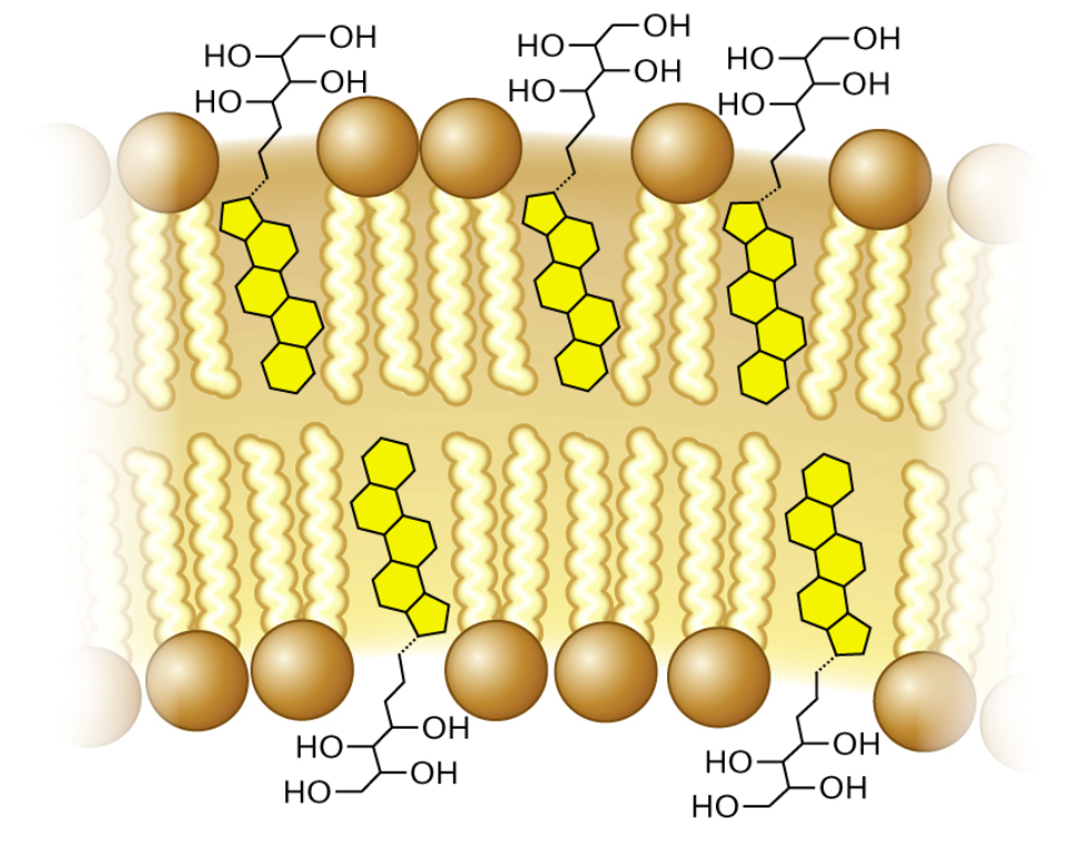
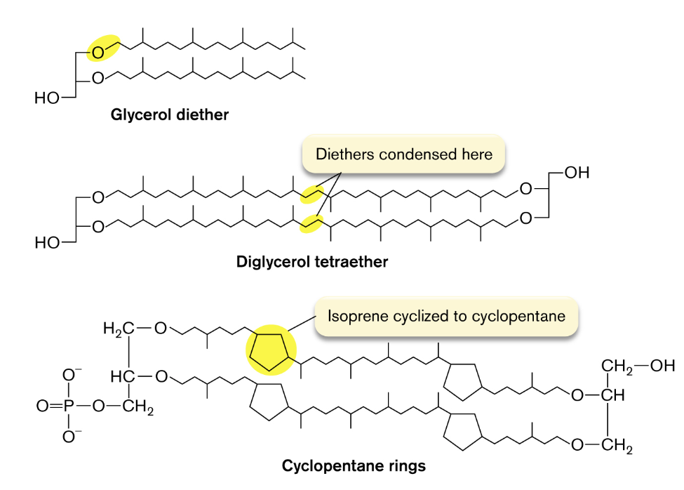
archaea have unique membrane lipids
these phospholipids replace the ester link between the glycerol and fatty acid with an ether link
ethers are more stable than esters which hydrolyze easily in water
archaeal phospholipids have hydrocarbon chains are branched terpenoids which limits the movement of the membrane
membrane proteins
structural support
these proteins can anchor together different layers of the cell envelope
these proteins can attach the membrane to the cytoskeleton or form the base of structures extending out from the cell
detection of environmental signals
secretion of virulence factors and communication factors
membrane protein complexes export toxins and cell signals across the envelope
ion transports and energy storage
transport of ions across a membrane generates a transmembrane gradient that stores energy
there is a requirement of a portion of hydrophobic amino-acid side chains that are soluble
molecules cross the cell membrane
because cell membranes act as a barrier to contain the cell contents and exclude extracellular material, selective transport is essential for cell survival
the ability to acquire nutrients, transport waste, and transmit signals to neighbor cells
passive diffusion
small, uncharged molecules like diatomic oxygen and carbon dioxide can easily permeate the membrane
large, strongly polar molecules like sugar and charged molecules like amino acids cannot penetrate the membrane and require transportation
water molecules can permeate the membrane, but their passage is increased by protein channels called aquaporins
osmosis
the internal concentration of water is lower than the concentration outside the cell
the solute concentration is higher inside the cell than outside
because of this, water tends to diffuse across the membrane into the cell which causes the cell volume to expand
osmotic pressure will cause a cell to lyse in the absence of a countering pressure such as that provided by the cell wall
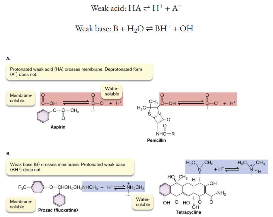
membrane-permeant weak acids and bases
these particles can only cross the membrane in their uncharged form which is HA for weak acids and B for weak bases
membrane-permeant acids conduct H+ ions across the membrane which can cause acidic stress (higher H+ concentration outside the cell drives weak acids into the cell)
membrane-permeant bases conduct OH- ions across the membrane which can cause alkali stress
transmembrane ion gradients
molecules that carry a fixed charge cannot cross the membrane like H+ and Na+
an ion gradient across the cell membrane can store energy for nutrition or to drive the transport of other molecules
inorganic and charged organic ions require transport proteins (passive | active)
a transport protein obtains energy for active transport by cotransport of another substance down its gradient from higher to lower concentration or by coupling transport to a chemical reaction
protective layers of the cell envelope
cell wall & some common structural support is an S-layer (outer membrane)
the bacterial cell wall, the sacculus, consists of a single interlinked molecule that envelopes the cell
typically encloses maximal volume with minimal surface area
the sacculus is a single molecule cage-like structure, highly porous to ions and organic molecules
the form is not rigid; it is a flexible mesh bag with unbreakable joints
turgor pressure within the enclosed cytoplasm fills the cell’s shape
peptidoglycan / murein structure
this component is unique to bacteria; the molecule consists of parallel polymers of disaccharides called glycan chains cross-linked with peptides of four to six amino acids
the peptide extension can form cross-bridges connecting parallel strands of glycan
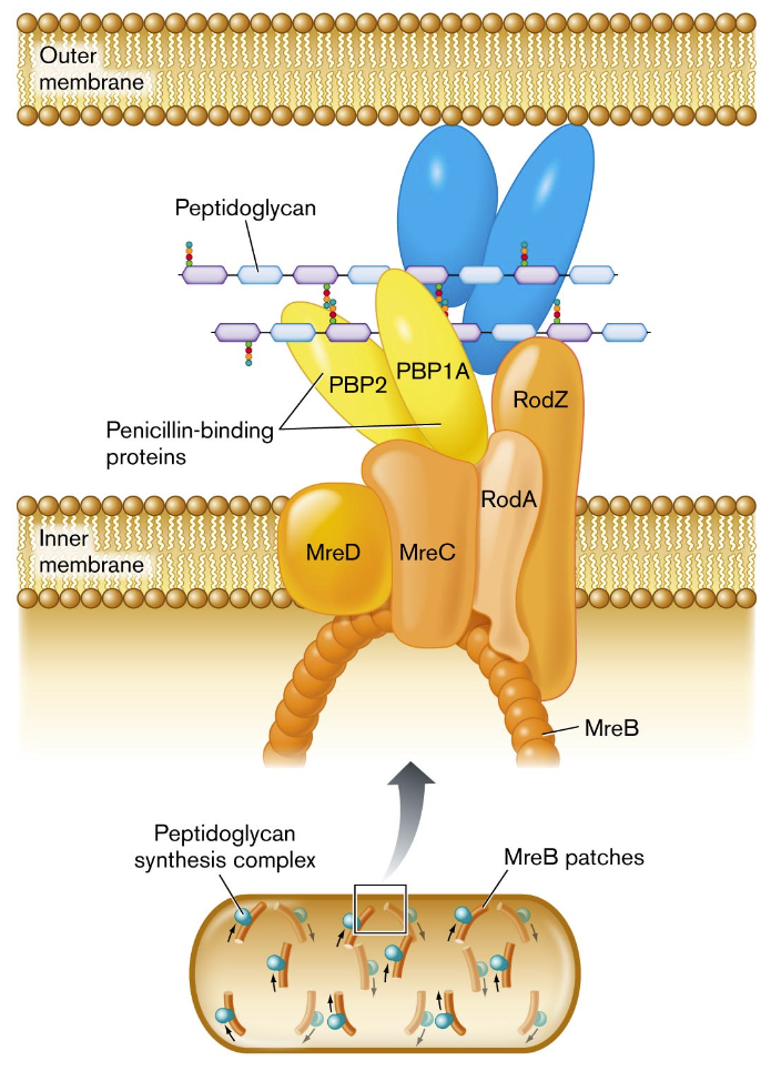
peptidoglycan synthesis as a target for antibiotics
the enzymes required to bind the antibiotic penicillin, termed penicillin-binding proteins, hold the function of building the peptide bonds and sealing the cross-bridges
peptidoglycan is a trait unique to bacteria which makes it a target for new antibiotics (despite the resistance that some strains of bacteria have formed against commonly prescribed antibiotics
the overall extension of the cell wall by the peptidoglycan layer is organized by a protein complex that includes MreB
this component polymerizes a helical direction along ana arc beneath the plasma membrane
cell envelope of bacteria
most bacteria have additional envelope layers that provide structural support and protection from predators and host defenses
additional molecules are attached to the cell wall and cell membrane and some thread through the layers
Gram-positive bacteria have a thick cell wall with 3-20 layers of peptidoglycan which are interpenetrated by teichoic acids
Gram-negative bacteria have a thin cell wall with 1-3 layers of peptidoglycan which are enclosed by an outer membrane
lipoproteins link the outer membrane to the peptidoglycan layer
they are bound to the periplasm before the inner membrane
firmicute cell envelope — Gram-positive
the multiple layer of peptidoglycan are reinforced by teichoic acids threaded through its multiple layers
the qualities of teichoic acids that help retain the Gram stain are the negatively charged cross-threads and the overall thickness of the Gram-positive cell wall
the cell wall attaches to extracellular structures through an enzyme, sortase, which forms a peptide bond from a cell wall cross-bridge to a protein extending from the cell
proteins attached by the sortases can help the cell acquire nutrients or help the cell adhere to a substrate
S-layer
this layer is composed of protein subunits that fit together like tiles which provides defense against phages or predators
this layer is rigid but it flexes and allows the passage of substances in either direction
capsule
a slippery outer layer composed of polysaccharides that surrounds the cell envelope of some bacteria
proteobacterial cell envelope — Gram-negative
the cell envelope of these bacteria includes 1-3 layers of peptidoglycan covered by an outer membrane
the outer membrane confers defensive abilities and toxigenic properties on many pathogens
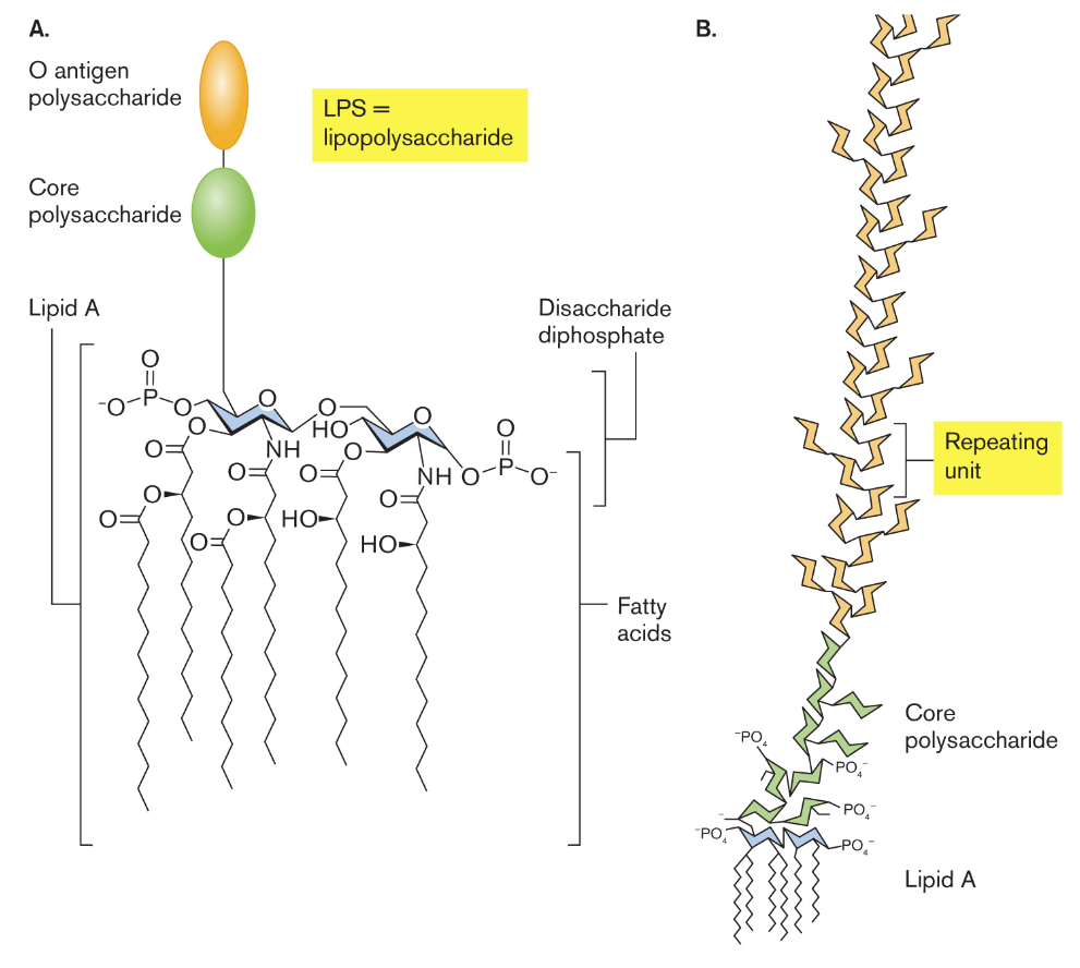
lipoprotein and lipopolysaccharide (LPS)
in Gram-negative bacteria, the inward facing leaflet of the outer membrane has a phospholipid composition similar to one of the inner membranes
the outer membrane’s inward-facing leaflet includes lipoproteins that connect the outer membrane to the peptide bridges of the cell wall
murein lipoprotein consists of a protein with an N-terminal cysteine attached to three fatty acid side chains
LPS’s act as endotoxins which are cell compartments that are harmless if the pathogen remains intact but when lysed, the endotoxins induce a potentially lethal shock to the host
outer membrane proteins
Gram-negative bacterial cells have porins that permit the entry of nutrients such as nutrients like sugars and peptides
outer membrane porins have limited specificity, allowing passive uptake of various molecules including antibiotics
to prevent the entry of dangerous molecules, cells express different outer membrane porins under different environmental conditions
in dilute environments, cells express porins of large pore size to maximize the uptake of nutrients
in rich environments, cells down-regulate the expression of large porins & express porins of smaller pore size to select only smaller nutrients as to avoid uptake of toxins
periplasm
this portion of the cell contains specific enzymes and nutrient transporters not found within the cytoplasm
these proteins in the periplasm are subjected to pH and salt concentration fluctuations because the outer membrane is porous to ions
capsule
some Gram-negative bacteria have capsules made of loose glycolipids
mycobacterial cell envelope
have effective defenses against host defenses & the gram stain is not applicable to use
the mycobacterial envelope includes features of both Gram-positive and Gram-negative cells
the peptidoglycan layer is linked to chains of galactose called galactans
the galactans are attached to arabinans
mycolic acids provide the basis for acid-fast staining due to the ester links that the arabinans form with mycolic acid
this function retains the dye carbolfuchsin
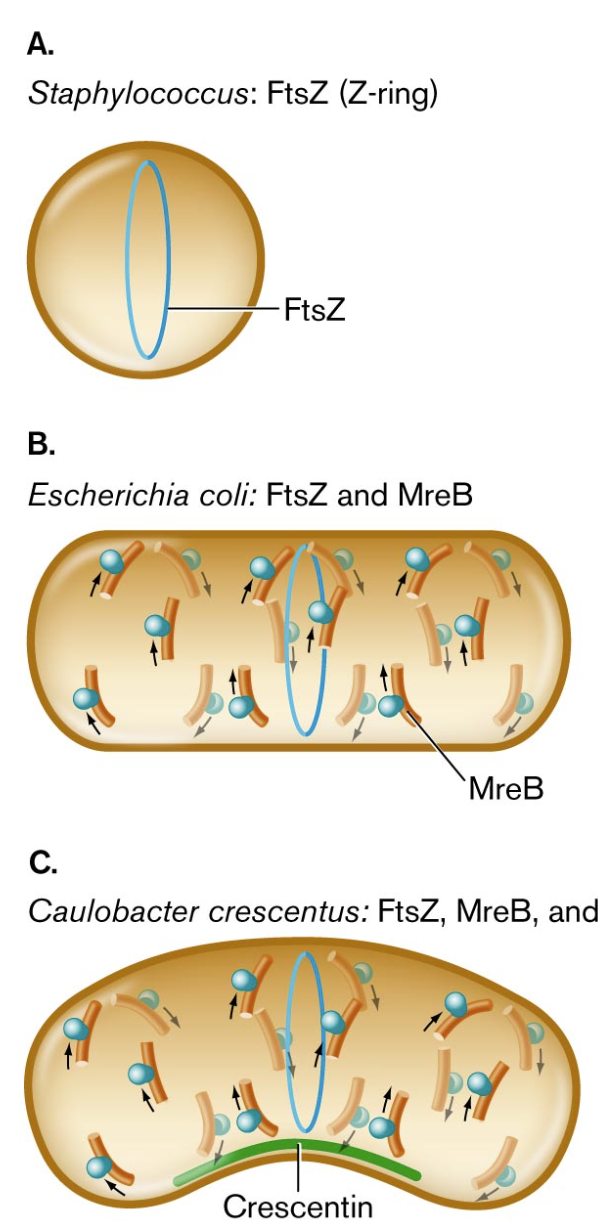
bacterial cytoskeleton
to determine the shape of bacteria, aside from turgor pressure, they possess protein cytoskeletal components
the functions of cytoskeletal proteins are probed by fluorescent protein fusions
in both spherical bacteria and rod-shaped bacilli requires cell division with the FtsZ protein
this encodes for a Z-ring to form and determine the cell diameter & manages the growth of the dividing partition - the septum
for rod-shaped bacteria, there is a requirement of elongation to polymerization of MreB where MreB travels in a helical arc beneath the cell membrane
if the rod-shaped bacteria is curved and forms a crescent shape, crescentin, polymerizes along the inner curve of the crescent
bacterial cell division by septation
in prokaryotes, the cell divides by a process called septation which forms a partition that divides the envelope
septation requires rapid biosynthesis of all envelope components including membranes and the cell wall
envelope expansion must coordinate the extension of all layers—and regulate the placement and timing of the septum
the overall process of septation is managed by a protein complex: divisome
this component manages assembly of the septum with its two envelopes back-to-back
FtsZ, a critical part to the divisome, polymerizes to form the Z-ring
FtsN helps regulate the timing of constriction of the septum
cocci shaped bacteria can split on any plane (diagonal, horizontal, vertical) but rod-shaped bacteria only split vertically
DNA is organized in the nucleoid by domains (loops)
the midpoint on the DNA is the origin of replication which is attached to the cell envelope at a point on the cell’s equator
the DNA may be looped back to the center of the cell, near the origin of replication
within the domains, the DNA is compacted by supercoils which causes portions of DNA to double back and twist upon themselves to result in compaction of the chromosome
DNA is also compacted by DNA-binding proteins
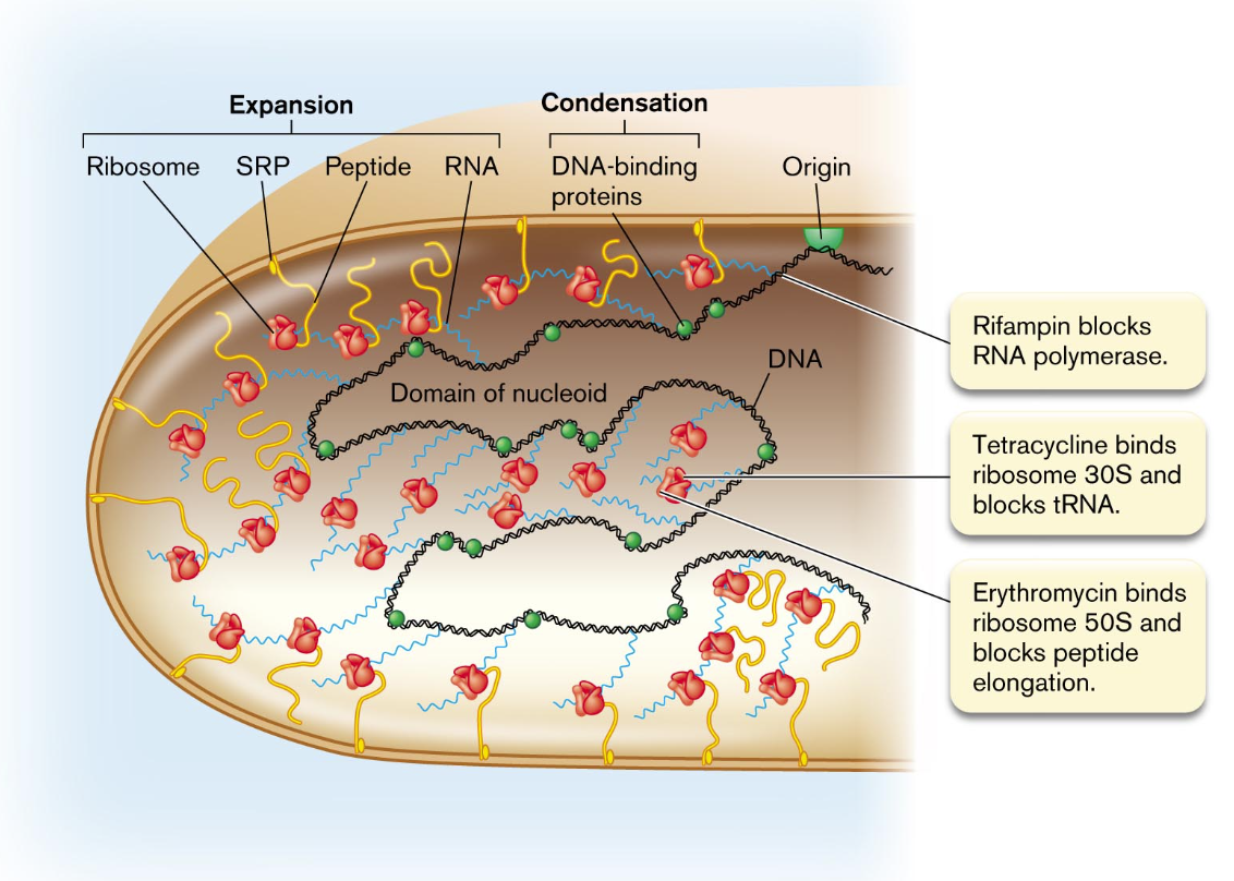
to initiate DNA replication, the DNA double helix at the origin is opened by binding proteins, and then DNA polymerase synthesizes new strands in both directions
in rapidly growing bacteria, the DNA is transcribed and the messenger RNA is translated to proteins while the DNA itself is being replicated
this phenomenon explains why bacterial cells can divide in as little as 10 mins
some of the newly translated proteins are made to function within the membrane and are synthesized in association with the membrane; they are directed there by signal recognition particles
DNA replication regulates cell division
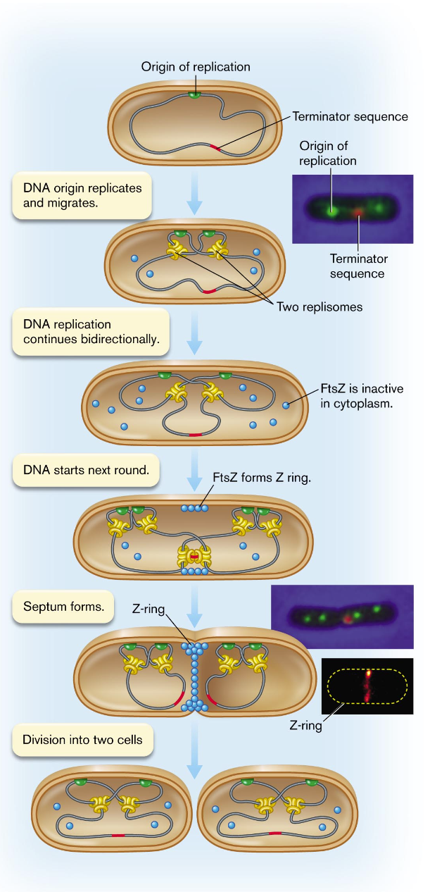
completion of replication triggers Z-ring formation
bacterial cell size
cell size depends on genetic regulators and environmental constraints
more resources will lead to cell elongation occurring quicker and reaching larger sizes before septation and division
with less resources, cell growth slows & early division produces smaller cells
bacterial cell differentiation
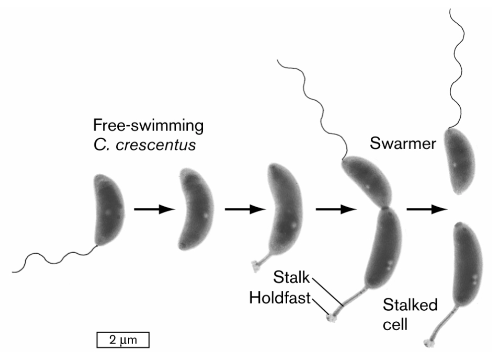
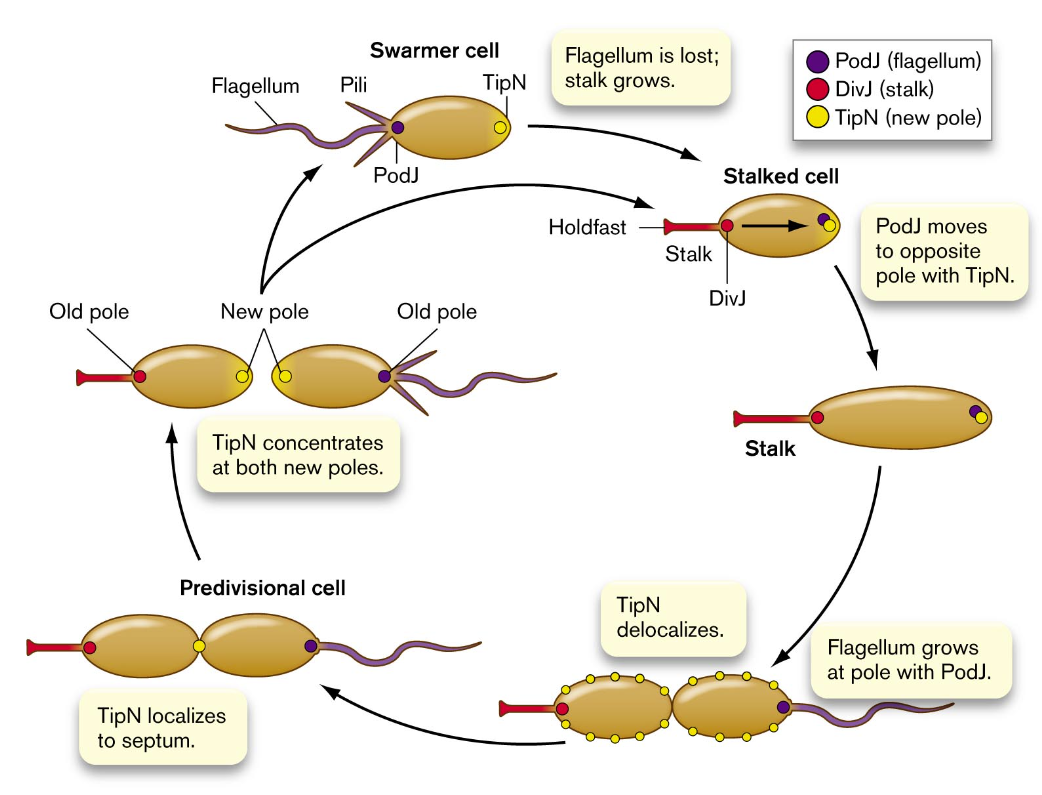
growth asymmetry and polar aging
cell division generates two daughter cell with chemically different poles
under environmental stress, at each cell division, some members of a population die of polar old age
the cause of this death is by the preferential accumulation of protein aggregates
a protein is more likely to aggregate under stress conditions such as low pH or antibiotic presence
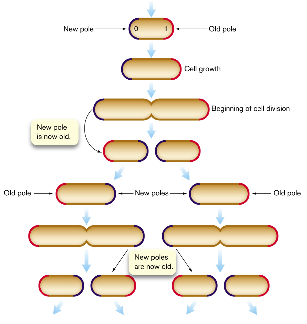
one consequence of polar aging is cells of different polar ages may differ in their resistance to antibiotics
an extreme form of asymmetrical growth is endospore formation
under starvation, desiccation, or other stress conditions, a bacterium can undergo an asymmetrical cell division to develop an endospore at one end
this requires an extreme form of cellular altruism where the mother cell sacrifices itself for the spore-forming cell to generate an endospore capable of remaining dormant but viable for years
membrane vesicles
functions that vesicle production has to outweigh the loss of resources
attraction of partner heterotrophs — because heterotrophs are attracted by the released carbon sources & consume excess oxygen and reactive oxygen species, for some bacteria, it is a requirement to have a partner for growth
phage decoys — the bacterial membrane vesicles have envelope receptors for phages which can trap the phages and prevent them from infecting cells
DNA transfer — the DNA released in cytoplasmic vesicles may provide useful information to encode for genetic traits for other members of the population as a form of horizontal gene transfer
membrane extensions and nanotubes
bacteria may possess cell extensions such as filaments and “pearling” chains of vesicles
nanotubes enable bacteria to directly share proteins and mRNA that encodes products useful under hostile conditions
another remarkable feat that derives from the presence of nanotubes is the fact that bacteria of different species can share beneficial components of cytoplasm
the nanotubes facilitate exchange of different amino acids between the two species
the nanotubes only form when the two types of cells each produce an amino acid lacking in the other which prompts metabolic cross-feeding
archaea show various kinds of intracellular nanotubes that are essential parts of the cell
thylakoids, carboxysomes, and storage granules
cyanobacteria need to maximize the amount of light that is necessary to drive photosynthesis & they do this with the presence of thylakoids
thylakoids consist of layers of folded sheets (lamellae) or tubes of membranes packed with chlorophylls and electron carriers
thylakoids conduct only the light reactions of photon absorption and energy storage
the energy obtained is spent fixing CO2
to stay at the top of water columns, some bacteria and archaea form gas vesicles to increase buoyancy
the gases are hydrogen or carbon dioxide produced by the cell’s metabolism
when light is scarce, cyanobacteria might digest their thylakoids for energy and as a source of nitrogen
they might also digest energy-rich materials from storage granules: PHB & PHA
these are polymers that of interest in biodegradable plastics since bacteria have been engineered to produce them industrially
another type of storage device is sulfur
granules of elemental sulfur produced by purple & green phototrophs through photolysis of hydrogen sulfide
the granules may be used as an oxidant when reduced substrates are available
the presence of potentially toxic sulfur granules may help cells avoid predation
pili and stalks
pili are constructed of straight filaments of protein monomers called pilin
short attachment of pili are called fimbriae
pili can provide a form of motility called “twitching” in which the pili act as limbs to “walk” the bacterium across a substrate
in Gram-negative enteric bacteria, pili of a different kind (sex pili) attach a donor cell to a recipient cell for transfer of DNA
the transfer of DNA is conjugation
stalks are another type of attachment organelle which is an extension of the envelope and cytoplasm
the tip of the stalk secretes adhesion factors that form a “holdfast” to firmly attach the bacterium in a favorable environment
a stalk and holdfast enable some types of bacteria to form large biofilms in streams
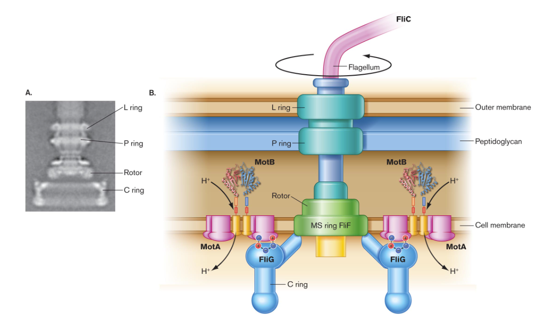
rotary flagella
many bacteria and archaea can swim by means of rotary flagella which can benefit the microorganism to disperse the population and decrease competition
movement can also prompt cells to swim towards a favorable habitat
flagellar movement
different bacterial species have different numbers and arrangements of flagella
how rotary flagellum work
each flagellum has a spiral filament of protein monomers called flagellin (protein FliC)
the filament rotates by means of a motor driven by the cell’s transmembrane proton current
the flagellar motor is embedded in the layers of the cell envelope & the motor possesses an axle and rotary parts
another protein, FliG, forms part of the device that generates torque (rotary force)
how to cells decide where to swim
most flagellated cells have an elaborate sensory system for taxis, the ability to swim towards favorable environments
taxis to specific chemicals is called chemotaxis which requires receptors that tell the bacterium when it is swimming toward a source of attractant or repellent
these molecules are detected by arrays of chemoreceptors that are located near a cell pole
another function of flagellum is the adherence of cells to a substrate to begin forming a biofilm