lecture 1
muscle cells are unique - many precursor cells fuse together into 1
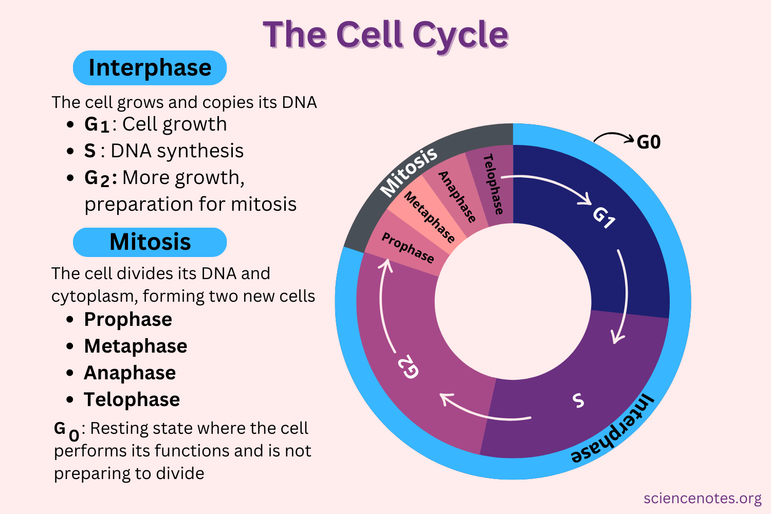
Mitosis - division of nuclear matter (not whole cell)
G1 (gap) - high level of biosynthesis activity (prep for DNA replication)
S - synthesis of DNA (replication)
G2 (prep for mitosis, cytokinesis) cytokinesis = division of whole cell
interphase = #2,3,4
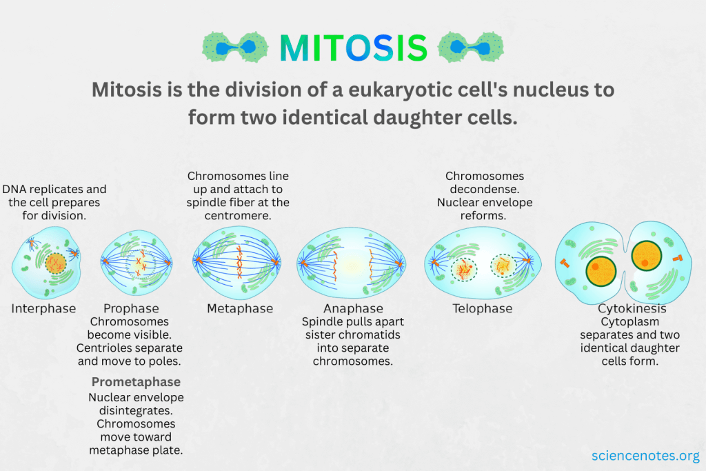
phases of mitosis
muscle tissue development
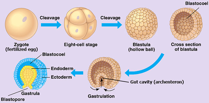
zygote (sperm and egg fuse)
| cleavage (the process of cell division that occurs after fertilization of an egg)
\/
blastula (just cell replication) (until it reaches a critical amount of cells)
| gastrulation - differentiation starts here
\/
germ layers (stem cells)
morphogenesis (from ball of cells → development of features? like head or feet
organogenesis (development of organs)
organogensis is the phase of embryonic development that starts at the end of gastrulation and continues until birth
during organogensis, three germ layers form called ectoderm, endoderm, and mesoderm
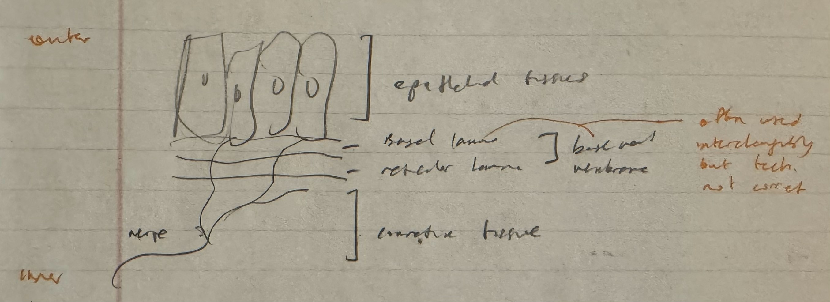
ectoderm (outer layer) → epidermis, nervous system
endoderm (innermost) → lining of digestive tubes, associated with organs (pancreas, liver, ect.)
mesodorm (middle) → heart, kidneys, gonads, blood cells, connective tissues (bone, tendons, cartilege, skeletal muscle)
**^everything is developing at the same time
spinal cord = bone
spinal collumn = neural tisssue
5 regions of mesoderm
chordamesoderm → notochord (becomes spine) formation of body structure and neural tube
intermediate mesoderm → urinary system, genital ducts
lateral plate mesoderm → heart, vasculature, blood cells
head mesoderm → muscles of the face
dorsal (paraxial) mesoderm → both side of neural tube produce connctive tissue including skeletal muscle
**when you reach critial number of cells, replication slows down -→ let cells develop
mesoderm develop into blocks of “sticky cells” called somites (cells cannot just be a jar of marbles; need to work together)
?? basal lamina (collagen, laminin, fibronectin)
join together with somites to form tissues
**Cells situated ventromedially in a somite differentiate into the sclerotome, which gives rise to cartilage, while the other part of the somite differentiates into dermomyotome which gives rise to muscle and dermis.
dorsal - back of body, ventral - front of body
ventral cells of somite (scelerotone) migrate ??(laterally?) - becomes chondrocytes (axiel skeleton - bones in head, neck, back, and chest)
remaining cells become bilayered, solid tube called dermomyotome
dermatome - dorsal layer, becomes dermis (skin)
myotome - ventral layer, becomes striated muscles of back and limbs (limb buds)
basal lamina - exterior of cells; allow cells to join together
like velcro
1 type of cells → tissue (no specific function)
(multiple) different cells → form organs (have a certain function)
Myogenesis - is the process of muscle tissue formation and development
myotome (from before) → premyoblasts (precursor to myoblasts that form skeletal muscle cells)
myoblasts charcteristics
bipolar spindle shaped cells
single large nucleus
many ribosomes → produce proteins (muscles need alot of proteins)
diffuse chromatin → can transcript and translate proteins (cannot with chromosomes)
myoblasts fuse together
process is calcium dependent and involves several steps
migration (to look for other myoblasts)
recognition (only other myoblasts) ****withdraws from cell cycle
alignment (guided by cell membrane glycoprotein - sugar protein base)
find bone to line up properly
as bone lengthens, muscle lengthens with it
fusion (reorganization of membrane components leading to tight junctions - make muscle cells stay together)
fused myoblasts (single nucleus) form nucleated myotubes (multinucleated) (immature myofibers)
different genes activated (muscle specific proteins i.e. creatine kinase, MHC, Mb, AChR)
increase in transcription and translation
fusion and muscle specific protein synthesis are separate but simultaneous events
nucleus migrate to perimeter of myotubes
Myotubes are long, cylindrical, multinucleated (syncytial) cells formed from the fusion of myoblasts. When their central nuclei are shifted to a subsarcolemmial position in the later stages of development, they are called myofibers.
not all myoblasts fuse, some form “satellite cells” - stem cells that help skeletal muscles grow, repair, and maintain themselves
**muscle developing with bones - 2 nervous systems
Muscle Cell Arrangment
early development of muscles related to skeletal and renal development
first myotubes formed (foundation) determine arrangment of all fibers in a muscle (collagen)
muscle development and arrangement of fetal animals dictated by skeletal growth and stretching
nervous system has no role in myotube alignment
proper alignment of myofibrils (actin and myosin) depends on proper attachment of whole cell
clusters of thick (myosin) and thin (actin) filaments first appear at the periphery and develop inward if sustained by stretching
longitudinal orientation of filaments follow membrane stretching
*muscle elongate with bone
**fibrils can still develop tension
********************** is this gonna be a short answer question
Primary, or 1 degree, myotubes (10%, form foundation)
formed and aligned above
show contractile activity before innervation
fiber type determined before innervation
secondary, or 2 degree myotubes (90-95%)
majority of final muscle mass
fiber type determined by innervation
innervation precedes contractile activity (don’t self contract)
process
strings of myoblasts adhere to exterior of primary/1degree myotube
contraction (fasciculations) of 1° myotube assists fusion of 2° myoblasts together
1° myotube provides architectural framework for proper alignment of 2° myofibrils
as 2° myotube matures, continued contraction of underlying myotube creates shearing force between associated myotubes
2° myotube freed from 1° myotube
newly developing 2° myotube pushes away older 2° myotube (may support other developing 2 ° myotubes)
**self contractile ability is important function of 1° and 2° myotubes
TERMINOLOGY
premyoblasts - cells capable of mitosis but not producing muscle proteins
myoblasts - cells no longer capable of mitosis but now starting to produce muscle proteins
myotube - a multinuclear myofibre produced by the fusion of myoblasts
secondary fibers - amultinuclear myofibre produced by the fusion of myoblasts on the surfaces of a myotube
myofiber (myocyte) - a muscle fiber matured from either a myotube or a secondary fiber
**myotube is not the same as a myofiber
**as fetal development completed, 2° fiber production decreased and distinction between 1° and 2° fiber obscured (myofibrils become centrally located and nuclei become peripheraly located)
Myostatin acts as a negative regulator of muscle growth by inhibiting the proliferation and differentiation of muscle cells (myoblasts)
Belgian blue cows → myostatin deletion
a lot more muscle but weaker specific force
not popular because causes a lot of damage to mothers birth canal