
A&P: Cellular Biology II
FALL SEMESTER 2024
Instructor: Anthony Uzwiak
uzwiak@dls.rutgers.edu

These notes dig deeper into the function of the nucleus and cell cycle.
Describe the structure and function of the nucleus and nuclear membrane
Describe the organization of the DNA molecule
Describe the stages of the cell cycle
Describe the differences between mitosis and meiosis
Describe the stages of mitosis and meiosis
Describe and classify the different types of neoplasia
Describe neoplastic transformation
Describe the epidemiology and etiology of cancer

Again—
Nucleus: Contains genetic material— directs cell activity
Located near the center of the cell
Consists of nucleoplasm surrounded by a nuclear envelope.
Nuclear envelope is composed of two membranes separated by a space
Nuclear envelope has nuclear pores, openings where molecules move between the nucleus and cytoplasm
Perinuclear cisterna is the liquid between membranes
Deoxyribonucleic acid (DNA) is mostly found within the nucleus
Most cells have a single nucleus— large cells are multinucleated. The red blood cell is the only cell that lacks a nucleus. |
Nucleolus is a dense region within the nucleus. Subunits for ribosomes are made in the nucleolus.
DNA are organized into chromosomes
Chromatin is composed of DNA and histones to form chromosomes
Nucleosomes are structural units of chromosomes, consisting of DNA wrapped around protein called histones
Cannot leave the nucleus— DNA directs protein synthesis by ribonucleic acid (RNA) which leaves nucleus through nuclear pores

The cell cycle, put simply, includes the changes a cell undergoes from the time it is formed until it splits into two cells. There are two major phases of a cell’s life— interphase and cell division (mitosis).
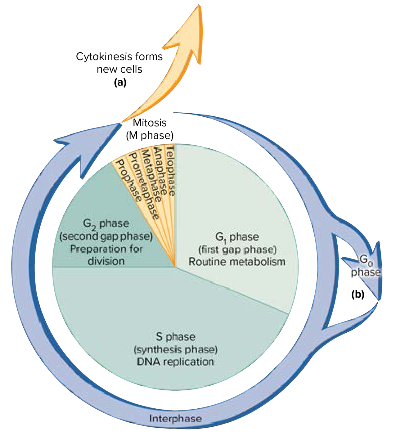
Interphase forms the cell formation up until cell division. Nearly all cell life is spent during interphase— during this phase, the cell carries out metabolic activities. There are three subphases to interphase.
G1: Growth phase with little cell division related activities
Carries out routine metabolic activities.
S: Synthetic phase— DNA replication occurs (synthesized).
DNA replication is when two DNA molecules separate, serving as the template strand for making new nucleotides.
One strand is continuous: called the leading strand, while the other is called the lagging strand
Each cell in the body (aside from sex cells) contains a specific number of chromosomes— diploid number
Diploid number in humans is 46
Sex cells have haploid number, 23, which is half of diploid number
G2: Brief growth period for synthesizing proteins necessary for division. Cell prepares for cell division.
Very brief
Cell division is necessary for tissue repair and growth. In cell division, a parent cell divides to form into two daughter cells, each having the same DNA as the parent cell. Cell division has two major events. Mitosis and cytokinesis.
Mitosis: division of chromosomes into new nuclei; has four phases
Daughter cells are identical to mother cell
After chromatids have been separated, daughter cells have received complementary chromosomes and are genetically identical
Cytokinesis: division of cytoplasm into new cells
Somatic (body) cells typically have 46 chromosomes (diploid), which exist in homologous pairs (23 chromosomes). The chromosome of each pair is inherited from one male parent and one female parent.
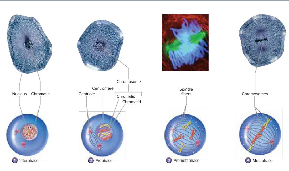
Interphase
Replication occurs. DNA organelles, and centrioles (microtubules) duplicate.
Prophase
Centrioles move to other ends of the cell
Chromatin becomes dense to make mitotic chromosomes
Each copy is a chromatid, attached to the centromere (center)
A protein called the kinetochore binds the centromere, providing a point of attachment for the microtubules to come and separate during mitosis
Sister chromatids are connected by the centromere
Nucleoli disappear
Centriole pairs are
Spindle fibers form and attach to kinetochore on chromosomes
Metaphase
Chromosomes align to the center of the cell
 |
Anaphase
Centromeres split, chromatids separate
Sister chromatids are pulled to opposite ends of the cell by the kinetochore fibers contracting
Resulting in a V-shape
Shortest stage of mitosis
Telophase
Chromosome movement halts
Nuclear envelope forms around the chromosomes
Chromosomes uncoil back to chromatin
Mitotic phase is completed
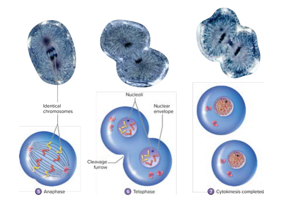 |
Cytokinesis is the division of the cell’s cytoplasm that begins and continues at the start of telophase (so, nearly at the end of the mitotic phase).
The first sign of cytokinesis comes from the formation of a cleavage furrow, an indentation of the plasma membrane that forms midway at the centrioles. Then, a contractile ring pulls the membrane inward forward and splits the cytoplasm into two.
Meiosis is a specialized type of cell division for gametes (reproductive sex cells) that occurs in the ovaries or testes. Remember how somatic cells are diploid (contain 46 chromosomes)? Gametes contain the haploid number of chromosomes (23).
🤔 Why is the reduced number of chromosomes important? This is because when a sperm cell and an oocyte fuse to form a fertilized egg, each have a haploid number of chromosomes. Both of them add together to create a diploid number of chromosomes in the fertilized egg. Excessive chromosomes can be lethal to offspring. So, 23 from the sperm cell plus 23 from the oocyte equals 46 chromosomes in the egg. |
The end result of meiosis are four daughter cells with half the number of chromosomes from the mother cell. There are two divisions of meiosis— meiosis I and meiosis II.
Prophase I
46 duplicate chromosomes (2 sister chromatids) connected by the centromere.
Chromosomes become visible
Nuclear membrane disappears
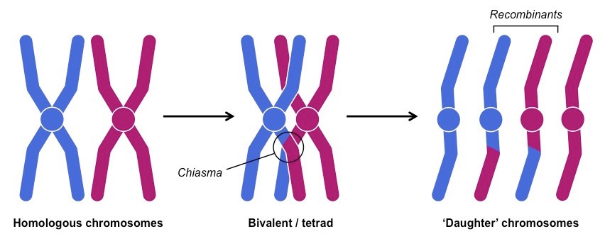
Homologous pairs come together in a process called synapsis— the homologous chromosomes form tetrads.
Part of the chromatid of one homologous chromosome breaks off and is exchanged with another chromatid. This is called crossing over, and results in a new genetic combination. Crossover points are called chiasmata.
 |
Metaphase I
Homologous pairs line up in the middle of the cell
However, alignment is random. Combined with crossing over, the genetic recombination is the cause of diversity of genetic composition in sperm cells and oocytes.
Anaphase I
Chromosomes of homologous pairs separate to opposite ends of the cell
Centromeres do not break, sister chromatids remain attached
Telophase I
New nuclei form
Meiosis I is completed
At the end of meiosis I, each daughter cell still has two chromatids. These chromatids separate in meiosis II.
Meiosis II is the same as mitosis, without interphase S, where chromosomes are duplicated. It contains the same stages, prophase, metaphase, anaphase, and telophase.

Cancer is the unchecked growth of genetically abnormal cells. Cancers are broken into different classifications— origin, characteristics, and prognosis or therapy.
Carcinoma
Originates from epithelial tissue
Glandular
Squamous
Melanocyte
Sarcoma
Originates from connective tissue
Cartilage
Bone
Fibrous connective
Meninges
Think: co- in connective with co- in sarcoma
It’s important to note that cancer will not occur in cells that do not replicate. For example, your muscle cells do not replicate. Therefore, there is no muscular cancer. |
Neoplasia is a term that refers to the increase of new cells. This can lead to the formation of tumors. There are two types of tumors— benign and malignant.
Benign Tumors
They resemble normal tumors, grow slowly, and do not invade other tissue
Malignant Tumors (Cancer)
These are poorly differentiated tissues that grow rapidly, destroy nearby tissue, and can metastasize (spread) to other parts of the body.
Dystrophy— a disorder that arises from abnormal cell size.
Hypertrophy is the increase of cell size.
Dysplasia— a disorder that arises from abnormal cell number.
Hyperplasia is the increase in the number of cells.
Aplasia is the decrease in the number of cells.
Both are normal during development
OK, what’s the difference between neoplasia and hyperplasia? Both refer to the increase of cell number. Neoplasia is usually used in the formation of tumors, while hyperplasia is benign and usually occurs in an organ or a tissue in response. |
Tumor mass
Size of the tumor indicates how advanced the cancer is
Lymph movement
Presence of cancer in nearby lymph nodes can indicate risk of metastasis
Metastasis
Spread of cancer to other parts of the body

Epidemiology is the study of disease— otherwise factors that lead to cancer. First, epidemiology focuses on risk factors.
Both host and environmental factors can lead to an increased risk of cancer.
Host Factors:
Age
Sex
Psychological Factors
Genetic Factors
Environmental/Lifestyle Factors:
Geographical location
Nutrition
Occupation (exposure to certain chemicals/pollutants)
Asbestos
Pesticides
Radiation
Cigarette smoking
Etiology of disease means that diseases are complex: oftentimes there is more than one single cause. It requires both:
DNA Damage
Inadequate physiological defense or repair
Cancer cells can form in numerous ways. Neoplastic transformation is the initiation of cancer cells.
Arise from a single cell
Cell suffers multiple transformation genetic mutations
Mutations are inherited or acquired
Acquired mutations
Random events during DNA replication
Induced by carcinogens (mutagens)
Initial DNA damage promotes further accumulation of future damage
Induce cell proliferation or growth (proto-oncogenes)
Inhibit growth of damaged cells (tumor suppressor genes)
There are multiple different ways of cancer treatment.
Surgery
First line of treatment
Resection of the tumor
Radiation therapy
X-rays or gamma rays are delivered to the tumor; kills cancer cells
Chemotherapy
Cytotoxic drugs damage the DNA of rapidly growing cancer cells
Bone Marrow Transplantation
Certain cancers may require high doses of radiation therapy or chemotherapy, which can be harmful to bone marrow
Biological response modifiers
Agents that boost the immune system response or antagonize tumor growth
Gene therapy
Modifies gene function to repair DNA or inhibit tumor growth

Tissue, as you may now, are a group of closely associated cells performing a similar function.
Tissue Type |
Nervous Muscular Epithelial Connective |
Primary Function Information Processing Contraction to Generate Force (movement) Cover exposed surfaces Structure and support |
Cell Types Neurons Smooth Cardiac Skeletal Squamous Cuboidal Columnar Transitional Glandular Fibroblasts WBCs Mast cells Plasma Cells Macrophages Adipohytes |
Fibers Minimal Minimal Basement Membrane Collagen Reticular Elastic |
Fluids Nutrient-rich, aqueous Minimal Limited Depends on the type of Connective Tissue |
Nervous tissues are responsible for the chemical and electrical transmission of information.
FALL SEMESTER 2024
Instructor: Anthony Uzwiak
uzwiak@dls.rutgers.edu
These notes dig deeper into the function of the nucleus and cell cycle.
Describe the structure and function of the nucleus and nuclear membrane
Describe the organization of the DNA molecule
Describe the stages of the cell cycle
Describe the differences between mitosis and meiosis
Describe the stages of mitosis and meiosis
Describe and classify the different types of neoplasia
Describe neoplastic transformation
Describe the epidemiology and etiology of cancer
Again—
Nucleus: Contains genetic material— directs cell activity
Located near the center of the cell
Consists of nucleoplasm surrounded by a nuclear envelope.
Nuclear envelope is composed of two membranes separated by a space
Nuclear envelope has nuclear pores, openings where molecules move between the nucleus and cytoplasm
Perinuclear cisterna is the liquid between membranes
Deoxyribonucleic acid (DNA) is mostly found within the nucleus
Most cells have a single nucleus— large cells are multinucleated. The red blood cell is the only cell that lacks a nucleus. |
Nucleolus is a dense region within the nucleus. Subunits for ribosomes are made in the nucleolus.
DNA are organized into chromosomes
Chromatin is composed of DNA and histones to form chromosomes
Nucleosomes are structural units of chromosomes, consisting of DNA wrapped around protein called histones
Cannot leave the nucleus— DNA directs protein synthesis by ribonucleic acid (RNA) which leaves nucleus through nuclear pores
The cell cycle, put simply, includes the changes a cell undergoes from the time it is formed until it splits into two cells. There are two major phases of a cell’s life— interphase and cell division (mitosis).
Interphase forms the cell formation up until cell division. Nearly all cell life is spent during interphase— during this phase, the cell carries out metabolic activities. There are three subphases to interphase.
G1: Growth phase with little cell division related activities
Carries out routine metabolic activities.
S: Synthetic phase— DNA replication occurs (synthesized).
DNA replication is when two DNA molecules separate, serving as the template strand for making new nucleotides.
One strand is continuous: called the leading strand, while the other is called the lagging strand
Each cell in the body (aside from sex cells) contains a specific number of chromosomes— diploid number
Diploid number in humans is 46
Sex cells have haploid number, 23, which is half of diploid number
G2: Brief growth period for synthesizing proteins necessary for division. Cell prepares for cell division.
Very brief
Cell division is necessary for tissue repair and growth. In cell division, a parent cell divides to form into two daughter cells, each having the same DNA as the parent cell. Cell division has two major events. Mitosis and cytokinesis.
Mitosis: division of chromosomes into new nuclei; has four phases
Daughter cells are identical to mother cell
After chromatids have been separated, daughter cells have received complementary chromosomes and are genetically identical
Cytokinesis: division of cytoplasm into new cells
Somatic (body) cells typically have 46 chromosomes (diploid), which exist in homologous pairs (23 chromosomes). The chromosome of each pair is inherited from one male parent and one female parent.
Interphase
Replication occurs. DNA organelles, and centrioles (microtubules) duplicate.
Prophase
Centrioles move to other ends of the cell
Chromatin becomes dense to make mitotic chromosomes
Each copy is a chromatid, attached to the centromere (center)
A protein called the kinetochore binds the centromere, providing a point of attachment for the microtubules to come and separate during mitosis
Sister chromatids are connected by the centromere
Nucleoli disappear
Centriole pairs are
Spindle fibers form and attach to kinetochore on chromosomes
Metaphase
Chromosomes align to the center of the cell
|
Anaphase
Centromeres split, chromatids separate
Sister chromatids are pulled to opposite ends of the cell by the kinetochore fibers contracting
Resulting in a V-shape
Shortest stage of mitosis
Telophase
Chromosome movement halts
Nuclear envelope forms around the chromosomes
Chromosomes uncoil back to chromatin
Mitotic phase is completed
|
Cytokinesis is the division of the cell’s cytoplasm that begins and continues at the start of telophase (so, nearly at the end of the mitotic phase).
The first sign of cytokinesis comes from the formation of a cleavage furrow, an indentation of the plasma membrane that forms midway at the centrioles. Then, a contractile ring pulls the membrane inward forward and splits the cytoplasm into two.
Meiosis is a specialized type of cell division for gametes (reproductive sex cells) that occurs in the ovaries or testes. Remember how somatic cells are diploid (contain 46 chromosomes)? Gametes contain the haploid number of chromosomes (23).
🤔 Why is the reduced number of chromosomes important? This is because when a sperm cell and an oocyte fuse to form a fertilized egg, each have a haploid number of chromosomes. Both of them add together to create a diploid number of chromosomes in the fertilized egg. Excessive chromosomes can be lethal to offspring. So, 23 from the sperm cell plus 23 from the oocyte equals 46 chromosomes in the egg. |
The end result of meiosis are four daughter cells with half the number of chromosomes from the mother cell. There are two divisions of meiosis— meiosis I and meiosis II.
Prophase I
46 duplicate chromosomes (2 sister chromatids) connected by the centromere.
Chromosomes become visible
Nuclear membrane disappears
Homologous pairs come together in a process called synapsis— the homologous chromosomes form tetrads.
Part of the chromatid of one homologous chromosome breaks off and is exchanged with another chromatid. This is called crossing over, and results in a new genetic combination. Crossover points are called chiasmata.
|
Metaphase I
Homologous pairs line up in the middle of the cell
However, alignment is random. Combined with crossing over, the genetic recombination is the cause of diversity of genetic composition in sperm cells and oocytes.
Anaphase I
Chromosomes of homologous pairs separate to opposite ends of the cell
Centromeres do not break, sister chromatids remain attached
Telophase I
New nuclei form
Meiosis I is completed
At the end of meiosis I, each daughter cell still has two chromatids. These chromatids separate in meiosis II.
Meiosis II is the same as mitosis, without interphase S, where chromosomes are duplicated. It contains the same stages, prophase, metaphase, anaphase, and telophase.
Cancer is the unchecked growth of genetically abnormal cells. Cancers are broken into different classifications— origin, characteristics, and prognosis or therapy.
Carcinoma
Originates from epithelial tissue
Glandular
Squamous
Melanocyte
Sarcoma
Originates from connective tissue
Cartilage
Bone
Fibrous connective
Meninges
Think: co- in connective with co- in sarcoma
It’s important to note that cancer will not occur in cells that do not replicate. For example, your muscle cells do not replicate. Therefore, there is no muscular cancer. |
Neoplasia is a term that refers to the increase of new cells. This can lead to the formation of tumors. There are two types of tumors— benign and malignant.
Benign Tumors
They resemble normal tumors, grow slowly, and do not invade other tissue
Malignant Tumors (Cancer)
These are poorly differentiated tissues that grow rapidly, destroy nearby tissue, and can metastasize (spread) to other parts of the body.
Dystrophy— a disorder that arises from abnormal cell size.
Hypertrophy is the increase of cell size.
Dysplasia— a disorder that arises from abnormal cell number.
Hyperplasia is the increase in the number of cells.
Aplasia is the decrease in the number of cells.
Both are normal during development
OK, what’s the difference between neoplasia and hyperplasia? Both refer to the increase of cell number. Neoplasia is usually used in the formation of tumors, while hyperplasia is benign and usually occurs in an organ or a tissue in response. |
Tumor mass
Size of the tumor indicates how advanced the cancer is
Lymph movement
Presence of cancer in nearby lymph nodes can indicate risk of metastasis
Metastasis
Spread of cancer to other parts of the body
Epidemiology is the study of disease— otherwise factors that lead to cancer. First, epidemiology focuses on risk factors.
Both host and environmental factors can lead to an increased risk of cancer.
Host Factors:
Age
Sex
Psychological Factors
Genetic Factors
Environmental/Lifestyle Factors:
Geographical location
Nutrition
Occupation (exposure to certain chemicals/pollutants)
Asbestos
Pesticides
Radiation
Cigarette smoking
Etiology of disease means that diseases are complex: oftentimes there is more than one single cause. It requires both:
DNA Damage
Inadequate physiological defense or repair
Cancer cells can form in numerous ways. Neoplastic transformation is the initiation of cancer cells.
Arise from a single cell
Cell suffers multiple transformation genetic mutations
Mutations are inherited or acquired
Acquired mutations
Random events during DNA replication
Induced by carcinogens (mutagens)
Initial DNA damage promotes further accumulation of future damage
Induce cell proliferation or growth (proto-oncogenes)
Inhibit growth of damaged cells (tumor suppressor genes)
There are multiple different ways of cancer treatment.
Surgery
First line of treatment
Resection of the tumor
Radiation therapy
X-rays or gamma rays are delivered to the tumor; kills cancer cells
Chemotherapy
Cytotoxic drugs damage the DNA of rapidly growing cancer cells
Bone Marrow Transplantation
Certain cancers may require high doses of radiation therapy or chemotherapy, which can be harmful to bone marrow
Biological response modifiers
Agents that boost the immune system response or antagonize tumor growth
Gene therapy
Modifies gene function to repair DNA or inhibit tumor growth
Tissue, as you may now, are a group of closely associated cells performing a similar function.
Tissue Type |
Nervous Muscular Epithelial Connective |
Primary Function Information Processing Contraction to Generate Force (movement) Cover exposed surfaces Structure and support |
Cell Types Neurons Smooth Cardiac Skeletal Squamous Cuboidal Columnar Transitional Glandular Fibroblasts WBCs Mast cells Plasma Cells Macrophages Adipohytes |
Fibers Minimal Minimal Basement Membrane Collagen Reticular Elastic |
Fluids Nutrient-rich, aqueous Minimal Limited Depends on the type of Connective Tissue |
Nervous tissues are responsible for the chemical and electrical transmission of information.

