Chapter 26 Disorders of Blood Flow and Blood Pressure Regulation
Chapter 26: Disorders of Blood Flow and Blood Pressure Regulation
Dr. Kelly Goebel DNP, ACNP-BC
Page 1: Overview
Focus on disorders affecting blood flow and blood pressure regulation.
Page 2: Vascular Structure
Components of Blood Vessels:
Collagen and internal elastic fibers
Smooth muscle cells
Connective tissue
Endothelial cells
Lumen
Tunica externa, tunica media, tunica intima
Page 3: Structure and Function
Endothelium→ single layer of cells
Controls vascular function, platelet adhesion, blood clotting, blood flow modulation, immune regulation.
Dysfunction caused by inflammation, hemodynamic stress, lipid molecules, hypoxia.
Releases cytokines and growth factors, influencing nitric oxide (relaxing) and endothelin (contracting).
Vascular Smooth Muscle Cells (SMCs):
Located in tunica media, respond to hormonal and neural stimulation to constrict/dilate blood vessels.
Page 4: Function of Blood Vessels
Arterioles:
Narrow lumen, regulates blood pressure via vasoconstriction/dilation.
Arteries:
Transport blood from heart to organs; no exchange of substances.
Capillaries:
Site of exchange for fluids, gases, nutrients; highly permeable.
Veins:
Transport blood back to heart; larger diameter, valvular structures prevent backflow.
Venules:
Collect fluid from capillaries; larger than capillaries, valves present.
Page 5: Blood Pressure Dynamics
Systole: - contraction
Blood pressure rises during left ventricle contraction.
Diastole:- Relaxation
Blood pressure falls as the heart relaxes.
Page 6: Regulation of Systemic Arterial Blood Pressure
Systolic Pressure: < 120 mm Hg
Diastolic Pressure: < 80 mm Hg
pulse pressure= systolic- diastolic
Mean Arterial Pressure (MAP):
Average pressure during ventricular contraction/relaxation; indicator of tissue perfusion (90-100 mm Hg).
Page 7: Mechanisms of Blood Pressure Regulation
Key Factors:
Cardiac output, peripheral vascular resistance, sympathetic activity, stroke volume, heart rate.
Hormonal Influences:
Angiotensin II, aldosterone, and the renin-angiotensin mechanism.
Page 8: Neural Regulation
Vagal Nerve Stimulation: Causes bradycardia. (rectal pressure stimulates vagal never)
Sympathetic Stimulation: Causes tachycardia.
Baroreceptors: Pressure-sensitive receptors (in the walls of blood vessels) affecting heart rate and vascular tone.→ contraction and heart rate increase
Page 9: Hormonal Regulation
Renin-Angiotensin-Aldosterone System (RAAS):
Involves angiotensinogen, renin, angiotensin I, and II, leading to vasoconstriction and sodium reabsorption→ increasing BP
Page 10: Systolic Blood Pressure Factors
Reflects:
Blood volume ejected, rate and force of ejection, elasticity of arteries.
Stroke volume = Volume of blood ejected from the ventricles with each beat
Cardiac output= SVx HR
Page 11: Diastolic Blood Pressure Factors
Reflects:
Closure of aortic valve, energy stored in elastic fibers, resistance to flow through arterioles.
Page 12: Disorders of Systemic Arterial Blood Flow
Impact of Arterial Disease:
Impairs blood flow, leading to ischemia (insufficient oxygen) and infarction (necrosis due to occlusion) → dead tissue within a living organism
Page 13: Dyslipidemia
Definition: Imbalance of lipid components in blood.
Types:
Hyperlipidemia(cholesterol, triglycerides, phospholipids), hypercholesterolemia, hypertriglyceridemia.
Lipoprotein Classifications:
VLDL, LDL (atherogenic)→ carry to the tissues, HDL (protective)→ remove to the tissue and take it to the liver
Page 14: Risk Factors for Dyslipidemia
Primary: Genetic basis (familial hypercholesterolemia).
Secondary: Dietary factors, obesity, Type II Diabetes, metabolic syndrome ( high glucose, BP, waist circunfe)
Page 15: Atherosclerosis
Definition: Formation of fibrofatty lesions in arteries.
Major Risk Factor: Hypercholesterolemia.
Traditional and Modifiable Risk Factors: Smoking, obesity, hypertension, diabetes.
screening risk: elevated CRP levels (c reactive protein -levels of inflammation)
Page 16: Pathogenesis of Atherosclerosis
Lesions:
Fatty streaks, fibrous atheromatous plaque(accumulation of lipid, proliferation of vascular smooth muscle cells, formation of scar tissue), complicated lesions, gets bigger→hemorrhage, ulceration)
Impact on Artery Lumen: Decreases as lesions grow.
Page 17-20: Development of Atherosclerosis
Stages:
Endothelial injury→ monocytes/ platelets adhere
inflammatory cell migration→monocytes into macrophages.
lipid accumulation→ Foam cells accumulation (macrophages that engulfs lipoproteins), necrotic debri in celular wall
plaque structure formation→ aggregation of macrophages, leukocytes, collagen→ formation of fibrous cap
Page 21: Clinical Manifestations of Atherosclerosis
Effects:
Ischemia, vessel obstruction, thrombosis, aneurysm formation(vessel weakening).
Commonly Affected Areas: Heart, brain, kidneys, lower extremities.
Page 22-24: Aneurysms
Definition: Abnormal localized dilation of blood vessels, most common in the aorta.
Etiology: Congenital, trauma, infection, atherosclerosis, elevated lipids, hypertension, smoking
Clinical Manifestations: Vary by size and location.
Thoracic
Abdominal (pulsating mass,
Classification: Ascending, arch, descending, thoracoabdominal, abdominal.
Page 25-26: Aortic Dissection
hemorrhage into vessel wall w/ tearing
Etiology: Hypertension, degeneration of medial layer of vessel wall
Clinical Manifestations of Dissection: Severe chest pain(tearing or ripping), elevated BP(will become unobtainable in one or both arms as the dissection disrupts arterial flow to arms) , potential for syncope,paralysis, and heart failure.
Page 27-30: Acute Arterial Occlusion
Etiology: Sudden interruption of arterial flow due to
embolus(is a thrombus piece that can travel in the body)→atrial fibrillation can cause emboli and make it travel in the body, ischemic heart; Emboli can lodge in major arteries
thrombus
Clinical Manifestations(6Ps): sudden onset of Pallor, pain, pulselessness, paresthesia(numbness, tingling) , paralysis, cold extremities.
Atherosclerotic Occlusive Disease (chronic occlusive arterial)
Common in lower extremity (femoral/popliteal)
Etiology:
advanced age
smoking
Manifestation
gradual
claudication (pain w/ walking) Calf pain
weak pedal pulses
ischemic pain at rest, ulceration, gangrene, tissue necrosis
Page 31-34: Disorders of Systemic Venous Circulation
Thromboembolism: Presence of thrombus in veins+inflammation,
Etiology - Virchow’s Triad
stasis of blood
hypercoagulability- Factor V Leiden
vessel wall injury
Manifestation- Pain, swelling(calf, posterior tibial), deep muscle tenderness, sometimes asymptomatic
Chronic Venous Insufficiency: Persistent venous hypertension leading to tissue congestion:edema and skin changes(brown pigmentation, stasis dermatitis →thin , shinny, bluish brown, irregularly pigment skin)
Etiology: prolonged standing, incompetent valves, DVT, inflammation, endothelial dysfunction.
Stages→ spider veins, varicose, swelling, skin changes, venous ulcer
Page 35-40: Disorders of Blood Pressure Regulation
Importance of Regulation: Ensures adequate tissue perfusion and prevents vessel damage.
Low BP→ don’t receive suff blood- don’t receive nutrients, oxygen and cell wate removal - hypoxic ischemia
HIgh BP- damage endothelial tissue, risk o atheosclerotic vascular disease and vascular rupture
Hypertension:
Primary: No specific cause; risk factors include genetics, age, gender, race, family history. modifiable- dietary, tobacco, obesity, diabetes
Secondary: Caused by medical conditions (e.g., renal failure). cocaine
Clinical Consequences: increase workload in LV hyperplasia(bc the the vessel is too vasoconstricted)
Blindness, chronic kidney disease, stroke, angina(chest pain), AMI,
Target organ damage, including heart, brain, kidneys, and vision.
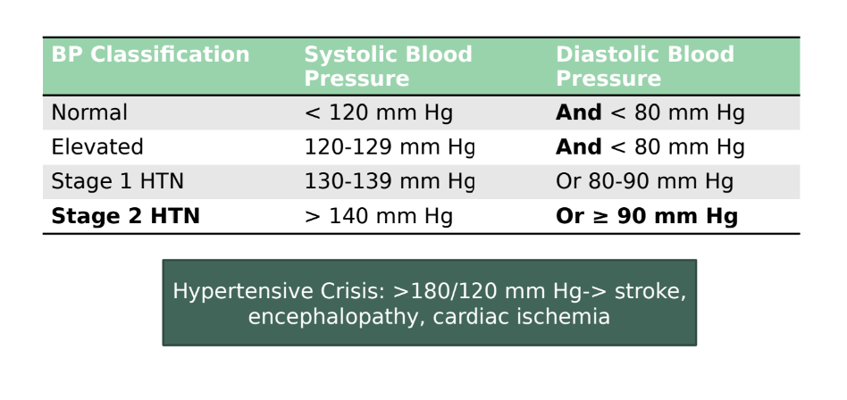
Classification Guidelines: Based on systolic and diastolic blood pressure readings.