Copy of Final Exam Topics List 2026
Final Exam Topics List 2026
Human Biology: Human Anatomy and Physiology
CH: 1 Human body orientation
Define the following organization of the human body
- Cell - the smallest units of living things
- Tissue - groups of similar cells that have a common function
- Epithelial: covers body surfaces and lines its cavities
- Muscle: provides movement
- Connective: supports and protects the body
- Nervous: communication by transmitting electrical impulses
- Organ - discrete structure composed of at least two tissue types and performs a specific function
- Organ system - Cells, Tissue, Organs, Systems
Define the following vocabulary words
- Metabolism - all chemical reactions that occur within body cells
- Catabolism - breaking down substances into simpler building blocks
- Anabolism - synthesizing complex substances from simpler building blocks
- Homeostasis - ability to maintain relatively stable internal conditions
CH: 2 Biochemistry
State why carbon makes up major compounds of life
- It can form four strong bonds; make long chains and rings, which are needed for things like DNA, proteins, and sugars.
- Its bonds are stable, so living organisms can rely on them.
Define the following vocabulary words
- Hydrophobic - water fearing
- Hydrophilic - water loving
- Peptide bond - bond between two amino acids by a dehydration reaction (poly = 10 or more)
List the characteristics of DNA
- Location:In the nucleus
- Function: genetic material, directs protein synthesis, replicates before cell division
- Structure: double strand coiled into a double helix
- Deoxyribose
- AGCT (AT & GC)
List the characteristics of RNA
- Location: Cytoplasm
- Function: Carries genetic instructions to make proteins
- Structure: Single strand either straight or folded
- Ribose
- AGCU (AU & GC)
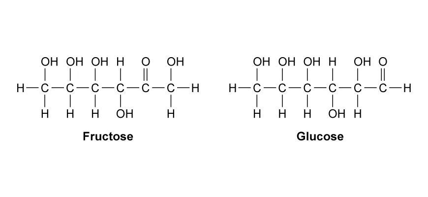
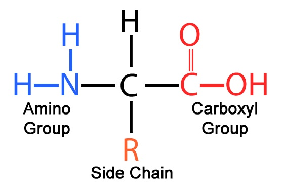
Determine the type of molecule (monosaccharide, fatty acid, or amino acid) from the molecular shape (picture)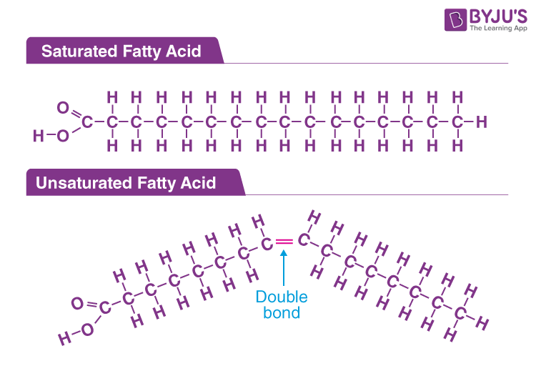
CH: 3 Cells
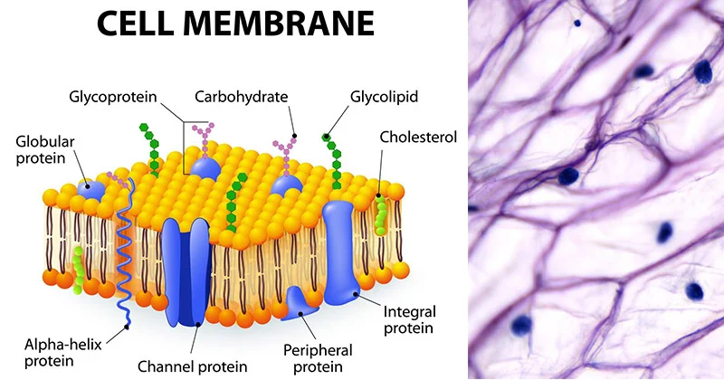
Label the structures of the plasma membrane
State the function of the following cell organelles
- Plasma membrane - separates the intracellular fluid within the cell and the extracellular fluid outside the cell
- Nucleus - control center of the cell, contains instructions to make all the body’s proteins and dictates the kinds and amounts of proteins to be made
- Cytoplasm -cellular material between the plasma membrane and the nucleus and is the site of most cellular activities
- Mitochondria - Produce ATP
CH: 4 Tissues
State the functions of the types of tissue
- Epithelial: covers
- Muscular: supports
- Connective: moves
- Nervous: controls
State the difference between voluntary and involuntary muscles
- Voluntary muscle: movement under conscious control (skeletal muscle)
- Involuntary muscle: movement not consciously controlled (cardiac and smooth muscle)
CH: 5 Integumentary system
State and define the two layers in the dermis
- Papillary - thin, superficial layer with fine interlacing collagen and elastic fibers that form a loosely woven mat with small blood vessels
- Reticular - deeper, coarse, dense irregular connective tissue
State the characteristics of the following sweat glands
- Eccrine - sweat glands (merocrine sweat glands)
- simple, coiled, tubular gland,
- Most numerous type of gland
- Abundant in palms, feet, and forehead
- Produces sweat (99% water, with some salts, and wastes)
- Apocrine - lies deeper in dermis and ducts empty into hair follicles
- Same components of sweat with added fatty substances and proteins
- Odorless, but when bacteria on the skin decomposes it is smells unpleasant (body odor)
- Sebaceous glands - (oil glands): found all over the body except palms and feet
- Sebum: oily substance that softens and lubricates the hair and skin and prevents water loss and is a bactericidal (kills bacteria)
- Develop as outgrowths of hair follicles or pores on the skin surface
List the functions of the skin
- Protection
- Sensation
- Absorption
- Heat regulation
- Exerection
- Secreation
CH: 6 Skeletal system
State the characteristic and examples of the following bone shapes
- Long - longer than they are wide (femur)
- Short - cube shaped (carpals)
- Flat - very flat & slight curve (ribs)
- Irregular - pelvis, vertebrae
- Compact - Dense outer layer
- Spongy - Internal layer
State the functions of the skeletal system
- Support
- Protection
- Anchorage
- Blood cell formation
- Mineral storage
- Fat storage
- Hormone Production
State the parts of the axial skeleton
- Skull
- Vertebral columns
- Thoracic cage
Describe the following bone disorders and prevention
- Osteomalacia - number of disorders in which the bones are poorly mineralized
- Bones are soft and weak
- Pain when weight is put on affected bones
- Caused by insufficient calcium in their diet or a vitamin D deficiency
- Increasing vitamin D by having exposure to sunlight can help cure the condition
- Rickets - similar to osteomalacia but in children
- Bones are still growing in children and it can be more severe
- Bowed legs and deformities of the pelvis, skull, and ribcage
- Caused by insufficient calcium in their diet or a vitamin D deficiency
- Increasing vitamin D by having exposure to sunlight can help cure the condition
- Osteoporosis - group of diseases in which bone resorption outpaces bone deposit
- Composition of the matrix remains normal but bone mass declines and bones become porous and light
- Bones become fragile and break very easily
- Risk Factors
- Decreased sex hormones: decrease in estrogen in older women
- Insufficient bone stress: low body weight or insufficient weight-bearing exercise slows bone deposit
- Diet poor in calcium, vitamin D, or protein
- Smoking: reduces estrogen and calcium absorption
- Genetics: runs in families
- Hormone-related conditions: diabetes or hyperthyroidism
- Alcohol and certain medications
- Preventing and treating: minimizing the risk factors and certain drugs
CH: 7 Muscular system
State characteristics of each of the three types of muscles
- Skeletal - voluntary and striated
- Cardiac - involuntary and striated
- Smooth - involuntary and nonstriated
State the overall result/function of the sliding filament model of contraction
- The sliding filament model explains how muscles produce force and shorten. It describes how actin (thin) filaments slide past myosin (thick) filaments, causing the sarcomere—the basic unit of a muscle fiber—to shorten. When many sarcomeres shorten together, the entire muscle contracts.
Define the following vocabulary words
- Sarcomere - smallest unit of a muscle fiber - region of a myofibril between 2 z discs
- Myosin - thick filament, rodlike protein
- Actin - thin filament, double strand of pearls
- Z disc - boundary of sarcomere
- Cross bridge - actin/myosin binding
Joints
Define and give an example of the following types of joints
- Fixed - A fixed joint is a joint that allows no movement. The bones are tightly connected by fibrous tissue.
- Example: structures in the skull
- Semimovable - A semimovable joint allows limited movement. Bones are connected by cartilage.
- Example: Joints between the vertebrae in the spine
- Synovial - A synovial joint is a freely movable joint that contains synovial fluid to reduce friction.
- Example: Knee joint
- Pivot - A pivot joint allows one bone to rotate around another.
- Example: shake your head no
- Hinge - A hinge joint allows movement in one direction (back and forth), like a door hinge.
- Example: Elbow joint
- Ball and socket - A ball and socket joint allows movement in many directions, including rotation.
- Example: Shoulder or hip joint
CH: 8 Nervous system
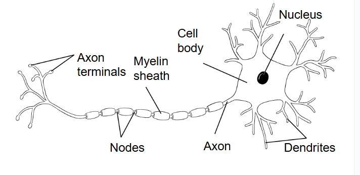
Label a neuron cell and state the function of each part
Cell body - contains nucleus and other organelles
Dendrites-to receive signals from other neurons and transmit
those signals to the cell body of the neuron
Axon- transmits the signal away from the cell body
Axon terminal- has branches and synaptic terminal buttons
Myelin sheath- covers the axon and insulates the axon to speed up
transmission of the signal
Nodes- serves to facilitate the rapid conduction of nerve impulses.
State the function of neurotransmitters - chemicals released by the synaptic terminal buttons across the cleft to sign the next neuron
State the divisions and the functions of the peripheral nervous system
- Somatic nervous system controls voluntary movements of skeletal muscles
State the function for the following parts of the brain: cerebrum, cerebral cortex, medulla oblongata, and cerebellum. -
- Cerebrum- largest portion of the human brain for thinking, judgement, and processing the senses
- Cerebral cortex- reasoning, language, complex thought, visual processing
- Thalamus- directs incoming sensory signals to correct region of the cerebral cortex
- Hypothalamus- maintains homeostasis by coordinating the nervous and the endocrine systems
- Medulla oblongata- swallowing center that coordinates the muscles of the mouth, so food can go down, controls breathing
- Cerebellum- coordination of muscle, posture and body position
List the sensory receptors, their function, and their stimuli.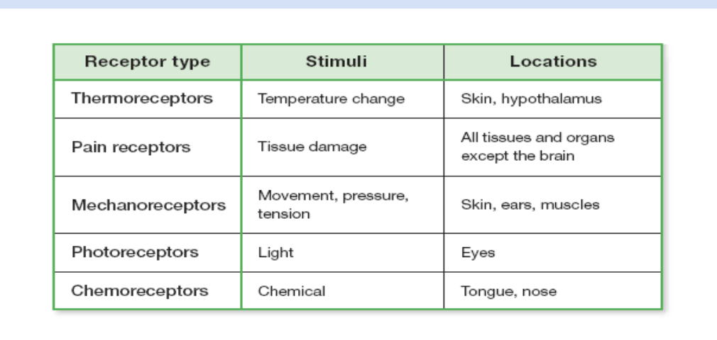
CH: 9 Cardiovascular System and Blood
State the functions of blood cells
- Red blood cells: Carry oxygen and carbon dioxide throughout the body.
- White blood cells: Fight infection and protect the body from disease.
- Platelets: Help blood clot to stop bleeding.
State the characteristics of arteries, veins, capillaries
- Arteries – Thick, muscular walls; carry blood away from the heart under high pressure.
- Veins – Have valves to prevent backflow; carry blood toward the heart under lower pressure.
- Capillaries – Walls are only one cell thick; site where oxygen, nutrients, and wastes are exchanged with tissues.
Blood pressure measurement: Diastolic and systolic pressure
- Systolic pressure- pressure of blood when ventricles contract usually 110-120 mmHg
- Diastolic pressure- pressure when ventricles relax usually 70-80mmHg
Blood types: antigens and antibodies
- Type A – A antigen, anti-B antibodies
- Type B – B antigen, anti-A antibodies

- Type AB – A and B antigens, no antibodies
- Type O – no antigens, anti-A and anti-B antibodies
Effect of epinephrine on the heart
- Adrenaline increases heart rate and force of contraction, so the heart pumps more blood per minute.
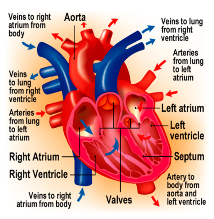
Label a diagram of the heart
Define
- Pacemaker (SA node)
- A group of cells in the right atrium that sets the heart’s rhythm by generating electrical impulses.
- Purkinje fibers
- Specialized fibers that quickly spread electrical signals through the ventricles, causing them to contract.
- Pulmonary circulation
- Movement of blood from the heart to the lungs and back to the heart for oxygenation.
- Systemic circulation
- Movement of blood from the heart to the rest of the body and back to the heart.
CH: 10 Respiratory System
Function of type I and type II pneumocytes
- Type I pneumocytes – Very thin cells with large surface area for gas exchange; adapted for rapid diffusion of gases; cannot undergo mitosis for repair.
- Type II pneumocytes – Cuboidal cells that produce surfactant to reduce surface tension and prevent alveolar collapse; can divide by mitosis to replace damaged alveolar cells.
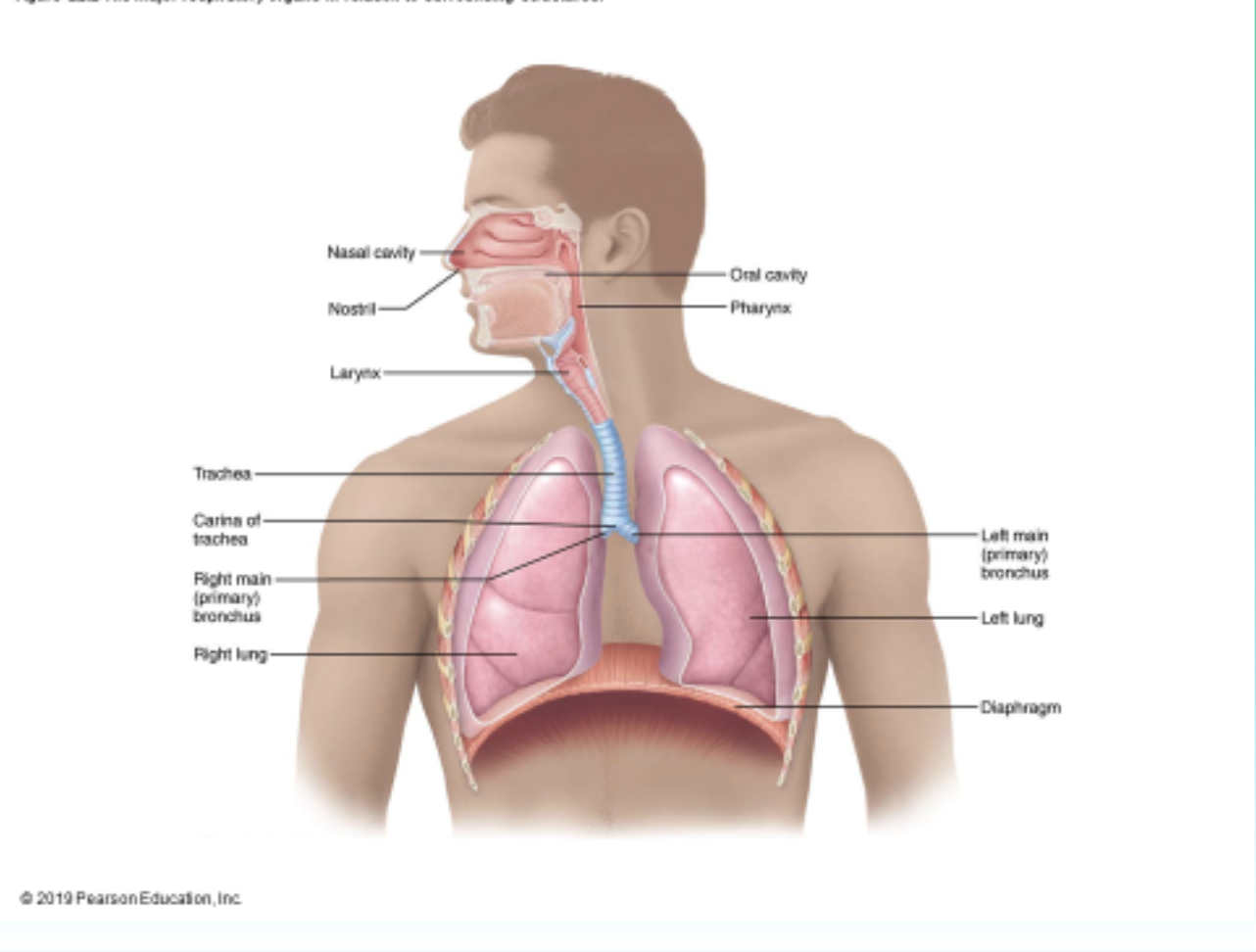
Label the structures of the respiratory system
State the function of the structures of the respiratory system
- Lungs: site of gas exchange between atmosphere and
blood and located in thoracic (thorax) cavity (closed to the outside air)
- Mouth and nose: air enters
- Pharynx: tube at the back of the nasal cavities and mouth and
is passageway for air and food
- Epiglottis: flap of cartilage that covers the opening to air passage
- Trachea: air passageway made of cartilage which contain cilia and mucus to trap particles
- Larynx: upper end of trachea that contains the vocal cords
- Bronchi: two branches that lead to the lungs from trachea
- Bronchioles: smaller tubes into the lungs
- Alveoli: clusters of tiny air sacs surrounded by capillaries
Explain the process of inhaling
- Inspiration- inhaling is the process of taking air into the lungs
Explain the process of exhaling
- Expiration- exhaling, a passive process of expelling air from lungs
State the changes to pressure and volume during inhaling
- As volume increases, pressure decreases.
State the characteristics of the following lung diseases: lung cancer and emphysema
Body adaptations at high altitudes
- Increase ventilation rate, Increase lung size, Increase number of capillaries, produce more red blood cells / hemoglobin
- By breathing faster and deeper or by producing more red blood cells to carry extra oxygen.
State the respiratory control center in the brain
- The respiratory control center is located in the medulla oblongata of the brainstem.
CH: 11 Digestive System and Nutrition
Define
- Digestion - an enzyme-facilitated chemical process. (series of chemical reactions, whereby the ingested food is converted into smaller and smaller molecular forms)
- Absorption - small molecular forms are absorbed through the cells of the digestive system and pass into the blood vessels
- Peristalsis - a series of rhythmic muscular contractions that move a bolus through the esophagus
State the order of the digestive system organs that food travels through
- Mouth
- Pharynx
- Esophagus
- Stomach
- Small intestine
- Large intestine (colon)
- Rectum
- Anus
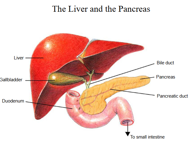
Label a diagram of the digestive system
State the function of the digestive system organs
- Liver- a large organ located to the right of the stomach
- Pancreas- an organ that lies behind the stomach
- Gallbladder- stores bile and releases it into the small intestine
- Mouth: Digestion begins here with chewing (mastication) and saliva secretion, which contains enzymes like amylase to start breaking down carbohydrates.
- Pharynx (throat): Serves as a passageway for food from the mouth to the esophagus.
- Esophagus: A muscular tube that moves food to the stomach using peristalsis.
- Stomach: A muscular organ that mixes food with gastric juices, breaking it down into a semi-liquid form called chyme.
- Small Intestine: The longest part of the GI tract, about 22 feet long, divided into the duodenum, jejunum, and ileum. It is the primary site for nutrient absorption.
- Large Intestine: Includes the cecum, colon, rectum, and anus. It absorbs water and electrolytes and stores waste before elimination.
State the function of hydrochloric acid in the stomach
- Lower pH to about 2
- Activates pepsin to digest proteins.
- Kills harmful bacteria in food
Function of the hormones of leptin and gastrin
State the function of the villi of the small intestine and identify it by a picture
- The small intestine is adapted for absorption through its structure. The mucosa forms the inner lining and contains many folds with villi, which increase surface area for absorption. Each villus has microvilli, which further increase surface area and speed up absorption. Inside the villi are blood vessels that absorb nutrients like glucose and amino acids, and a lacteal that absorbs fats. The basal labyrinth helps move absorbed substances into the blood and lymph, and pinocytosis allows cells to take in small droplets of nutrients. Together, these features make absorption very efficient.
State the cause of scurvy and rickets
- Vitamin C - Important for protection against infections, helping in wound healing, maintaining healthy gums, teeth, bones, muscle contractions and blood vessels.
- A lack of vitamin C causes Scurvy.
- Vitamin D - (sun, and milk) proper formation of bones
- Children: rickets
- Lead to irregular, thick, and wide bone growth
CH: 12 Urinary System and Liver
Label a diagram of a kidney
State the functions of the different sections of the kidney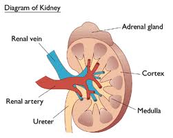
- Renal cortex
- Outer layer
- Filters blood to form urine (first step)
- Renal medulla
- Inner layer
- Reabsorbs water and salts to concentrate urine
- Renal pelvis
- Collects urine
- Sends it to the ureter
- Ureter
- Carries urine to the bladder
ADH function and structure that it targets
- ADH controls how much water is reabsorbed in the collecting duct of the kidney.
- Collecting ducts (and distal convoluted tubules)
Components of the filtrate
- fluid that is filtered from the glomerulus passes through the basement membrane; prevents large molecules like proteins from becoming part of the filtrate
State the substances that are reabsorbed in the proximal convoluted tubule
- Glucose
- Amino acids
- Most mineral ions (salts like sodium and chloride)
- Most water
- Some urea (small amount)
List the structures that urine flows through
- Kidney → collecting ducts → pelvis → ureter → bladder → urethra
State the function of bile
- Emulsifies fats in the small intestine, breaking large fat droplets into smaller ones to increase surface area for digestion by lipase.
State the functions of the liver
- Regulates blood glucose by converting excess glucose into glycogen and storing it
- Stores nutrients such as glycogen, vitamins (A, D, B12), and iron
- Detoxifies the blood, breaking down harmful substances like alcohol and drugs
- Produces bile, which helps break down fats during digestion
- Deaminates amino acids, removing excess nitrogen and converting it into urea for excretion
State the nutrients that are stored in the liver
- Glycogen (stored form of glucose for energy)
- Vitamins (especially A, D, and B12)
- Iron (stored as ferritin for making hemoglobin)
Define
- Hepatocytes (liver cells) - function is to remove or add substances to the blood
- Kupffer cells - line the inside of sinusoids and use phagocytosis to remove old erythrocytes and bacteria from the blood
- a) Contain many lysosomes
- b) Specialized leukocytes (white blood cells)
- Infant jaundice - happens because a newborn’s liver is not mature enough to properly process bilirubin.
- This causes a yellowing of the skin and eyes due to buildup of bilirubin in the blood.
CH: 13 Immune System
Define
- Antibodies - protein molecules produced by plasma cell leukocytes in response to a specific pathogen
- a) Each antibody is different because it responds to a different pathogen
- b) Y-shaped protein
- Antigens - proteins embedded on the outer surface of the invader (bacteria), “not-self” proteins that trigger an immune response
- Invader
- Pathogen -any living organism or virus that is capable of causing a disease
- a) Include viruses, bacteria, protozoa, fungi, and worms
- b) Exposure to majority of pathogens does not result in disease since we are well defended
State that the skin is the first line of defense
State the difference between the primary and secondary immune response.
- Primary immune response - first encounter with a particular pathogen
- Takes a week or more to be successful and symptoms of the disease will be experienced as the immune system works to eliminate the pathogen
- Secondary immune response (include memory cells) - second or third encounter with a particular pathogen
- Quicker and more intense response so symptoms are rarely experienced
Explain how vaccines work and give pros and cons of vaccinations
- Vaccines expose the body to a safe part of a germ so the immune system learns to fight it. The body makes antibodies and memory cells, so if the real germ shows up later, it can respond quickly and prevent disease.
- Pro
- No die from bad disease
- Heard immunity (protect most of community)
- Con
- Side effects
- Child's body may not be able to handle
State the function of helper T cells and B cells
- Helper T cells
- Activate and coordinate the immune response.
- They release chemicals called cytokines that stimulate other immune cells, including B cells and killer T cells.
- B cells
- Produce antibodies that bind to specific antigens on pathogens.
- Some B cells become memory cells, allowing a faster response if the pathogen enters again.
State the chemical that is released during and allergic reaction and where that chemical is released from
- Allergen causes IgE antibodies to attach to the mast cell. Next time it binds and the mast cell releases histamines and they bind to blood vessels and make them release fluid.
Explain the blood clotting process and the four proteins that are involved
- When a blood vessel is damaged, platelets gather at the site and stick together to form a temporary plug. They also release chemicals that activate the clotting cascade.
- Prothrombin – inactive protein in plasma
- Thrombin – active enzyme formed from prothrombin
- Fibrinogen – soluble plasma protein
- Fibrin – insoluble fibers that form the clot
CH: 14 Endocrine System
Define
- Endocrine system - consists of numerous glands that produce a wide variety of hormones
- Hormones - chemical messengers that usually have a physiological effect far from the gland of origin and are transported by the blood
- Glands - structure that makes and secretes hormones
- Peptide hormones - composed of amino acids (proteins)
- Steroid hormones - made from cholesterol and classified as lipids
- Steroid hormones control the production of proteins within the target cell
State the function, gland of production, and target cells for the following hormones
- Thyroid hormone
- Thyroid - most body cells - Regulates metabolic rate & temperature
- ADH
- Hypothalamus posterior pituitary - kidney collecting ducts - reabsorb water from urine
- Oxytocin
- Hypothalamus posterior pituitary -uterus & mammary glands - contractions during labor and release of milk
- Growth hormone
- Anterior pituitary - liver, bones, muscles - tissue growth
- Glucagon
- Pancreas (alpha cells) - cells of liver and muscles - converts glycogen into glucose to increase blood sugar levels
- Insulin
- Pancreas (beta cells) - all body cells, liver, and muscle cells - converts glucose into glycogen to decrease blood sugar levels, body cells to take in glucose
- Prolactin
- Anterior pituitary - mammary gland - milk production
- Leptin
- Adipose fat tissue - hypothalamus - decrease appetite
CH: 15 Reproductive System
State the function, gland of production, and target
cells for the following hormones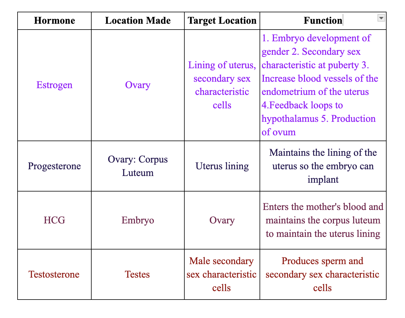
- Estrogen
- Progesterone
- Testosterone
- HCG
State the characteristics of sperm cells
- Spermatozoa
- flagellum (movement)
- Miitochondira for ATP
- Produced starting at puberty
- Small cell
State the characteristics of egg cells
- Ovum
- Many organelles
- Has yolk
- Stays in (doesn't move)
- Large cell
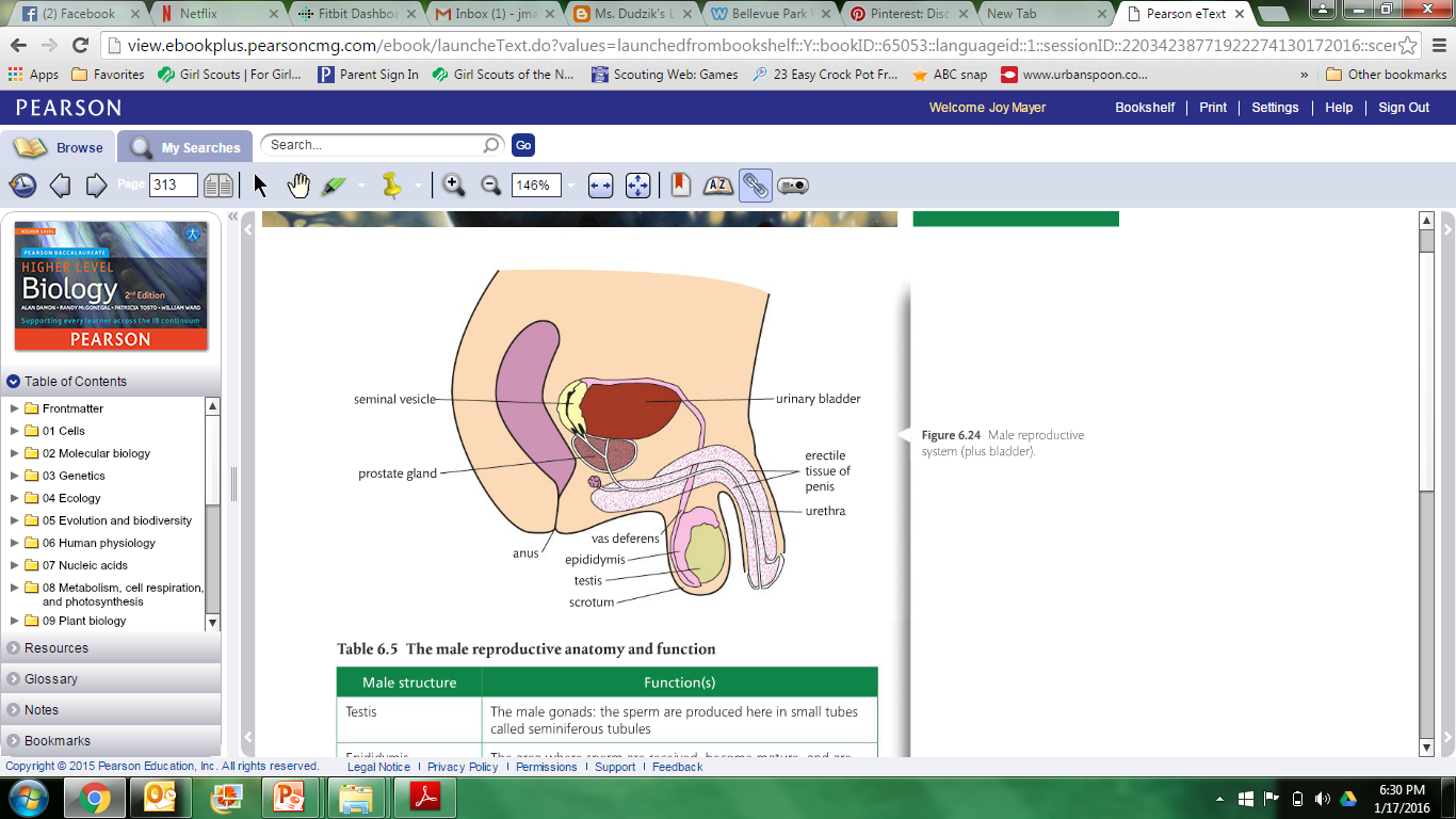
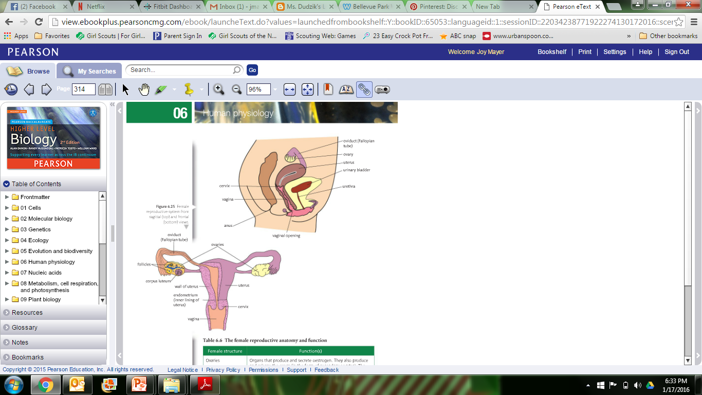
Label a diagram of the female and male reproductive systems
State the substances that pass from the mother to the fetus through the umbilical cord
- Oxygen
- Water
- Nutrients
- BAD - alcohol or medication
- FETUS TO MOTHER?
- Carbon dioxide, Urea, other metabolic wastes
State the function of spermatogenesis and oogenesis
- Spermatogenesis
- Meiosis I produces 2 cells
- Meiosis II produces 4 haploid spermatozoa cells
- Oogenesis
- Making eggs in the ovaries
- Before birth:
Cells (oogonia) start meiosis → become primary oocytes but pause. - At puberty (each cycle):
One primary oocyte finishes meiosis I → forms:
- Before birth:
- Making eggs in the ovaries
- Secondary oocyte (big cell)
- Polar body (small, not used)
- Ovulation: Secondary oocyte is released and pauses again (meiosis II).
- If fertilized: It finishes meiosis II → becomes a mature egg (ovum).
- If not fertilized: It breaks down and is lost during menstruation.
State the steps of fertilization
- Sperm reaches egg and attaches
- Acrosome reaction for the sperm to get into the egg
- Plasma membranes of sperm and egg fuse
- Cortical reaction: chemicals released that blocks all other sperm from entering
- Sperm’s nucleus and egg’s nucleus join to form the zygote
Define: Blastocyst
- Trophoblast: placenta
- Inner cell mass: embryo
- Fluid filled cavity
CH: 16 Pregnancy and Labor
Define
- Amnion - transparent membranous sac that surrounds the embryo
- Amniotic fluid “bag of water”
- Yolk sac - sac that hangs from the surface of the embryo and gives nutrients during week two and three
- Forms the digestive tube
- Source of earliest blood cells
- Germ cells that will form sperm and ova
- Allantois - base for the umbilical cord and eventually becomes the urinary bladder
- Chorion - outermost layer that helps to form the placenta and encloses the embryo and other membranes
- Gastrulation - process of forming the primary germ layers of the embryo
- Ectoderm - “outer skin” structures that become the nervous system and skin epidermis
- Endoderm - “inner skin” forms the linings of the digestive, respiratory, and urinary system and glands
- Mesoderm - “middle skin” forms everything else
List characteristics of first trimester
1. Head is still dominant, but body elongates
2. Eyes are present
3. Skin epidermis and dermis, facial features present but in a crude form
4.Liver producing bile
5. Heartbeat detected
6. Blood cells forming in bone marrow
7.Lungs begin to develop and fetus inhales and exhales amniotic fluid
8.Gender of fetus can be determined
9. Length is 3.5 inches
List characteristics of second trimester
- Sensory organs differentiated and blinking of eyes
- Sucking motion with lips
- Growth of body outpaces head
- Digestive tract glands form
- Kidneys full structure
- Bones are distinct
- Vernix caseosa (fatty secretions) cover body
- Lanugo (silklike hair) covers body
- Fetal position assumed due to space restrictions
- Limbs reach near-final portions
- Quickening: mother can feel fetal movement
- Length: 7.5 inches
List characteristics of third trimester
- Increase in weight
- Can survive outside mother
- Myelination of spinal cord
- Skin is wrinkled, fingernails and toenails complete
- Body is lean and well proportioned
- Bone marrow becomes only site of blood cell formation
- Fat accumulates
- Skin thickens
- 14 inches and 7 pounds
State the three stages of labor
- Dilation stage
- Expulsion stage
- Placental stage
State the difference between identical and fraternal twins
- Identical (monozygotic)
- Fertilization of one egg & sperm
- Splits into two cells
- Same DNA
- Same gender
- Fraternal (dizygotic)
- 2 separate eggs & 2 sperm cells
- Two sets of DNA
- Could be same or different gender
- Just siblings born at the same time
Final Exam
- 20% of grade
- 100 multiple choice from chapters 1-16
- 8 short answer questions from chapters 9-16
- Bold topics on this list are short answer questions
- Bring pencil for multiple choice scantron