RBM1518 Human Physiology - Week 2, Session 4 Notes
Learning Objectives
Describe how meninges, cerebrospinal fluid, and the blood brain barrier protect the CNS.
List the major lobes and functional areas of the cerebral cortex.
Describe the location of the diencephalon and name its subdivisions and functions.
Identify the three major regions of the brain stem and note the functions of each area.
Describe the general function of the basal nuclei (basal ganglia).
Describe the location and function of the cerebellum.
Locate the limbic system and the reticular formation and explain the role of each functional system.
Define the role of spinal cord
Distinguish between ascending and descending pathways
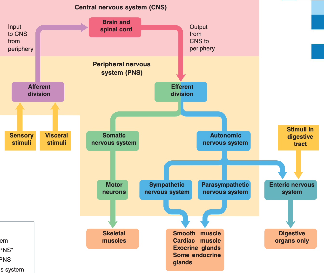
Organization of the Nervous System
Central Nervous System (CNS): Brain and spinal cord
Peripheral Nervous System (PNS): Afferent and efferent divisions
Afferent Division: Input to the CNS from the periphery
Sensory stimuli
Visceral stimuli (stimuli in the digestive tract)
Efferent Division: Output from the CNS to the periphery
Somatic Nervous System: Motor neurons that control skeletal muscles
Autonomic Nervous System: Controls smooth muscle, cardiac muscle, exocrine glands, and some endocrine glands
Sympathetic nervous system
Parasympathetic nervous system
Enteric nervous system (digestive organs only)
Effector Organs: Made up of muscle and gland tissue
Key Concept: Central Nervous System
Brain:
Control center of the nervous system
Occupies the cranial cavity
Spinal Cord:
Continuous with the medulla oblongata (an inferior aspect of the brain stem)
Exits the skull through the foramen magnum (hole in the base of the skull)
Consists of 31 segments corresponding to the 31 pairs of spinal nerves
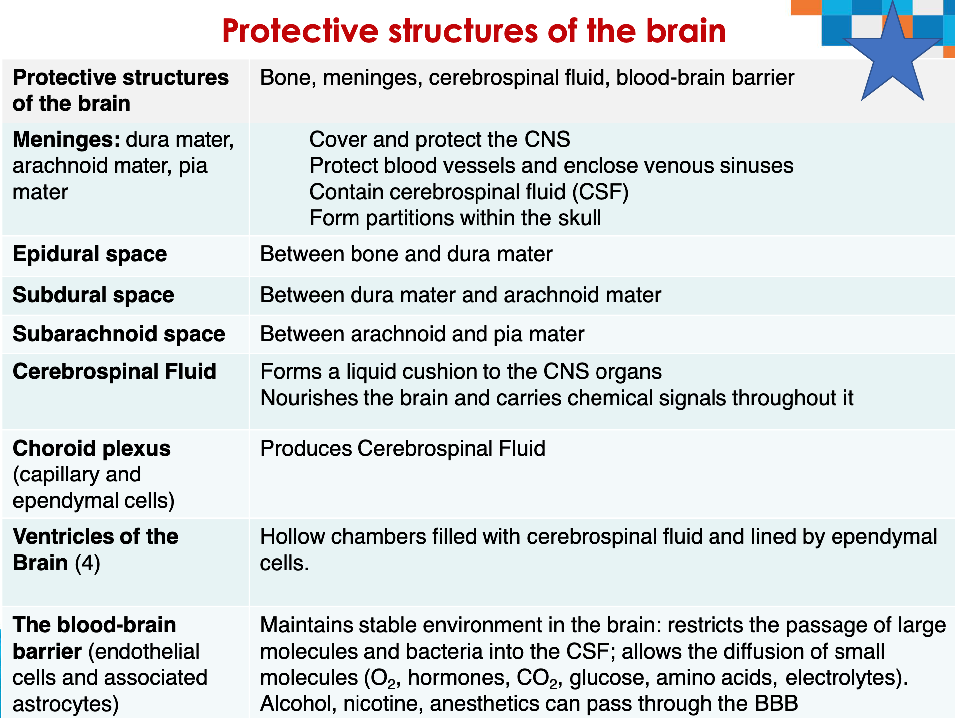
Covering of the Brain: Meninges and CSF
The brain is protected by bone, meninges, and cerebrospinal fluid.
Harmful substances are shielded from the brain by the blood-brain barrier.
The blood-brain barrier (endothelial cells and associated astrocytes) maintains a stable environment in the brain.
Protective Structures of the Brain
Grey matter & white matter
Grey matter contains neuron soma (cell body), synapses, dendrites, and non-
myelinated axons.White matter contains mostly myelinated axons that implement global
communicationNucleus: cell bodies imbedded in the white matter in the brain (e.g. basal nuclei)
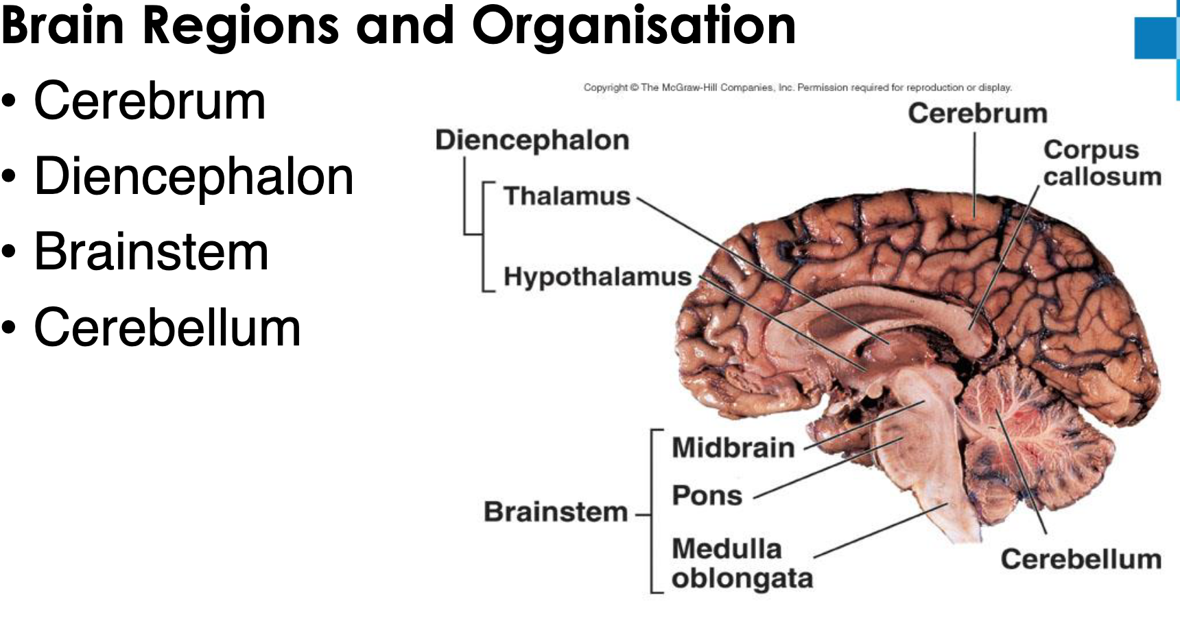
Central Nervous System - Brain Regions and Organisation
The Cerebral Cortex
Three types of functional areas
Sensory areas—conscious awareness of sensation
Association areas—integrate diverse information
Motor areas—control voluntary movement
Each hemisphere concerned with contralateral (opposite) side of body
Left hemisphere controls language, math, and logic
Right hemisphere controls visual-spatial skills, intuition, emotion, and artistic and musical skills
Hemispheres communicate almost instantaneously via fiber tracts and functional integration – corpus callosum
Cerebral dominance: the hemisphere that is dominant for language
90% of humans have left-sided dominance which results in right-handedness
In other 10%, roles of hemispheres are reversed
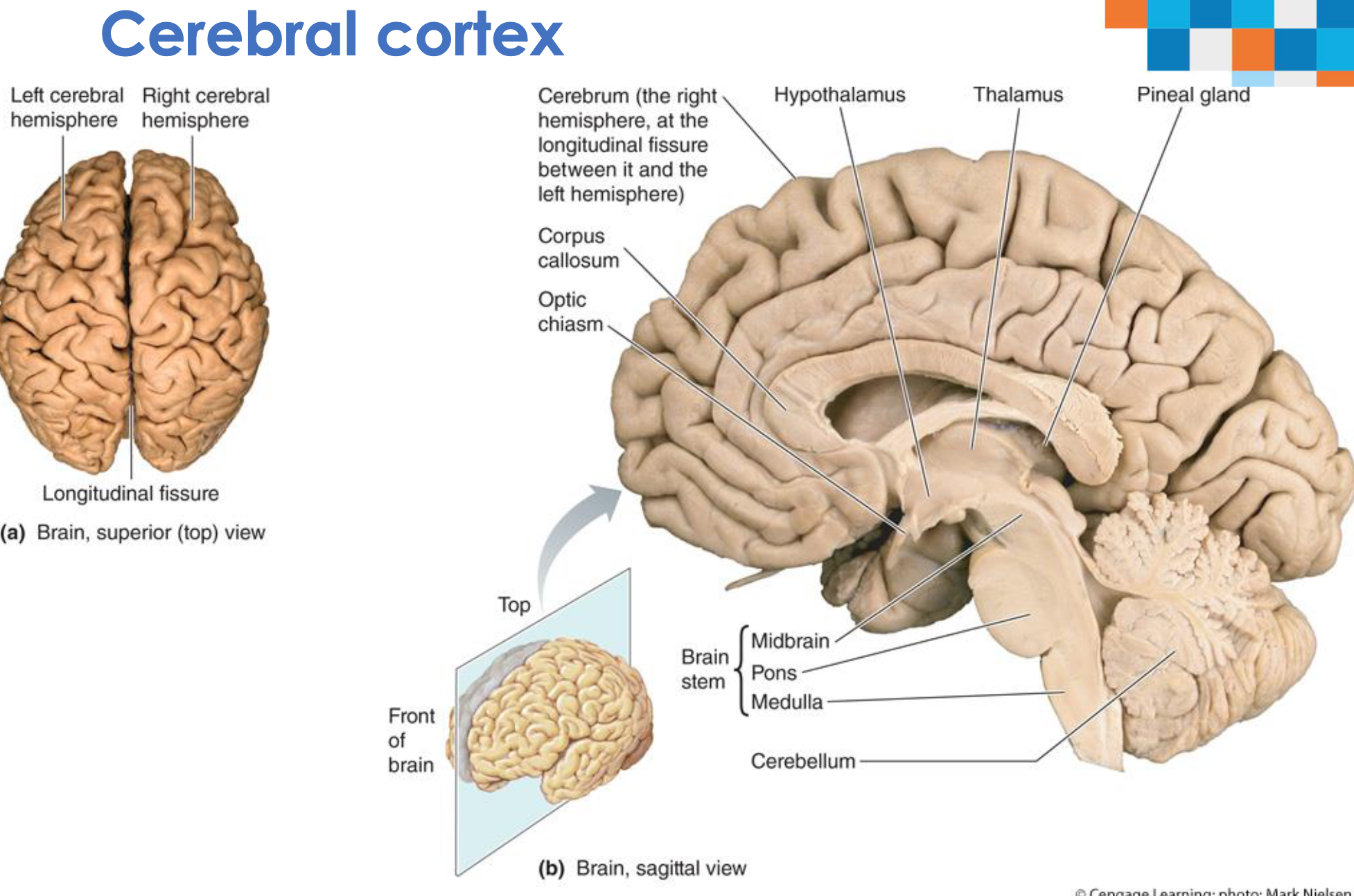
Cerebral Cortex - Hemispheres
*Left Cerebral Hemisphere
*Right Cerebral Hemisphere
*Longitudinal fissure
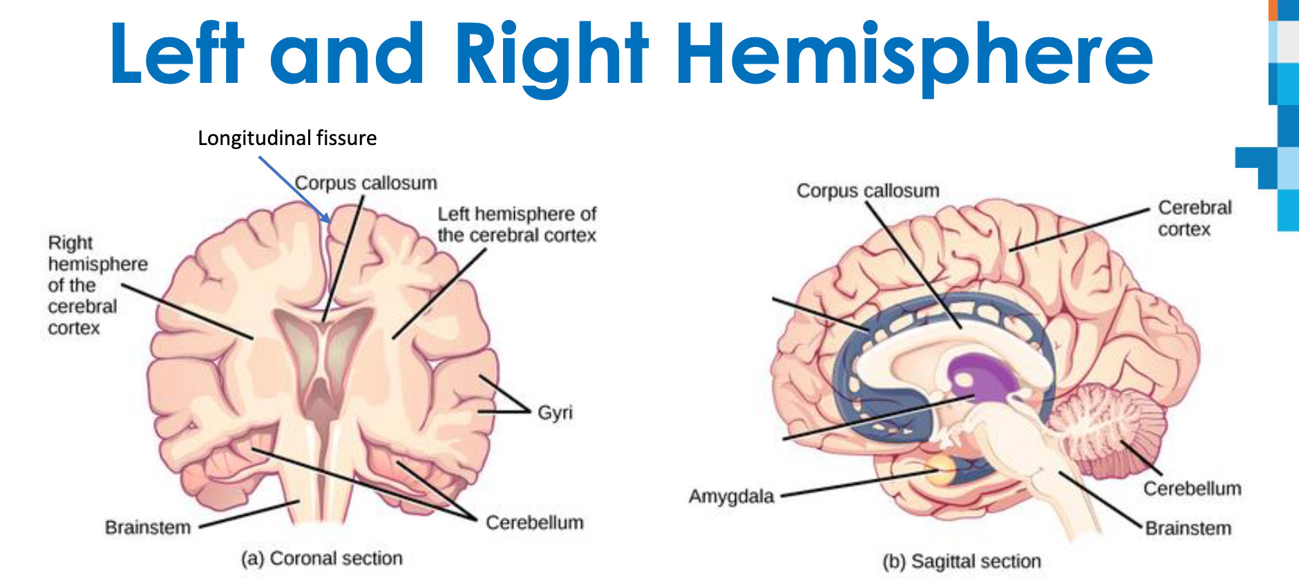
Left and Right Hemisphere
Longitudinal fissure Cerebrum is divided into left and right hemisphere by the longitudinal fissure
It is connected by interconnecting axons called corpus callosum
Cortex of the hemispheres is wrinkled in order to increase area (can fit more neurons)
Folds are called gyri; grooves are called sulci
Left Brain/Right Brain
Left Brain: Logical, sequential, rational, analytical, objective, looks at parts
Right Brain: Random, intuitive, holistic, synthesizing, subjective, looks at wholes
Brain Lobes
Cerebral cortex is 3mm layer of gray matter with extensive folds to increase the surface area.
Contains elevated ridges (gyri) and shallow grooves (sulci).
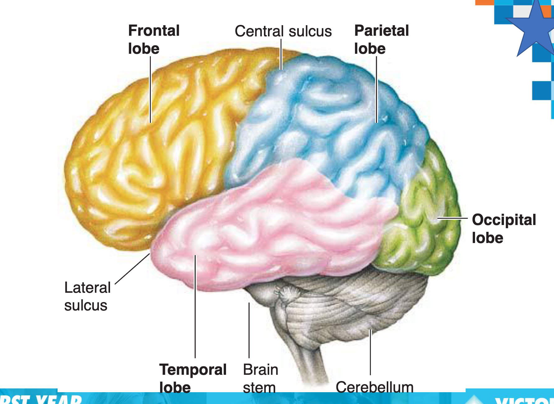
Deep sulci divide the hemispheres into 5 lobes:
Frontal – voluntary motor function, planning, mood, social judgment, intellect, speech
Parietal – somatic sensory reception and integration
Temporal – hearing, smell, learning, memory
Occipital – visual centre
Insula – taste (gustatory cortex) deep to the temporal lobe
Brain Parts
Functional Areas of the Cerebral Cortex
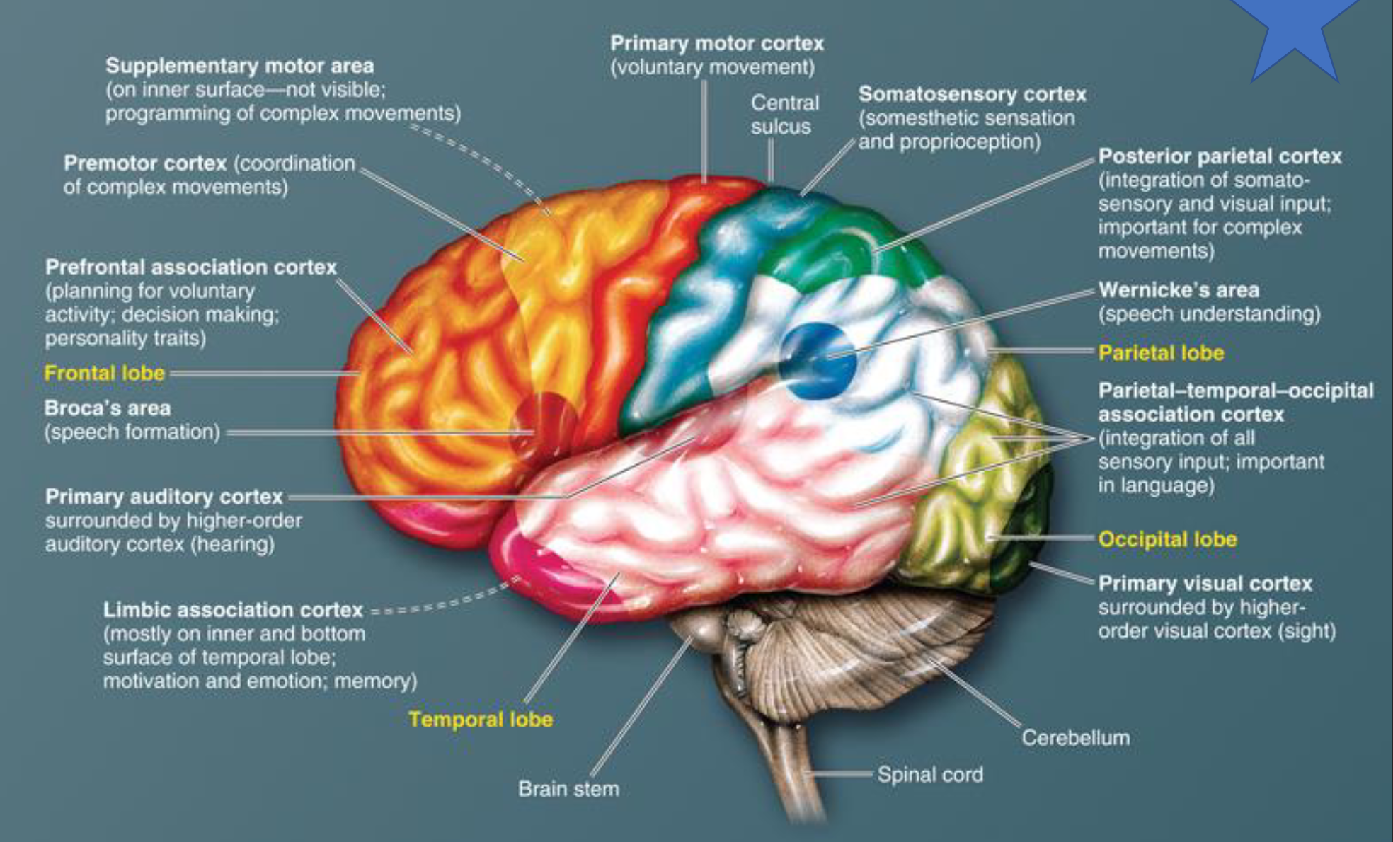
Frontal lobe: Prefrontal association cortex (planning for voluntary activity; decision making; personality traits), Premotor cortex (coordination of complex movements), Supplementary motor area (programming of complex movements - on inner surface-not visible), Broca's area (speech formation), Primary motor cortex (voluntary movement)
Parietal lobe: Somatosensory cortex (somesthetic sensation and proprioception), Posterior parietal cortex (integration of somatosensory and visual input; important for complex movements), Parietal-temporal-occipital association cortex (integration of all sensory input; important in language), Central sulcus
Occipital lobe: Primary visual cortex surrounded by higher-order visual cortex (sight)
Temporal lobe: Primary auditory cortex surrounded by higher-order auditory cortex (hearing), Limbic association cortex (mostly on inner and bottom surface of temporal lobe; motivation and emotion; memory), Wernicke's area (speech understanding)
Parietal and Frontal Lobes
The parietal lobes accomplish somatosensory processing
Somatosensory cortex – found in the postcentral gyrus
Sensations from the surface of the body, such as touch, pressure, heat, cold, and pain (and proprioception)
The primary motor cortex – found in the precentral gyrus
located in the frontal lobes controls the skeletal muscles
Stimulation of different areas of the primary motor cortex brings about movement in different regions of the body
Higher motor areas are also important in motor control
Supplementary motor area
Premotor cortex
Posterior parietal cortex
Brain has a body map
The distorted human figure drawn to reflect the relative sensory space our body parts occupy on the cerebral cortex is called a homunculus
Sensory homunculus is in the postcentral gyrus ( parietal lobe)
Motor homunculus looks distorted and is placed in the precentral gyrus ( frontal lobe)
Cerebral Hemispheres: Lobes and Their Functions
Lobe | Areas included | Functions |
|---|---|---|
Frontal | Primary motor cortex; Premotor cortex; Broca’s area; Frontal eye field; Prefrontal cortex. | Voluntary motor control of skeletal muscle, skilled motor activities. Broca’s area (left hemisphere) – speech. Higher intellectual processes (task management, problem solving etc.) |
Parietal | Primary somato- sensory cortex (PSC) Somatosensory association cortex | Somatic sensation: SENSES PSC – spatial discrimination – identifies the stimulated body region. SAC -comprehensive understanding of the stimulus. |
Temporal | Auditory areas Hippocampus Wernicke’s area Olfactory cortex | Auditory areas: hearing, perception of sound Hippocampus: Emotion & memory; Wernicke’s area - language Olfactory cortex -Smell |
Occipital | Visual cortex | Primary visual cortex - Receives visual information from the retinas. Visual association area - Interprets visual stimuli. |
Insula | Gustatory cortex | Taste |
Basal Nuclei
Masses of gray matter found deep within the cortical white matter.
Modify ongoing activity in motor pathways
Modulate thalamus by inhibiting antagonistic and unnecessary movement.
Help coordinate slow sustained contractions necessary for posture.
Parkinson's disease
*Degeneration of dopamine neurons in substantia nigra is hallmark sign of Parkinson's Disease
Diencephalon, Brainstem and Cerebellum
Cerebral Cortex
Thalamus (medial)
Basal nuclei (lateral to thalamus)
Sensory perception
Voluntary control of movement
Language
Personality Traits
Sophisticated mental events, such as thinking, memory, decision making, creativity, and self-consciousness
Basal Nuclei
Inhibition of muscle tone
Coordination of slow, sustained movements
Suppression of useless patterns of movement
Thalamus
Relay station for all synaptic input
Crude awareness of sensation
Some degree of consciousness
Role in motor control
Hypothalamus
Regulation of many homeostatic functions, such as temperature control, thirst, urine output, and food intake
Important link between nervous and endocrine systems
Extensive involvement with emotion and basic behavioral patterns
Role in sleep-wake cycle
Cerebellum
Maintenance of balance
Enhancement of muscle tone
Coordination and planning of skilled voluntary muscle activity
Brain Stem
Midbrain
Pons
Medulla - Spinal cord
Origin of majority of peripheral cranial nerves
Cardiovascular, respiratory, and digestive control centers
Regulation of muscle reflexes involved with equilibrium and posture
Reception and integration of all synaptic input from spinal cord; arousal and activation of cerebral cortex
Role in sleep-wake cycle
Limbic System: Emotional Brain
Consists of a ring of forebrain structures that surround the brain stem Parts especially important in emotions, basic behavioural patterns, motivation.
Amygdala – deals with anger, danger, and fear responses
Hippocampus – converts new information into long-term memories.
Emotional memory;
Most limbic system outputs relay through Hypothalamus (visceral control) and Thalamus (gate to the cerebral cortex)
Cerebral Hemispheres: Lobes and their Functions - Lesions
Lobe | Function | Effect of lesion |
|---|---|---|
Frontal | Primary motor cortex - Voluntary motor control of skeletal muscle; Premotor cortex- skilled motor activities. Broca’s area (left hemisphere) – speech production, planning to speak. Higher intellectual processes (task management, problem solving etc.) | Primary motor cortex – paralyses skeletal muscles on the opposite side of the body; Premotor cortex – loss of the motor skills programmed on that region. Broca’s area – loss of ability to speak. Prefrontal cortex – mental and personality disorders, loss of judgement and attentiveness. |
Temporal | Auditory areas - hearing, perception of sound Hippocampus - Emotion & memory; Wernicke’s area - language Olfactory cortex - Smell Gustatory cortex (insula) - taste | Damage to auditory areas – inability to interpret/discriminate and localize sounds. Hippocampus – damage to one side results in anterograde amnesia – inability to form new memories, bilateral damage – total amnesia. Damage to Wernicke’s area – inability to interpret spoken language. Olfactory cortex – inability to interpret different types of smell. Gustatory cortex – inability to recognise different tastes. |
Parietal | Somatic sensation: PSC – spatial discrimination – identifies the stimulated body region. SAA - comprehensive understanding of the stimulus. Wernicke’s area - language | Damage to PSC – inability to localize stimulated areas of the body, damage to SAA – inability to recognize objects (size, texture, shape, temperature, etc) without looking at them. |
Occipital | Primary visual cortex - Receives visual information from the retinas. Visual association area - Interprets visual stimuli. | Damage to primary visual cortex – functional blindness. Patients with damage to visual association area can see, but cannot comprehend what they are looking at. |
Spinal Cord
Conduction pathway between the body and the brain
Bundle of nerve tissue within the vertebral canal
Protected by vertebral column
42-45 cm long
Extends from foramen magnum of skull to the level of L1/2 vertebra in adults
Tapered inferior end: conus medularis
Cervical and lumbar enlargements: thicker due to dense motor and sensory innervation of limbs
Spinal Cord Cross Section
Grey Matter
Looks like the letter H or a butterfly
Contains the:
Dorsal horn
Ventral horn
Lateral Horn (in thoracolumbar region)
White matter
Composed of predominantly myelinated, but also non-myelinated, fibres that allow communication between different segments/areas of the spinal cord, and the spinal cord and the brain
They run in 3 directions
Ascending
Descending
Transverse
White Matter of Spinal Cord
Myelinated and non-myelinated nerve fibers allow communication between parts of spinal cord, and spinal cord andbrain Organized into tracts (bundles of nerve fibres with a similar function)
Run in three directions:
Ascending: up to higher centers (sensory inputs); carry impulses to the brain
Descending: from brain to cord or lower cord levels (motor outputs); carry impulses from brain to effectors
Transverse: from one side to other (commissural fibers)
Ascending Pathways
Conduct sensory pathways upward through a chain of three neurons:
First-order neuron ➢Conducts impulses from cutaneous receptors and proprioceptors ➢Branches diffusely as it enters spinal cord or medulla ➢Synapses with second-order neuron
Second-order neuron ➢Interneuron ➢Cell body in dorsal horn of spinal cord or medullary nuclei ➢Axons extend to thalamus or cerebellum
Third-order neuron ➢ Also an interneuron ➢ Cell bodies in thalamus ➢ Axon extends to somatosensory cortex ➢ No third-order neurons in cerebellum
Descending tracts
*Lateral corticospinal tract
*Ventral corticospinal tract