Respiratory System
 Function:
Supplies the blood with oxygen so it gets to all parts of the body. It also removes waste such as carbon dioxide.
How
Through breathing. When you breathe, you take in oxygen and breathe out carbon dioxide (gas change).
Breathing in is known as inspiration
Breathing out is known as expiration
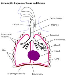
The pharynx
As the air passes through the nasal cavity, the air is warmed slightly. The air meets the pharynx, a junction at the back of the oral cavity.
Air must pass through the trachea.
Cartilage in trachea
Trachea is a tube lined C-shaped supporting rings.
These rings are made of cartilage. They help to hold the tube open.
When we breathe in the trachea will expand to allow more air in.
The Trachea
Goblet cells and cilia are found in trachea and bronchi.
They protect alveoli in the lungs from inhaled dust and microbes.
Goblet cells produce sticky mucus which traps them.
Cilia beat in a rhythmic action to move them up and out of the airways
Smooth muscle
The walls of the trachea, bronchi and bronchioles are lined with smooth muscle which can contract and relax.
During exercise, smooth muscle will relax which causes airway to expand and allows more air to flow through them.
Smooth muscle will contract if toxic gas is present to restrict airflow by narrowing the airways.
Contracting smooth muscle is a cause of asthma.
Ventilation
The breathing system does not have a fixed shape. It has the ability to move, whilst remaining enclosed within protection of the ribcage. This means the ribcage must move.
When you breathe in, your rib moves up and out. The overall effect of this is that your chest (thoracic cavity) expands.
When you breathe out, your ribs move down and inwards. The overall effect of this is that your chest gets smaller.
The Diaphragm
It is a dome shape and as we inhale it contracts and flattens. This results in a change in volume in the thorax. (gets bigger)
volume increases = pressure decreases
pressure increases = volume decreases
Alveoli and gas exchange
Gas exchange happens in the alveoli by diffusion, from high concentration to low concentration.
Cells need oxygen for respiration and remove carbon dioxide.
Oxygen and Carbon dioxide are transported to the lungs by the circulatory system.
Gas exchange takes place between the alveoli in the lungs and the capillary network to close them.
The alveoli membrane and pulmonary capillaries are very thin and very close to each other (short diffusion distance).
Oxygen dissolves in the moist lining, so it can diffuse in the blood.
Efficient gas exchange requires
large surface area
large concentration gradients (low oxygen in capillary, high oxygen in alveoli)
short diffusion pathway (thickness of the membrane that the molecules must diffuse across)
Alveoli adaptions
thin walls = short diffusion distance
large surface area = increases diffusion
secrete surfactant = keeps alveoli moist to stop them sticking together
large capillary network = maintains diffusion and concentration gradient
Restrictive lung diseases
-Lung volumes which are abnormally low (particularly residual volume) indicate restrictive lung disease e.g. pulmonary fibrosis, lung cancer, and pneumonia. Vital capacity will also be low.
restrictive = less residual volume
Obstructive lung disease
People suffering from obstructive lung diseases such as COPD e.g. bronchitis, and emphysema have residual volume values that are greater than normal.
It is important to remember however that the residual volume cannot be measured by spirometry.
Asthma is an obstructive lung disease. Asthmatics usually have normal lung volumes (except for residual volume) but their flow rate is reduced.
obstructive = more residual volume
Residual volume is the oxygen you can’t get out of your lungs.
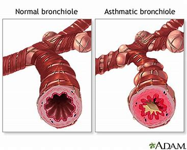
The asthmatic bronchiole has thicker walls (smooth muscle contracts, makes airway narrower which reduces airflow)