Exam 2 Notes
Chapter_4_Book
Overview
This chapter focuses on the development of the brain, exploring the processes from a single fertilized egg to a complex organ with billions of neurons connected through trillions of synapses.
1. Development Stages
Timeline of Brain Development:
Growth marked by significant milestones in brain development through various decades: 2010, 2020, 2030, and projections for 2050.
2. Case Study: Michael May
Michael May's experiences illustrate the challenges of blindness and brain development:
Accident at Age 3: Lost sight in left eye and severely damaged the right.
Early Life: Achieved proficiency in activities like Ping-Pong and ski despite challenges.
Restoration of Sight at Age 46: Successful surgery enabled vision, yet faced difficulties in recognizing faces or static three-dimensional objects.
This led to considerations of neural plasticity and pathways for visual processing.
3. Brain Structure and Function
A. Early Embryonic Development
Ectoderm: Outermost layer of the developing embryo that gives rise to skin and nervous system.
Neural Tube: Forms from the ectoderm; precursor to the central nervous system (CNS), dividing into forebrain, midbrain, and hindbrain.
Milestones of Development:
By 10 weeks, most body organs begin to take shape, and an embryo shows significant brain development, including structures in the neural tube.
B. Cellular Processes of Brain Development
Six Stages:
Neurogenesis: Division to produce neurons.
Cell Migration: Movement of nerve cells to establish populations.
Cell Differentiation: Refinement of cells into distinct neuron types.
Synaptogenesis: Formation of synaptic connections.
Neuronal Cell Death: Survival of the fittest neurons leading to selective cell death.
Synapse Rearrangement: Continuous refinement of synaptic connections throughout life.
4. Unique Characteristics of Human Brain Development
Humans display prolonged brain growth after birth, leading to a greater brain-to-body weight ratio compared to other primates.
Increased dendritic growth and synaptic complexity are noted from birth into early childhood.
Neurogenesis continues into adulthood, particularly in regions such as the hippocampus, contributing to learning and memory.
5. Role of Experiences on Brain Development
A. Impact of Experiences
Early experience significantly shapes synaptic connections and influences the brain's plasticity.
Neurotrophic Factors: Chemicals that help neurons survive and thrive, directly impacting synapse formation and maintenance.
The presence of active synapses influences survival; inactive synapses tend to retract.
B. Visual System Development and Disorders
Early life experiences can result in amblyopia (dull vision), where visual deprivation during sensitive periods leads to long-lasting impairment.
Monocular Deprivation: Keeping one eye deprived affects visual cortical responses and can lead to permanent blindness in that eye if not corrected early.
Ocular Dominance Histogram: Illustrates the shift in response from both eyes when one eye is deprived, leading to changes in visual perception and behavior.
6. Neurotransmitters and Alzheimer’s Disease
The aging brain experiences various changes:
Alzheimer's Disease: Associated with cognitive decline, characterized by neurofibrillary tangles and amyloid plaques which disrupt neuronal function.
Importance of lifestyle choices to potentially mitigate risks includes physical and mental activity.
7. Lasting Implications of Brain Development
Understanding that although much of the brain’s structural development is dictated by genetics, experiences and environment profoundly shape its functioning.
Epigenetic influences show how experiences, such as maternal care, can lead to differential gene expression affecting stress responses and behavior throughout life.
Conclusion
Brain development is a meticulous interplay of intrinsic genetic information and extrinsic environmental factors. The cumulative effects of early experiences contribute to an individual’s neurodevelopmental trajectory, impacting lifelong cognitive functions.
Chapter 4: Brain Development
Embryonic Disk
Layers of the Embryonic Disk:
Endoderm (inner layer): Develops into the digestive and respiratory systems.
Mesoderm (middle layer): Forms muscles, skeleton, and circulatory system.
Ectoderm (outer layer): Develops into sensory and nervous systems.
Neural Tube Development: Marks the beginning of the central nervous system (CNS).
The Neural Tube
Brain Divisions from the Neural Tube:
Forebrain: Includes the cortex, thalamus, and hypothalamus.
Midbrain: Comprised of the tectum, tegmentum, and periaqueductal gray.
Hindbrain: Contains the cerebellum, pons, and medulla.
Interior Structure: The neural tube houses ventricles and the central canal of the spinal cord.
Making New Neurons
Neurogenesis: The production of new neurons through mitosis.
Cell Migration: Movement of nerve cells or precursors to establish different brain regions.
Cell Differentiation: Process where cells develop into specific types of neurons or glial cells.
Creating Synaptic Connections
Synaptogenesis: The formation of new synaptic connections.
Stem Cells: Undifferentiated cells located in embryonic tissues.
Continued Neurogenesis: Occurs throughout adulthood, especially in hippocampal regions.
Factors Affecting Neurogenesis: Ongoing research into environmental and biological influences.
Cell Death
Neuronal Cell Death: Selective death of nerve cells that begins before birth, known as apoptosis.
Factors for Neuron Survival:
Importance of synaptic connections.
Neurotrophic Factors: Nutrients that support neuron viability and survival.
Synaptic Formation & Rearrangement
Synaptic Rearrangement: The loss and refinement of synaptic connections experienced throughout life.
Driving Forces: Various factors motivate this synaptic rearrangement, including experience.
Synaptic Rearrangement Through Early Adulthood
Maturation Timeline: This process continues into adolescence with a posterior to anterior maturation pattern.
Prefrontal Cortex (PFC): Recognized as the last brain area to mature, following the "use it or lose it" principle.
Why Are Synapses Pruned?
Reason for Pruning: Evidence suggests excessive connections correlate with intellectual disability.
Example: Fragile X Syndrome: Impacting 1 in 11,000 females and 1 in 7,000 males.
Objective of Pruning: To prevent system overload and promote efficiency.
Sensitive Periods
Definition of Sensitive Period: A developmental phase during which an organism is highly susceptible to external factors.
Impact of Experience:
Binocular Deprivation: Can cause irreversible changes in the visual system.
Experience-Expectant Development: The brain's reliance on specific stimuli at certain developmental stages for normative growth.
Experience-Dependent Development: Individual experiences shaping neural connections, leading to variability.
Trait Expression
Genotype vs. Phenotype:
Genotype: All genetic material in a person.
Phenotype: Observable characteristics influenced by the environment.
Example with Twins: Identical genotypes can result in different phenotypes due to environmental factors.
Epigenetics
Epigenetics Defined: The investigation of how factors influence gene expression without altering the nucleotide sequence.
Methylation: A form of chemical modification of DNA, affecting gene expression likelihood without changing genetic information.
Environment & Heredity
Range of Reaction: Variation in traits due to environmental influences.
Canalization: The concept where certain traits are less impacted by environmental changes.
Example: Eye color versus musical ability often shows high heritability.
Changes in Cognition
Cognitive Changes Over Time: Many abilities exhibit changes later in life, depending on the specific ability.
Cortical Volume: Generally decreases with age, but not all cognitive decline is typical with aging.
Hippocampal Loss: Associated with memory decline, yet memory issues should not be seen as an inevitable aging process.
Cognitive Ability Trajectories
Research Findings:
Stability in most intellectual competencies in adulthood, particularly in crystallized abilities.
Fluid Abilities: These tend to decline steadily through the later adult years.
Cognitive Abilities Across the Lifespan
Fluid Abilities: Basic cognitive processing skills- working memory, processing speed, pattern discovery.
Crystallized Abilities: The knowledge built through experiences and learning over time.
Dementia
Description of Dementia: A broad term for various conditions marked by progressive cognitive decline due to brain changes.
Symptoms: Impacts memory, reasoning, behavior, emotional stability, and daily functioning.
Alzheimer’s Disease
Nature of Alzheimer's: A neurodegenerative disorder that leads to significant cognitive and behavioral changes and ultimately death within 1-10 years.
Age Relation: Risk increases with age as neurodegeneration progresses.
Plaques & Tangles in Alzheimer’s Disease
Diagnostic Approach: Alzheimer's diagnosed by exclusion with the aid of brain imaging to rule out other causes.
Amyloid Plaques: Clusters of substance beta-amyloid located in key brain areas.
Neurofibrillary Tangles: Abnormal proteins within neurons, correlating with degrees of cognitive impairment.
Neurotransmitter Impact: Loss of neurons in the basal forebrain decreases acetylcholine levels, exacerbating symptoms.
Alzheimer's Brain Characteristics
Neurodegeneration Effects:
Severe shrinkage of cerebral cortex and hippocampus.
Enlarged ventricles observed in brains affected by Alzheimer's disease.
Risk Factors for Alzheimer’s
Gender: Women show a higher risk than men.
Age: Advancing age increases the likelihood of developing Alzheimer's.
Genetics: Similar factors that heighten cardiovascular risks also apply to Alzheimer's risk.
Protective Factors Against Alzheimer’s
Education: Learning fosters neuron activity, enhancing neural connections and developing cognitive reserve.
Exercise: Promotes growth in the hippocampal area and positively influences overall cognitive function.
Chapter_7_Book
Vision: From Eye to Brain
1. Overview of Vision
Importance of Vision: Crucial for finding food, avoiding predators, and locating shelter.
Visual Overload: The visual system processes a vast amount of information, likened to drinking from a waterfall.
2. Case Study of D.F.
Incident: D.F. was exposed to carbon monoxide from a malfunctioning heater, leading to unconsciousness.
Symptoms: Post-incident, D.F. could not recognize objects but could interact with them appropriately. For example, she could identify a flashlight by its features but not recognize it as a flashlight.
D.F.'s ability to perform tasks without recognition suggests dichotomy between visual perception and visual action, indicating the complex nature of visual processing.
3. Visual System Adaptations
Species-Specific Adaptations: Different species have evolved visual capabilities fitting their ecological needs.
Nocturnal species exhibit better night vision than diurnal species.
Creatures like hawks possess keen long-distance vision, while rodents have better close-range vision.
Many insects detect ultraviolet light, which reveals floral patterns invisible to humans.
4. Basic Anatomy of the Eye
Retina: Contains photoreceptors that convert light into neural signals via a process called transduction.
Key Components:
Cornea: Transparent outer layer that refracts light.
Lens: Further refines light focus.
Ciliary Muscles: Control lens shape for focusing (accommodation).
Photoreceptors: Rods (sensitive to low light, no color) and Cones (responsible for color vision).
Nerve Pathway: Signals travel from the retina to the brain via the optic nerve, passing through the optic chiasm where visual information is processed.
5. Visual Processing Stages
Retinal Processing: Involves conversion of light signals into electrical signals by photoreceptors, which stimulate subsequent bipolar and ganglion cells in the retina.
Convergence: Multiple rods converge onto a single ganglion cell, enhancing light sensitivity but reducing acuity (detail).
Retinal Cell Types:
Rods: 100 million, active in dim light (scotopic vision).
Cones: 4 million, require more light, responsible for color (photopic vision).
6. Vision and Light Sensitivity
Adjustments to Light Levels:
Pupil Adjustment: The pupil's size changes based on light exposure (dilation and constriction).
Photoreceptor Adaptation: Sensitivity of cones and rods adjusts to ambient light levels, allowing vision across a wide intensity range.
7. Visual Acuity and Field
Fovea: Center of the retina, where visual acuity is highest due to the dense packing of cones. Objects are focused here for clear vision.
Blind Spot: Lacks photoreceptors where the optic nerve exits the eye but is compensated by the brain, filling in gaps in visual perception.
8. Neural Pathways to the Brain
Optic Tract: Carries visual information from the optic chiasm to the thalamus and then to the primary visual cortex (V1).
Cortical Processing: V1 processes basic visual information; surrounding extrastriate areas further analyze form, color, and motion.
9. Receptive Fields
Ganglion Cells: Have concentric receptive fields (on-center/off-surround and off-center/on-surround), crucial for contrast detection.
Contrasting the Two Systems:
Scotopic System: Operates in dim light, high sensitivity.
Photopic System: Operates in bright light, supports color vision and detail recognition.
10. Color Vision
Trichromatic Theory: States three types of cones (short-wavelength, medium-wavelength, and long-wavelength) respond to different colors.
Color perception results from the combined activation of these cones.
Opponent-Process Theory: Suggests there are opposing pairs of colors (red-green, blue-yellow) processed in different ways.
11. Visual Streams in Cortex
Dorsal Stream: Processes object location and movement for spatial awareness ("where" pathway).
Ventral Stream: Processes object identity ("what" pathway), including facial recognition.
D.F.'s case exemplifies dissociation where she retained motor skills for grasping but lost object recognition ability.
12. Vision Improvement and Impairments
Myopia: Caused by an elongated eyeball, making distant objects blurry; linked to increased time indoors.
Amblyopia: Poor vision in one eye due to misalignment, can be improved with treatment (patching the dominant eye).
Macular Degeneration: Leading cause of vision loss, impacts sharp central vision.
SENSORY SYSTEMS
EXTEROCEPTIVE SENSORY SYSTEMS
Visual: Related to sight.
Auditory: Related to hearing.
Somatosensory: Related to touch and bodily sensations.
Olfactory: Related to smell.
Gustatory: Related to taste.
SENSORY SYSTEM ESSENTIALS
Discrimination of Energy Forms: Sensory systems must be capable of distinguishing between various forms of energy, such as light, sound, chemical signals, and mechanical pressure. This discrimination is crucial for the brain to interpret and respond appropriately to different environmental stimuli.
Reliability and Speed of Response: Sensory systems should respond quickly and consistently to incoming stimuli. This rapid signaling allows organisms to react to changes in their environment almost instantaneously, which is essential for survival, such as escaping predators or catching prey.
Discrimination of Stimulus Intensities: Sensory systems must possess the ability to detect differences in the intensity of stimuli. For example, the ability to sense variations in brightness, volume, or taste intensity allows for nuanced perceptions that inform more complex decision-making.
Suppression of Extraneous Information: Effective sensory systems should filter out irrelevant or distracting sensory information to focus on pertinent stimuli. This selectivity prevents overload and allows for more efficient processing of crucial information, ensuring organisms respond to the most immediate and important aspects of their environment.
SENSORY SYSTEM COMMONALITIES
All sensory systems have:
Specialized organ(s).
Specialized receptor cells.
Transduce information (energy) from the environment.
A specific pathway from receptor to cortex.
Includes a relay center (nuclei) in the thalamus.
SENSORY SYSTEM ORGANIZATION
Primary Cortex:
Receives input directly from the thalamus.
Processes elementary information (basic form).
Secondary Cortex:
Receives input from the primary cortex.
Processes higher-order information (specific form, motion).
Association Cortex:
Receives input from more than one sensory system and interacts with the motor system.
THE VISUAL SYSTEM
VISION: ENERGY
The energy experienced is light.
If there is no light, there is no vision.
Visible Light Spectrum:
Electromagnetic energy between 380 and 760 nanometers.
Follows the VIBGYOR sequence.
WAVELENGTHS AND FREQUENCY
High Frequency:
Short wavelengths (Gamma rays, X-rays, Ultraviolet rays).
Low Frequency:
Long wavelengths (Infrared, radio waves).
Visible Spectrum:
Ranges from approximately 380 nm (violet) to 760 nm (red).
THREE DIMENSIONS OF COLOR PERCEPTION
Brightness: How light or dark something is.
Hue: The color of an object (e.g., blue, green, red).
A continuous scale.
Saturation: How rich a color is.
As saturation decreases, we see more shades of grey.
VISION: ORGAN
The Eye:
Iris: Eye color and contractile tissue.
Pupil: Hole in the iris that allows light to enter.
Lens: Focuses incoming light onto the retina using ciliary muscles.
Retina: Back of the eye where signal transduction occurs.
Fovea: Region of highest visual acuity.
Optic Nerve: Transmits visual information to the brain.
Sclera: The white protective covering of the eye.
Cornea: The outer layer that protects the eye.
RETINA DETAILS
Fovea:
Indentation at the center of the retina specialized for high acuity vision.
Blind Spot:
Gap in the retinal tissue where the optic nerve exits; undetectable due to visual completion.
VISION: SIGNAL PROCESSING
Light energy is transduced to neural signals for transmission to the brain.
Primary types of photoreceptors: Rods and Cones.
VISION: SIGNAL PROCESSING FLOW
Light energy transduced into neural signals passes through:
Photoreceptors
Horizontal Cells: Lateral communication.
Bipolar Cells
Amacrine Cells: Lateral communication.
Retinal Ganglion Cells: Relay visual information to the brain.
DUPLEXITY THEORY OF VISION
Scotopic Vision:
Mediated by rods.
Low acuity and detail.
High sensitivity in dim light, lacks color vision.
Photopic Vision:
Mediated by cones.
High acuity, fine detail, and color vision in bright light.
CONVERGENCE IN THE VISUAL SYSTEM
High convergence in rod-fed circuits leads to low acuity; low convergence in cone-fed circuits leads to high acuity.
PHOTORECEPTOR FUNCTION
Rhodopsin: Photopigment in rods.
In the dark, sodium channels remain open, resulting in partial depolarization.
In light, rhodopsin is bleached, sodium channels close leading to hyperpolarization and blocking glutamate release.
VISION: PATHWAY
Pathway involves the retina, optic nerve, and lateral geniculate nucleus (LGN) in the thalamus, which relays information to the primary visual cortex.
VISUAL PROCESSING IN THE CORTEX
Primary Visual Cortex: Processes basic visual inputs and some depth.
Secondary Visual Cortex: Handles higher-order processing, including form and motion.
"What" Stream: Identifies objects; runs from primary to inferotemporal cortex.
"Where" Stream: Determines spatial location and guides movement; runs from primary to prestriate cortex.
ASSOCIATION CORTEX
Located in the parietal lobe; integrates information across senses and with the motor system.
VISION: DAMAGE
At Retina Level:
Myopia: Nearsightedness; trouble seeing distant objects, corrected with lenses.
Macular Degeneration: Leads to retinal detachment and death of photoreceptors.
Color Deficiency: Unable to perceive all colors, with normal acuity.
At Secondary/Association Cortex:
Optic Ataxia: Difficulty in using vision to reach for objects, but can identify them.
Prosopagnosia: Inability to recognize faces.
Akinetopsia: Inability to perceive movement smoothly.
SEEING COLOR
Reflectance: Color perception depends on the wavelength of light reflected by objects.
COLOR PERCEPTION THEORIES
Trichromatic Hypothesis: Color perception based on the ratio of activity in three kinds of receptors (blue, green, red-sensitive).
Opponent Process Hypothesis: Colors perceived in opposing pairs; cells excite one color and inhibit the complementary.
COMPATIBILITY OF THEORIES
Both theories coexist in the visual system, allowing for complex color perception.
Chapter_6_Book
Overview of Sensory Systems
This chapter covers the senses of hearing, balance, taste, and smell.
Hearing
Historical Perspective
Georg von Békésy worked in the 1920s to understand why human ears are more sensitive than microphones, leading to insights on hearing restoration.
His experiments on cadavers provided fundamental knowledge on how auditory data is translated into neural activity.
Structure of the Ear
The ear is divided into three main parts:
External Ear: Captures and channels sound into the ear canal.
Middle Ear: Contains ossicles (malleus, incus, stapes) which amplify sound vibrations.
Inner Ear: Houses the cochlea, where sound is transduced into neural signals.
Sound Characteristics
Decibel (dB): Measures sound intensity as perceived loudness.
Hertz (Hz): Measures frequency of sound, defining pitch.
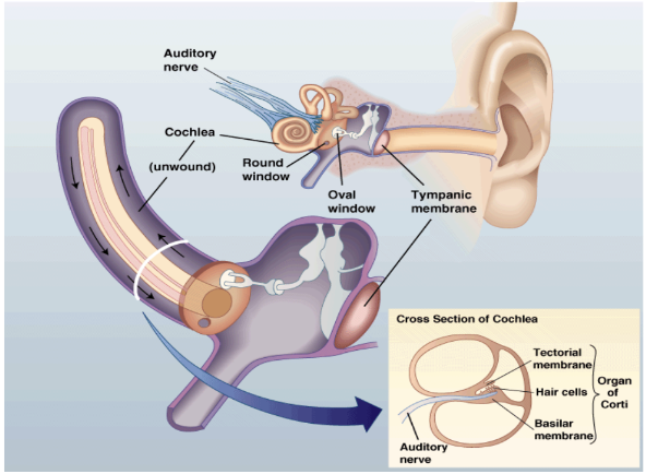
Sound Transduction Process
Capturing Sound Energy: The external ear funnel sound waves to the tympanic membrane.
Amplification: Sound vibrations are transmitted from the tympanic membrane through the ossicles to the oval window of the cochlea.
Cochlear Mechanics: The cochlea contains the organ of Corti, which converts vibrations into electrical signals via hair cells.
Signal Transmission: Auditory nerve fibers carry signals to the brain, with distinct pathways for sound processing.
Auditory Localization
The brain uses interaural intensity differences and interaural temporal differences for sound localization.
Factors like skull shape and ear position impact sound perception.
Frequency Encoding
Place Coding: Different frequencies activate specific locations on the basilar membrane.
Temporal Coding: The firing rate of neurons conveys frequency information, especially effective at lower frequencies.
Balance
Vestibular System
The vestibular system, located in the inner ear, detects the body's position and movement via the semicircular canals and otoliths.
Semicircular Canals: Detect rotation; each canal is oriented at right angles to efficiently track head movements.
Saccule and Utricle: Detect linear accelerations and static head positions.
Vestibular Functionality
Maintains balance and coordinates motor functions, informing responses to head orientation and movement.
Vestibular pathways have strong connections to the brain areas responsible for movement control.
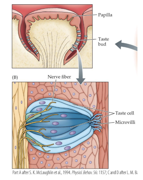
Taste (Gustation)
Components of Taste
Taste Buds: Clusters of taste receptor cells located within papillae.
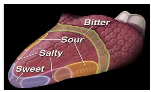
The primary tastes include:
Sweet: Detected by GPCRs involving T1R family receptors.
Sour: Associated with proton ion channels.
Salty: Primarily through sodium ion channels.
Bitter: Detected by numerous T2R family receptors.
Umami: Related to savory flavors; receptors include certain T1R variants.
Taste Transduction Process
Taste cells release neurotransmitters upon binding with tastants, sending information to the brain via cranial nerves.
Misconceptions about localized taste maps have been debunked; all tastes can be detected throughout the tongue.
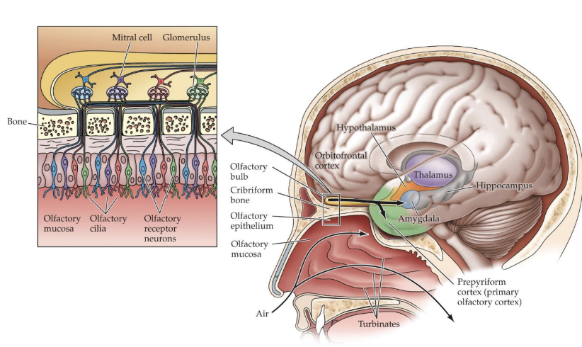
Smell (Olfaction)
Olfactory System
The olfactory epithelium contains three types of cells including olfactory receptor neurons, which detect odorants.
Each receptor neuron expresses one of about 400 functional olfactory receptors, allowing for vast odor discrimination.
Olfactory Transduction Process
Odorants interact with receptors on olfactory cilia, initiating a second-messenger system to transduce the signal.
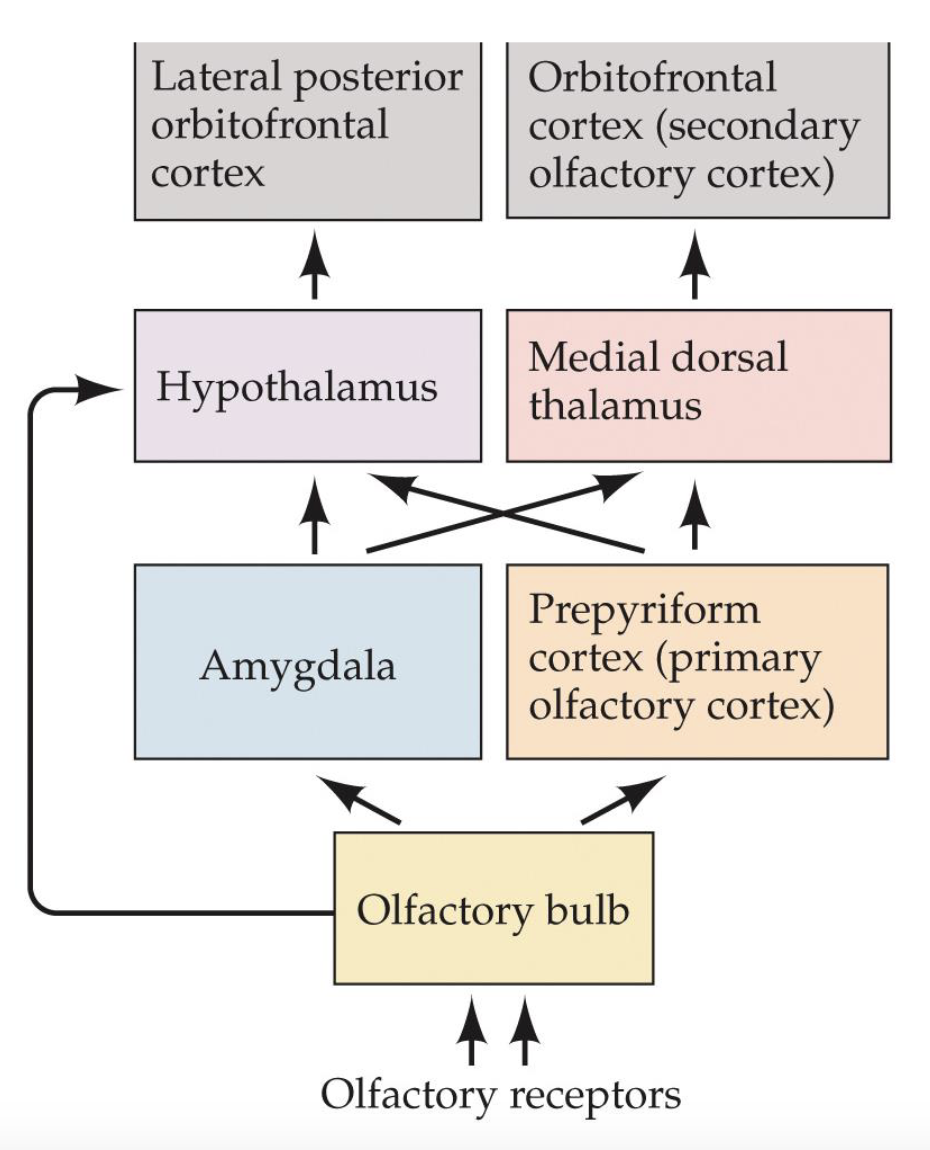
Axons from these neurons synapse in the olfactory bulb, which organizes information for processing in various brain regions like the amygdala and hippocampus.
Vomeronasal System
This secondary olfactory system detects pheromones and communicates with the brain to influence reproductive and social behaviors.
Humans possess only vestigial components of this system, relying on the main olfactory pathways for similar functions.
Conclusion
The senses of hearing, balance, taste, and smell allow for a complex interaction with the environment, guiding behaviors critical for survival and interaction.
SENSORY SYSTEMS
Overview of sensory systems, covering audition and chemical senses.
AUDITION
Sounds produce vibrations in air molecules, which create sound waves.
From sound waves, we detect two main aspects:
Loudness: Measured in decibels (dB).
Pitch: Measured in hertz (Hz).
Timbre: Quality of sound that distinguishes different types of sound production.
Humans can hear sounds between 20 Hz and 20,000 Hz.
ORGAN: The Ear
External ear (pinna): Gathers sound and plays a role in transducing auditory signals.
Ear canal: Leads sound to the tympanic membrane (eardrum).
Tympanic membrane: Vibrates in response to sound.
Middle ear: Contains ossicles; amplifies vibrations. Includes:
Ossicles: Malleus (hammer), Incus (anvil), Stapes (stirrup)
Oval window: Connects middle ear to inner ear.
Round window: Absorbs sound waves.
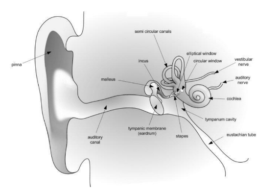
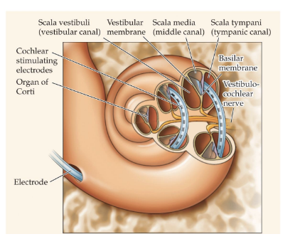
Inner ear:
Cochlea: Snail-shaped structure housing the primary receptor cells for hearing.
Contains three parallel canals: Scala vestibuli, Scala media, and Scala tympani.
Organ of Corti: Converts vibrations to neural activity.
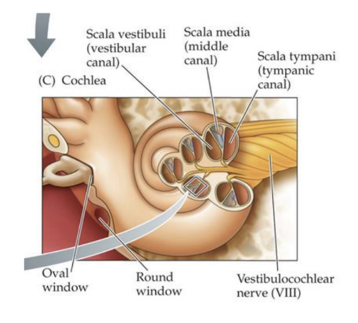
SIGNAL PROCESSING
Transduction: Happens in the organ of Corti within the cochlea.
Basilar membrane: Bottom membrane of the cochlea; hair cells are embedded in it.
Tectorial membrane: Rests on the hair cells.
Hair cells: Function as receptors in the auditory system.
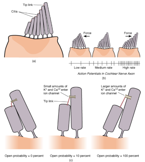
RECEPTOR
Inner hair cell:
Contains stereocilia that detect sound vibrations.
Has afferent (incoming) and efferent (outgoing) nerve endings.
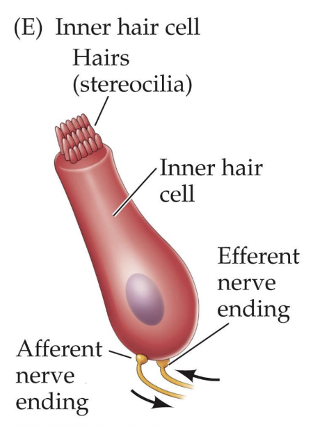
HAIR CELL TRANSDUCTION
Requires mechanical energy.
Tip link stretches when stereocilia bend, opening K+ or Ca++ channels.
Leads to depolarization, vesicle fusion, and neurotransmitter (NT) release.
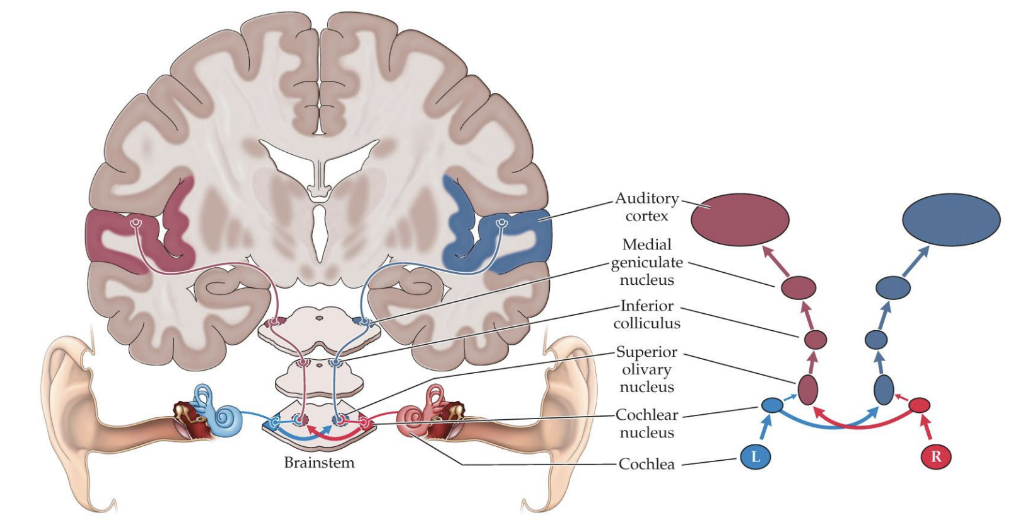
AUDITION PATHWAY
Key structures in sound processing:
Cochlea: Initiates auditory signal processing.
Cochlear nucleus: First relay in the brainstem.
Superior olivary nucleus: Involved in sound localization.
Inferior colliculus: Integrates auditory information.
Medial geniculate nucleus: Thalamic processing area for auditory signals.
Auditory cortex: Processes sound information.
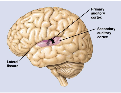
AUDITION CORTEX
Primary auditory cortex: Located in the superior temporal gyrus, processes basic sound information.
Secondary auditory cortex: Also located in the superior temporal gyrus, processes detailed sound and localizes it.
Association areas: Located in the posterior parietal lobe, integrate with other sensory information.
AUDITION DEAFNESS
Affects approximately 37.5 million individuals.
Three major types of deafness:
Conduction deafness: Damage to ossicles, often due to fusion.
Sensorineural deafness: Damage to or destruction of hair cells.
Central deafness: Damage to the auditory cortex, leading to processing issues.
VESTIBULAR SYSTEM
Responsible for balance, movement planning, and directing sensory organs.
Integrates with other brain regions involved in motor control.
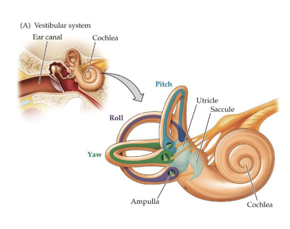
SEMICIRCULAR CANALS
Crucial for balance.
Three axes of rotation: Yaw, Pitch, Roll.
CHEMICAL SENSES
Gustation (taste) and Olfaction (smell) are two chemical senses.
GUSTATION (Taste)
Involves chemicals in solution in the oral cavity.
Taste buds located around papillae on the tongue.
OLFACTION (Smell)
Involves airborne chemicals in the nasal passages.
CHEMICAL SENSES COOPERATION
Taste and smell work together when eating to create flavor.
DAMAGE TO SENSES
Anosmia: Inability to smell, often due to trauma.
Ageusia: Inability to taste, can be observed after damage to ear on the same side.
THE GUSTATORY SYSTEM
Taste receptors found in clusters called taste buds on the tongue.
PRIMARY TASTES
Five primary tastes:
Sweet
Sour
Bitter
Salty
Umami
Originally believed each had distinct receptors, but recent findings suggest:
Sweet, bitter, and umami are coupled to G-proteins (metabotropic).
Salty and sour interaction acts on ion channels (ionotropic).
THE OLFACTORY SYSTEM
Olfactory receptors: About 6 million receptor neurons.
Contain hundreds to thousands of receptor proteins.
Operates through G-protein-coupled receptors (GPCRs).
SMELL: UNIQUE FEATURES
Olfactory receptor cells are continually replaced as they deteriorate.
These receptors can project to multiple brain regions and often bypass the thalamus.
Chapter_5_Book
The Sensorimotor System
1. Introduction to Sensory Systems
Ian Waterman's experience: After a viral infection at age 19, he lost his sense of light touch below the neck but retains pain and temperature sensations.
He adapted to use visual feedback for motor control, learning how to walk by watching his movements.
Challenges faced: Difficulty with balance, walking in darkness, and maintaining calm to avoid unexpected movements.
2. Basics of Sensory Processing
All species respond to environmental cues, using specialized senses to interpret stimuli:
Olfaction (smell), auditory perception (sound), and vision (light).
This chapter covers:
Principles of sensory processing
The role of touch and pain as sensory modalities
Integration of sensory inputs for efficient movement.
3. Sensory Processing and the Somatosensory System
3.1 Key Definitions
Receptor Cell: Specialized cells converting stimuli into electrical signals.
Stimulus: A physical event triggering sensory responses.
Labeled Lines: Concept where specific nerve pathways report distinct types of information.
3.2 Sensory Processing Steps
Transduction: Conversion of stimulus energy into electrical signals (action potentials).
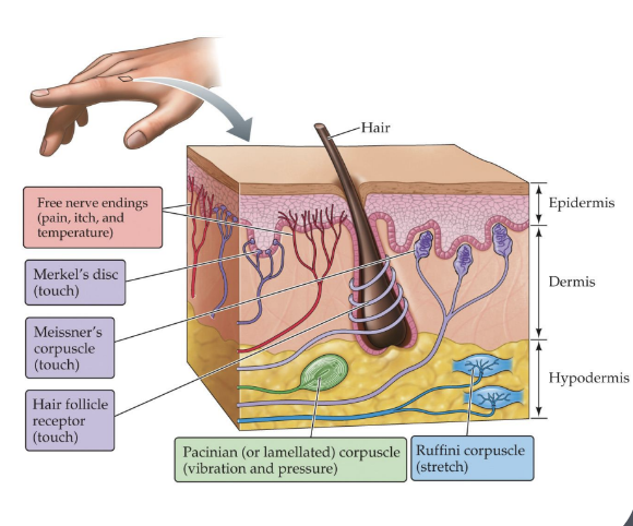
Sensory receptor diversity in the skin includes receptors for touch, pain, and temperature:
Pacinian Corpuscles: Respond to vibration/pressure.
Meissner's Corpuscles: Light touch detection.
Merkel's Discs: Edges and texture sensitivity.
Ruffini Corpuscles: Stretch detection.
Free Nerve Endings: Pain, temperature, and itch.
3.3 Response Coding
Each sensory pathway has distinct processing pathways that filter and encode the location and intensity of the stimulus.
Receptive Fields: Areas that affect a neuron's firing rate upon stimulation.
Adaptation: Decreased response to constant stimuli facilitates focus on critical changes.
3.4 Pain and Its Significance
Pain drives behavioral adaptations to avoid harmful stimuli, promoting recovery behaviors.
Classification of pain:
Sensory-Discriminative Dimension: Pure sensory experience.
Motivational-Affective Dimension: Emotional response.
Cognitive-Evaluative Dimension: Interpretation of pain intensity.
4. Pain Pathways and Modulation
4.1 Pathways of Pain
Nociceptors detect damage and transmit signals via A-delta fibers and C fibers:
A-delta fibers: Fast, sharp pain sensation.
C fibers: Slow, dull pain response.
Anterolateral System: Carries pain and temperature information to the brain.
4.2 Modulation of Pain
Pain perception can be altered by the brain's inhibitory processes:
Endogenous Opioids: Natural pain relief chemicals (e.g., endorphins).
Gate Control Theory: Pain modulation at the spinal cord level.
5. Motor Control and Sensory Feedback
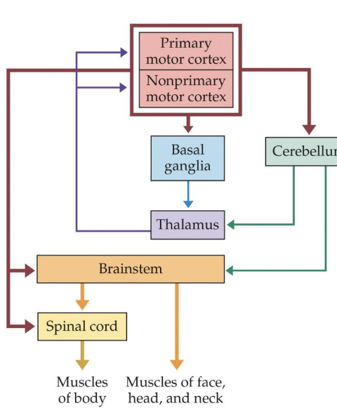
5.1 Motor Hierarchy
Hierarchical organization:
Skeletal System and Muscles: Determine possible movements.
Spinal Cord: Integrates sensory inputs and motor outputs.
Brainstem: Coordinates commands.
Primary Motor Cortex: Executive region for initiating movements.
Nonprimary Motor Cortices: Supplementary areas for refining movement.
Cerebellum & Basal Ganglia: Modulate ongoing motor commands and learning.
5.2 Proprioception
Proprioceptors (muscle spindles and Golgi tendon organs) provide feedback about muscle state, aiding in movement control.
Muscle Spindles: Signal muscle length/stretch.
Golgi Tendon Organs: Monitor tension and protect muscles from overload.
5.3 Pathways from Motor Control
Pyramidal System: Controls fine motor movements; fibers run from the primary motor cortex to spinal motors.
Extrapyramidal System: Modulates movement, involving basal ganglia and cerebellum, affecting muscle tone and coordination.
6. Neuroplasticity in Motor Systems
Motor areas in the cortex can change with experience (training, rehabilitation).
Example: Musicians develop larger cortical areas for finger control.
7. Coordination and Interaction of Systems
Understanding motor behavior involves integrating sensory feedback with planned actions to adapt to external responses.
Mirror Neurons: Fire in response to personal movement or observation of movement, linked to empathy and learning.
8. Movement Disorders
Parkinson’s Disease: Loss of dopaminergic neurons causing tremors, rigidity, and slow movement.
Huntington’s Disease: Genetic disorder leading to involuntary movements and cognitive decline due to basal ganglia damage.
9. Conclusion
Sensory and motor systems are intricately connected, working in concert to allow adaptive behaviors and responses to the environment.
Ch_5_Sensorimotor_system
SENSORIMOTOR SYSTEMS
SOMATOSENSORY PROCESSING
Components: Touch, temperature, and pain
SOMATOSENSORY: ENERGY AND ORGAN
Energy
Types of stimuli processed:
Pressure
Vibration
Tickle
Smoothness
More complicated stimuli
Organ
Main organ involved: Skin
SOMATOSENSORY: RECEPTOR CELLS
Cutaneous Receptors
Free Nerve Endings
Carry information about temperature change, pain, itch
Merkel’s Discs, Meissner’s Corpuscles, Hair Follicle Receptors
Carry information about touch (skin indentation and displacement)
Pacinian Corpuscles
Carry information about vibration and pressure (texture)
Largest and deepest receptors
Ruffini Endings
Carry information about skin stretch
SOMATOSENSORY: PATHWAYS
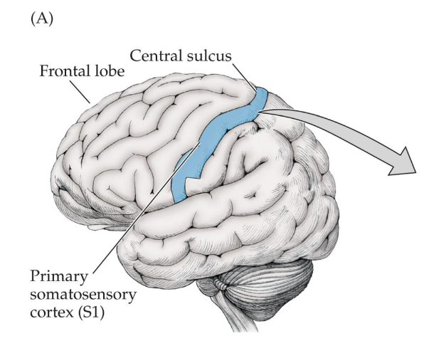
Primary Somatosensory Cortex
Located in the post central gyrus of the parietal lobe
Positioned posterior to frontal lobe and anterior to central sulcus
Known as Primary Somatosensory Cortex (S1)
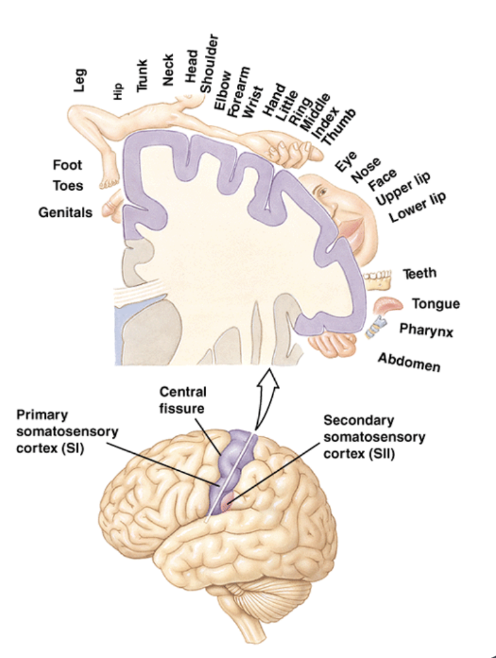
Somatosensory Homunculus
Map representing sensory processing in the primary cortex
Exhibits disproportionate representation of different body areas
Input primarily from the contralateral side of the body
SOMATOSENSORY: PATHWAYS (Continued)
Secondary Somatosensory Cortex
Located ventral to primary cortex in post central gyrus along lateral fissure
Projections extend to the posterior parietal lobe
PAIN
Definition
“An unpleasant sensory and emotional experience associated with actual or potential tissue damage”
Dimensions of Pain
Sensory-discriminative: Throbbing, gnawing, shooting, etc.
Motivational-affective: Tiring, sickening, fearful, etc.
Cognitive-evaluative: No pain, mild, excruciating
PAIN: PURPOSES
Short-lasting pain: Encourages withdrawal from harmful stimuli
Long-lasting pain: Promotes recuperative behaviors
Expression of pain: Serves as a social signal to other animals
PAIN: RECEPTORS AND FIBERS
Nociceptors
Evolutionary perspective: Pain responds to harmful stimulation without a specific stimulus
Types of Pain Fibers
A Delta Fibers: Large-diameter, myelinated; responsible for sharp pain
C Fibers: Small-diameter, unmyelinated; responsible for dull pain
PAIN: TREATMENT
Analgesia
Common treatments include:
Drugs: Opioids and NSAIDs
Opioids: Bind to receptors in the brain, primarily in the periaqueductal gray area, activate endogenous opioids (endorphins)
Transcutaneous Electrical Nerve Stimulation
Placebo Effects
Acupuncture
MOTOR PROCESSING
GENERAL MODEL/HIERARCHY
Overview of top-down signaling from the brain to muscles
Muscles of head and neck controlled by cranial nerves
Spinal cord manages skeletal muscle responses to sensory inputs
MOTOR CORTICES
Lateral View
Includes Premotor Cortex, Supplementary Motor Area, Primary Motor Cortex

Modern View of Primary Motor Cortex
Supports initiation of complex species-typical movements (motor plans)
Examples: Feeding, walking, dressing
Neurons direct movements toward target/endpoint rather than predetermined directions
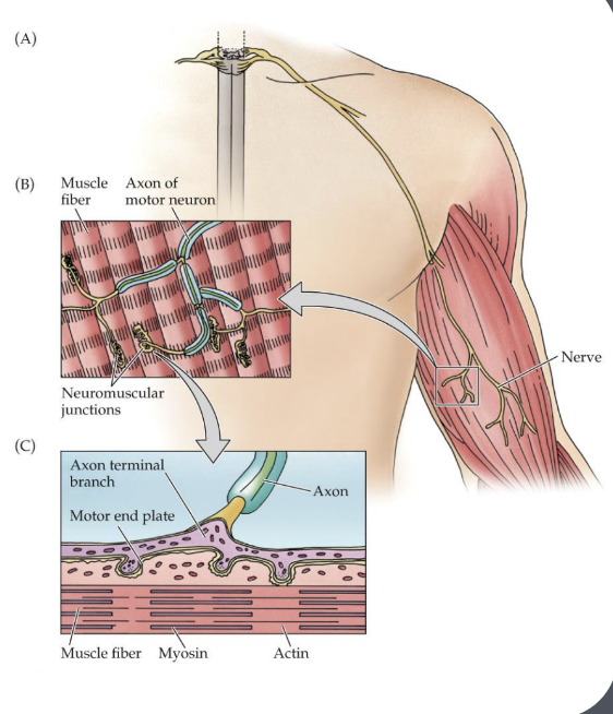
SKELETAL MUSCLE
Structure
Muscle fibers bound together and attached to bones via tendons
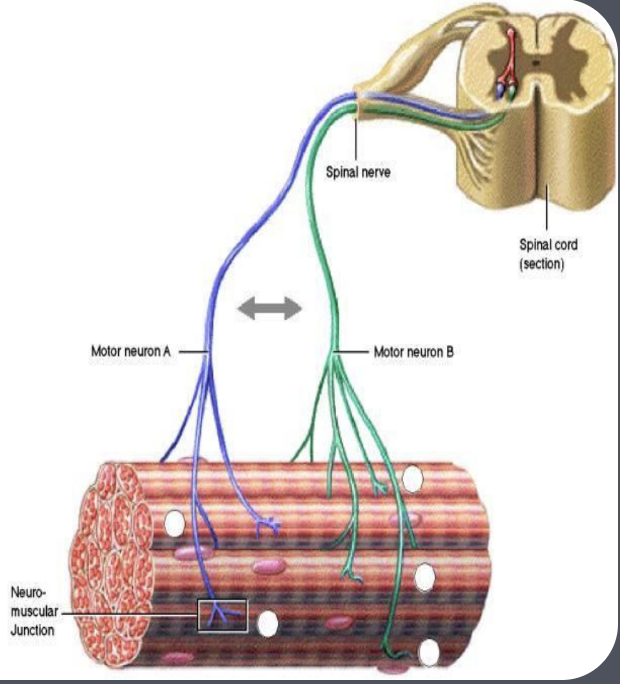
Motor Pool
Collection of all motor neurons that innervate a specific muscle
SKELETAL MUSCLE: Motor Unit
Defined as a single motor neuron and the muscle fibers it innervates
Smallest functional unit of motor activity
All fibers contract upon firing of the motor neuron
There’s variability in fiber count based on control needs (fewer fibers for fine control)
SKELETAL MUSCLE: Contraction
Triggered by motor neuron releasing Acetylcholine at the neuromuscular junction
Contractions occur in one direction only
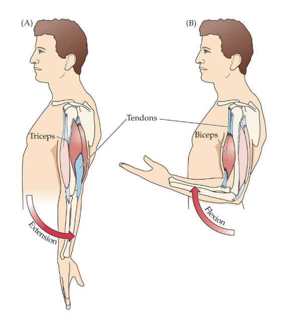
SKELETAL MUSCLE: Antagonistic Muscle Groups
Flexors: Bend or flex a joint
Extensors: Straighten or extend a joint
SKELETAL MUSCLE TYPES
Fast-Twitch Muscle Fibers
Characteristics:
Respond quickly, generate great force, fatigue quickly, fewer blood vessels (pale color)
Slow-Twitch Muscle Fibers
Characteristics:
Respond slower, weaker, capable of sustained contraction, highly vascularized (more red)
SENSORY FEEDBACK REGULATES MOVEMENT
Importance of constant monitoring for coordinated movements
Relies on understanding muscle state and limb positioning
Proprioception
SENSORY FEEDBACK REGULATES MOVEMENT (Continued)
Receptors Monitoring Muscle Activity
Muscle Spindles: Detect changes in muscle length
Golgi Tendon Organs: Detect muscle tension
SPINAL REFLEXES
Stretch Reflex
Activated by sudden external stretching of a muscle
Functions to maintain limb stability
Example: Patellar tendon reflex
SENSORIMOTOR SYSTEM: DAMAGE
Cerebellum Functions
Involved in timing, fine-tuning, and motor learning
Damage Consequences
Impairment in movement control, stability, posture, visual tracking
Decomposition of movement and balance issues (e.g. Ataxia)
Difficulty learning new movements
PROGRESSIVE MOTOR DISORDERS
Parkinson’s Disease
Caused by the loss of dopamine neurons in the substantia nigra projecting to the basal ganglia
Symptoms: Slow movement, tremors, diminished facial expression, and difficulty initiating movements
Huntington’s Disease
Damage to basal ganglia due to dominant genetic contribution
Symptoms: Clumsiness, involuntary movements, aimless jerks, writhing, and eventual cognitive decline