knowt logo

5: Cell Structure

Functional Anatomy of Cells

  • Almost all human cells are microscopic in size.

  • Their diameters range from 7.5 micrometres to about 150μm.

Units of Size

UNIT

SYMBOL

EQUAL TO

USED TO MEASURE

Centimetre

cm

1/100 metre

Objects visible to the eye

Millimetre

mm

1/1,000 metre (1/10 cm)

Very large cells; groups of cells

Micrometre (micron)

μm

1/1,000,000 metre (1/1000 mm)

Most cells; large organelles

Nanometre

nm

1/1,000,000,000 metre (1/1,000 μm)

Small organelles; large biomolecules

Cell Types

TYPES

FEATURES

FUNCTIONS

Nerve Cells

  • Surface that is sensitive to stimuli

  • Long extensions

  • Detect changes in internal or external environment

  • Transmit nerve impulses from one part of the body to another

Muscle Cells

  • Elongated, threadlike

  • Contain tiny fibres that slide together forcefully

  • Contract (shorten) to allow movement of body parts

Red Blood Cells

  • Contain haemoglobin, a red pigment that attracts, then releases, oxygen

  • Transport oxygen in the bloodstream (from lungs to other parts of the body)

Gland Cells

  • Contain sacs that release a secretion to the outside of the cell

  • Release substances such as hormones, enzymes, mucus, and sweat

Immune Cells

  • Some have outer membranes able to engulf other cells

  • Some have systems that manufacture antibodies

  • Some are able to destroy other cells

  • Recognise and destroy “nonself” cells such as cancer cells and invading bacteria

Cells

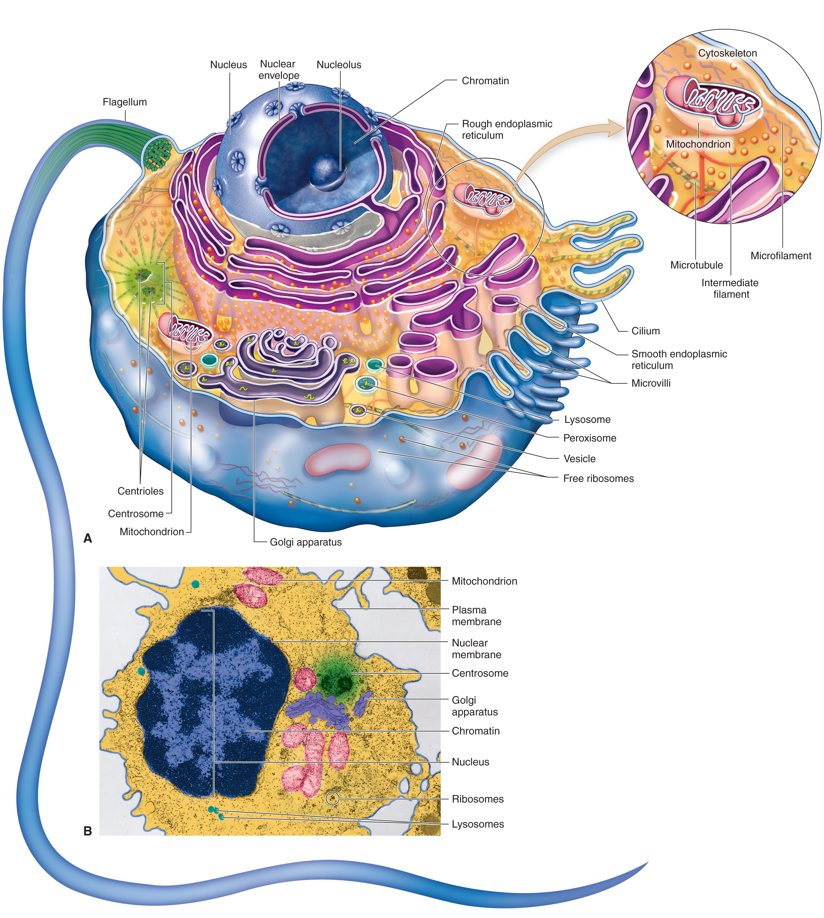
Major Cell Structures and Their Functions

CELL STRUCTURE

DESCRIPTION

FUNCTIONS

Membranous

Plasma membrane

Phospholipid bilayer reinforced with cholesterol and embedded with proteins and other organic molecules

Serves as the boundary of the cell, maintains its integrity; protein molecules embedded in plasma membrane perform various functions— they serve as markers that identify cells of each individual, as receptor molecules for certain hormones and other molecules, and as transport mechanisms

Endoplasmic reticulum (ER)

Network of canals and sacs extending from the nuclear envelope; may have ribosomes attached

Ribosomes attached to rough ER synthesise polypeptides that enter rough ER for folding and finishing, then move on to smooth ER; ER synthesises IMPs and membrane lipids incorporated in cell membranes, steroid hormones, detoxification enzymes, glycogen-regulating enzymes, and carbohydrates used to form glycoproteins—also removes and stores Ca ++ from the cell’s interior.

Rough ER: produce proteins in the cells.
Smooth ER: produce lipids and also detoxify toxins in the body

Golgi apparatus

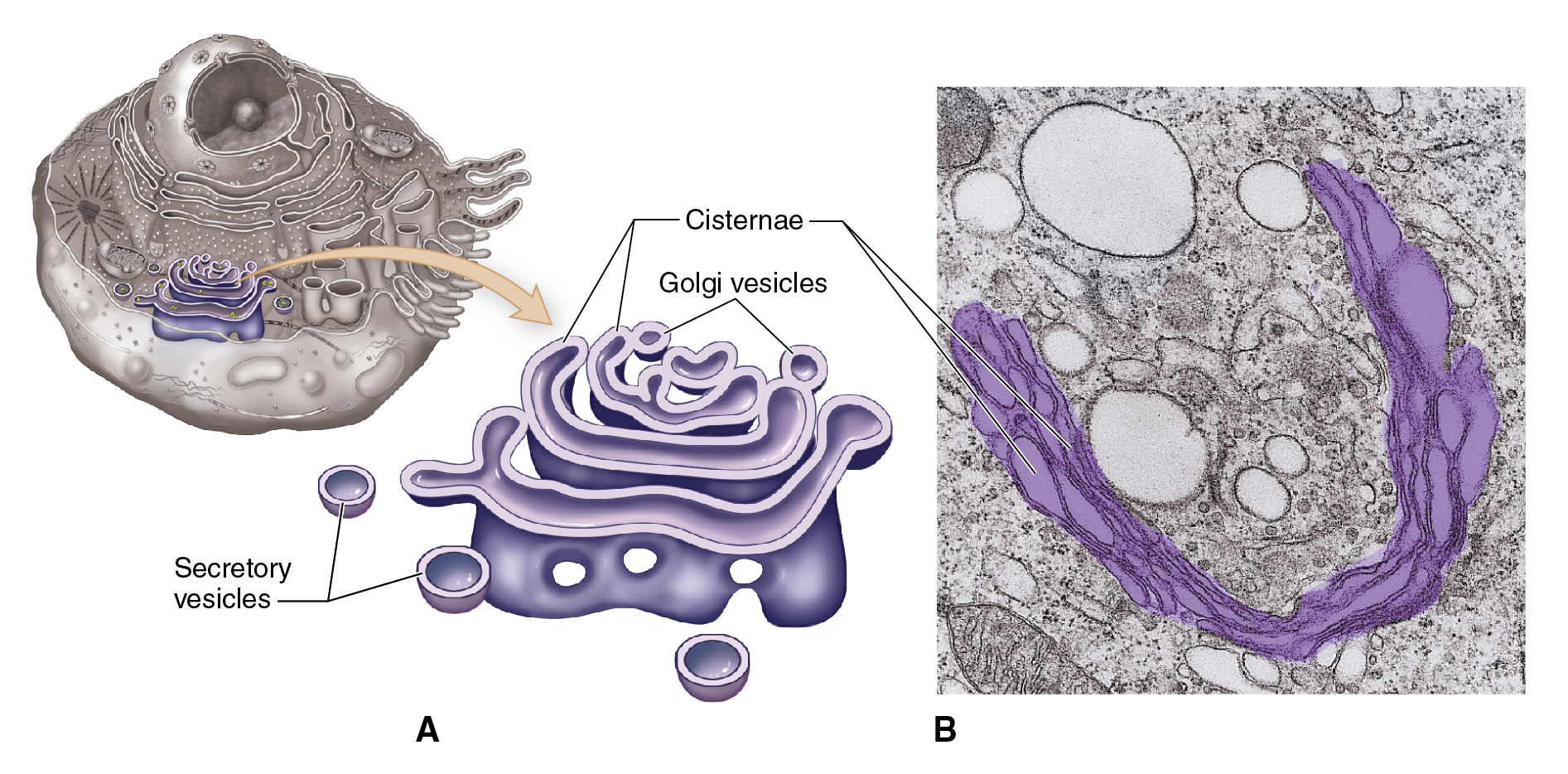
Stack of flattened sacs (cisternae) surrounded by vesicles

Synthesises carbohydrate, combines it with protein, and packages the product as globules of glycoprotein.

transport, sorting, and modification of both protein and lipids.

Vesicles

Tiny membranous bags

Temporarily contain molecules for transport or later use

move molecules, secrete substances, digest materials, or regulate the pressure in the cell

Lysosomes

Tiny membranous bags containing enzymes

Digestive enzymes break down defective cell parts (autophagy) and ingested particles; a cell’s “digestive system”; some lysosomes are involved in membrane repair or secretion

Peroxisomes

Tiny membranous bags containing enzymes

Enzymes detoxify harmful substances in the cell.

They break down molecules through oxidation; they are not part of the endomembrane system.

Mitochondria

Tiny membranous capsule surrounding an inner, highly folded membrane embedded with enzymes; has small, ringlike chromosome (DNA)

Catabolism; adenosine triphosphate (ATP) synthesis; a cell’s “power plants”

Nucleus

A usually central, spherical double-membrane container of chromatin (DNA); has large pores

Houses the genetic code, which in turn dictates protein synthesis, thereby playing an essential role in other cell activities, namely, cell transport, metabolism, and growth

Nonmembranous

Ribosomes

Small particles assembled from two tiny subunits of rRNA and protein

Site of protein synthesis; a cell’s “protein factories”

Proteasomes

Hollow protein cylinders with embedded enzymes

Destroys misfolded or otherwise abnormal proteins manufactured by the cell; a “quality control” mechanism for protein synthesis

Cytoskeleton

Network of interconnecting flexible filaments, stiff tubules, and molecular motors within the cell

Supporting framework of the cell and its organelles; functions in cell movement (using molecular motors); forms cell extensions (microvilli, cilia, flagella)

Centrosome

Region of cytoskeleton that includes two cylindrical groupings of microtubules called centrioles

Acts as the microtubule-organizing centre (MTOC) of the cell; centrioles assist in forming and organizing microtubules

Microvilli

Short, fingerlike extensions of plasma membrane; supported internally by microfilaments

Tiny, fingerlike extensions that increase a cell’s absorptive surface area

Cilia and flagella

Moderate (cilia) to long (flagella) hairlike extensions of plasma membrane; supported internally by cylindrical formation of microtubules, sometimes with attached molecular motors

Cilia move substances over the cell surface or detect changes outside the cell; flagella propel sperm cells

Nucleolus

Dense area of chromatin and related molecules within nucleus

Site of formation of ribosome subunits

Cell Structures

  • Main Cell Structure:

    • Plasma Membrane

    • Cytoplasm

    • Organelles

    • Nucleus

  • Each cell is surrounded by a plasma membrane that separates it from the environment.

  • Cytoplasm — gel-like substance inside the cell; it is made up of various organelles and molecules suspended in cytosol.

  • Cytosol — also known as the intracellular fluid; the fluid present in the cell and is a constituent of the cytoplasm.
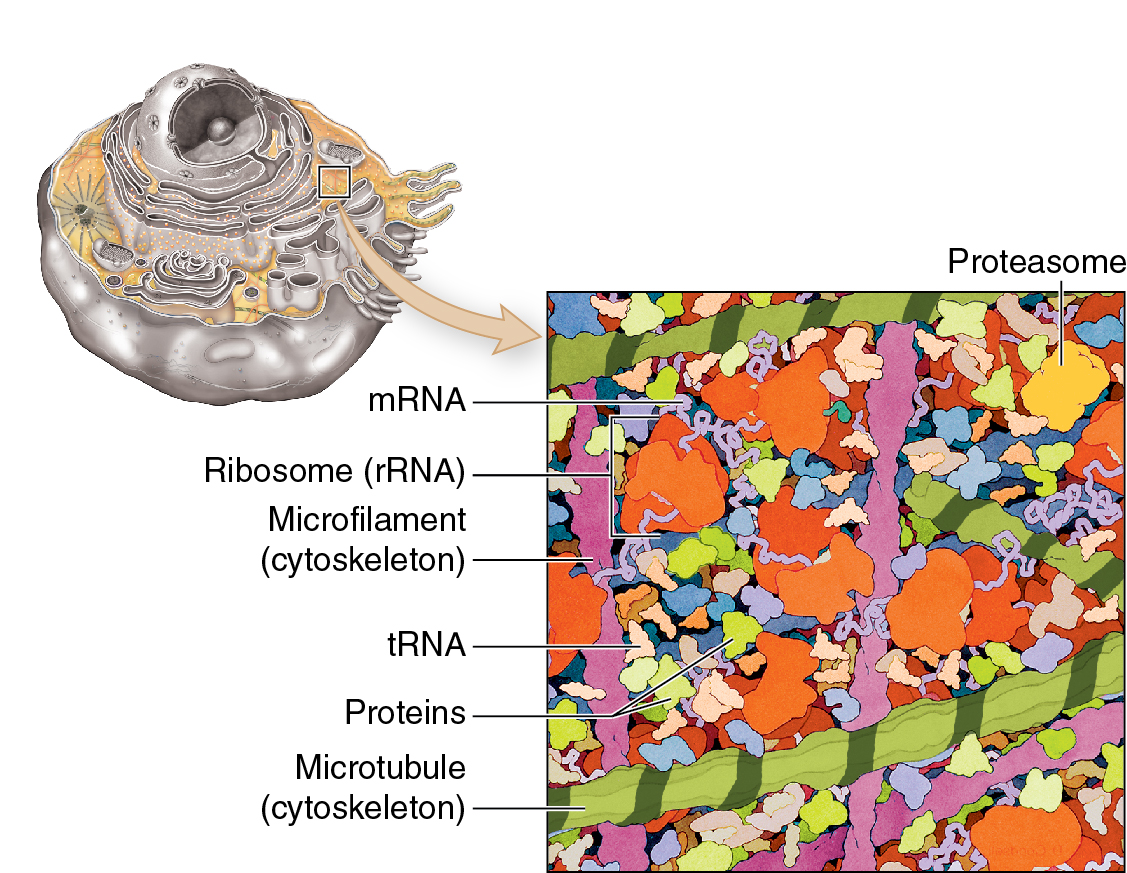
Cytoplasm

Cell Membranes

  • Membranous organelles — sacs and canals made of the same type of membrane material as the plasma membrane.

    • This material is a very thin sheet (about 7.5 nm thick), made of lipid, protein, and other molecules.

Membrane Structure

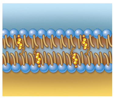
  • Fluid Mosaic Model — concept of cell membranes, like the times in an art mosaic, the different molecules that make up a cell membrane are arranged in a sheet.

    • It shows that the molecules of a cell membrane are bound tightly enough to form a continuous sheet but loosely enough that the molecules can slip past one another.

Plasma Membrane
  • Chemical attractions — the forces that hold a cell membrane together.

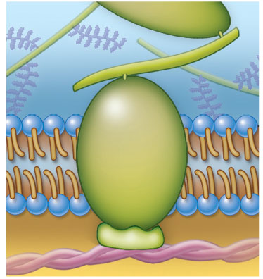
  • The primary structure of a cell membrane is a double layer of phospholipid molecules.

    • Because their heads are hydrophilic and the tails are hydrophobic, these molecules naturally arrange themselves into bilayers in water.

    • Phospholipid bilayers appear wherever phospholipid molecules are scattered among the water molecules.

  • Cholesterol — a steroid lipid that mixes with phospholipid molecules to form a blend of lipids that stays just fluid enough to function properly at body temperature.

    • Without this, the cell membranes would break far too easily.

  • Phospholipids and cholesterol serve as a ‘fence’ that fits together to form a barrier.

    • Water and water-soluble things can’t easily pass through because most of the molecules are hydrophobic.

  • Different parts of the membrane can be stiffer or more flexible, depending on what the cell needs. Many of these are packed more densely with proteins, while others have fewer proteins.

  • Some membrane lipids combine with carbohydrates to form glycolipids.

    • Some membrane lipids combine with protein to form lipoproteins.

  • Lipid rafts — stiff groupings of membrane molecules that travel together around the membrane.

    • It helps organize things in the membrane and can even help the cell take substances.

  • Caveolae — tiny pits of the plasma membrane like tiny caves.

    • They form from lipid rafts and protein molecules that pinch in and move inside the cell.

    • It can capture extracellular material and shuttle it inside the cell.

    • Some of these may have CD36 cholesterol receptors that attract low-density lipoproteins.

    • When LDLs attach to these receptors, the caveola closes and brings the LDL inside the cell. The LDLs then build up behind the lining of the blood vessel, making it narrower and blocking blood flow. This can lead to serious problems like stroke and heart disease.

Membrane Function

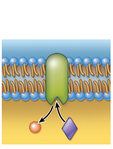
  • Integral Membrane Proteins (IMPs) — protein integrated into the structure of the membrane itself.

Membrane Function

Description

Transport

IMPs act as transporters, forming gates that allow specific water-soluble molecules to pass through the membrane. Cells can control when these gates open or close

Identification

Some IMPs, in the form of glycoproteins, act as markers to help the immune system identify "self" from "nonself" cells, aiding in immune responses and compatibility

Signalling

IMPs function as receptors, reacting to hormones or chemicals to trigger metabolic changes through signal transduction, playing a vital role in cellular communication

  • Signal Transduction — the process which cells translate the signal received by a membrane receptor into a specific chemical change in the cell.

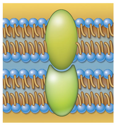
Connection

IMPs connect the cell membrane to other cells, membranes, or to internal/external structures like the cytoskeleton or extracellular matrix, supporting tissue formation

Functional Anatomy of Cell Membranes

ANATOMY

STRUCTURE

FUNCTION

Sheet (bilayer) of phospholipids stabilised by cholesterol

Maintains boundary (integrity) of a cell or membranous organelle

Integral membrane proteins that act as channels or carriers of molecules

Controlled transport of water-soluble molecules from one compartment to another

Receptor molecules that trigger metabolic changes in membrane (or on other side of membrane)

Sensitivity to hormones and other regulatory chemicals; involved in signal transduction

Enzyme molecules that catalyze specific chemical reactions

Regulation of metabolic reactions

Integral membrane proteins that bind to molecules outside the cell

Form connections between one cell and another

Integral membrane proteins that bind to support structures

Support and maintain the shape of a cell or membranous organelle; participate in cell movement; bind to fibres of the extracellular matrix (ECM)

Glycoproteins or proteins in the membrane that act as markers

Recognition of cells or organelles


Cytoplasm and organelles

Cytoplasm

  • Early scientists viewed cytoplasm as a uniform fluid. Advances in microscopy revealed its complexity, including organelles previously considered inclusions due to their undetected roles.

  • Components:

    • Cytosol

    • Organelles

Organelles

  • Membranous Organelles — Structures made of cell membranes (sacs or canals).

  • Nonmembranous Organelles — Structures made of microscopic filaments or other particles, not enclosed by membranes.

Endoplasmic Reticulum

  • Endoplasmic Reticulum — A network within the cytoplasm, consisting of membranous canals and sacs.

    • It is found throughout the cytoplasm and is involved in various cellular functions.

  • Types of ER

    • Rough Endoplasmic Reticulum (RER)

      • It is composed of flattened sacs with ribosomes on their surface, giving it a "rough" appearance.

      • Synthesizes proteins as new polypeptides enter the RER's lumen (internal cavity).

      • Helps fold these proteins with the assistance of chaperone molecules.

      • Produces phospholipids that contribute to the ER's membrane.

    • Smooth Endoplasmic Reticulum (SER)

      • It is more tubular in shape and lacks ribosomes, giving it a "smooth" appearance.

      • Continues chemical processing initiated in the RER.

      • Synthesizes lipids, carbohydrates, and steroid hormones.

      • Contains enzymes that detoxify harmful substances (like drugs) and regulate glycogen breakdown into glucose.

      • Synthesizes most phospholipids and cholesterol for cell membranes.

      • Stores calcium ions (Ca²⁺) to maintain low internal concentrations, which is important for various cellular functions.
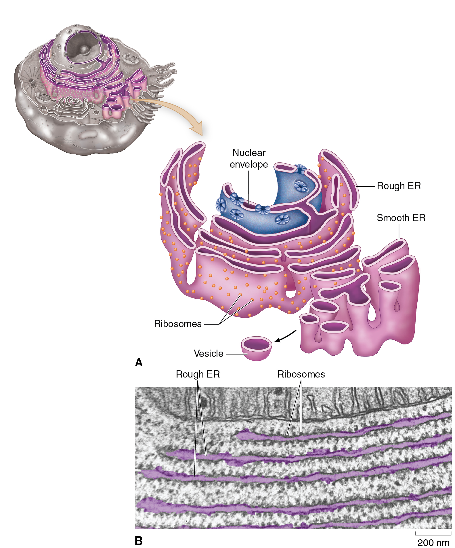
ER

Ribosomes

  • Ribosomes — nonmembranous structures essential for protein synthesis, composed of two subunits (large and small).

  • The cell’s protein factories.

  • Found attached to the rough ER and free in the cytoplasm.

  • They translate the genetic code (from mRNA) to synthesize proteins.

  • They assemble amino acids into polypeptide chains as directed by mRNA, which contains the "recipe" for proteins.

  • Polyribosomes — groups of ribosomes that work together on the same mRNA strand, appearing like strings of beads under an electron microscope.

  • The two subunits temporarily join when mRNA is present and separate once the protein synthesis is complete.

  • Types of RNA in Ribosomes

    • Ribosomal RNA (rRNA) — Forms the core of ribosomes.

    • Messenger RNA (mRNA) — Carries genetic instructions from DNA for protein synthesis.

    • Transfer RNA (tRNA) — Transfers specific amino acids to the ribosome based on the mRNA sequence.
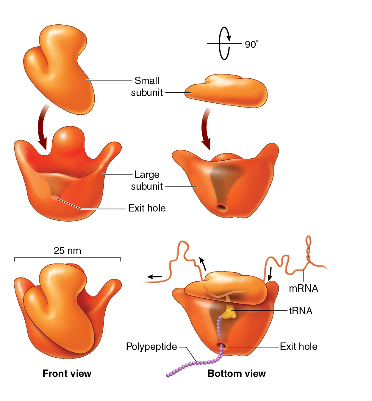
Ribosome

Golgi Apparatus

  • Golgi apparatus — a membranous organelle made of small sacs called cisternae. It's located near the nucleus and is also known as the Golgi complex.

  • It processes, modifies, and packages proteins and glycoproteins (proteins with attached carbohydrates) for export or use in the cell. The proteins come from the endoplasmic reticulum (ER) and pass through the Golgi apparatus for final modification before they leave the cell or become part of the plasma membrane.

  • Proteins are packaged in small vesicles from the ER, moved through the Golgi apparatus, and eventually secreted out of the cell through exocytosis.

    • Exocytosis — the process of vesicles fusing with the plasma membrane and releasing contents.
Golgi Apparatus

Lysosomes

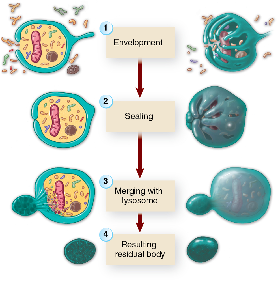
  • Lysosomes — membranous vesicles that pinch off from the Golgi apparatus and contain digestive enzymes.

  • They break down unnecessary proteins and other cellular waste, a process called autophagy (self-eating).

  • It also help destroy bacteria or other harmful particles, earning them the nickname "digestive bags" or "suicide bags."

  • Lysosomes help recycle old organelles or proteins by breaking them down into basic components, which the cell can reuse.
Proteasomes

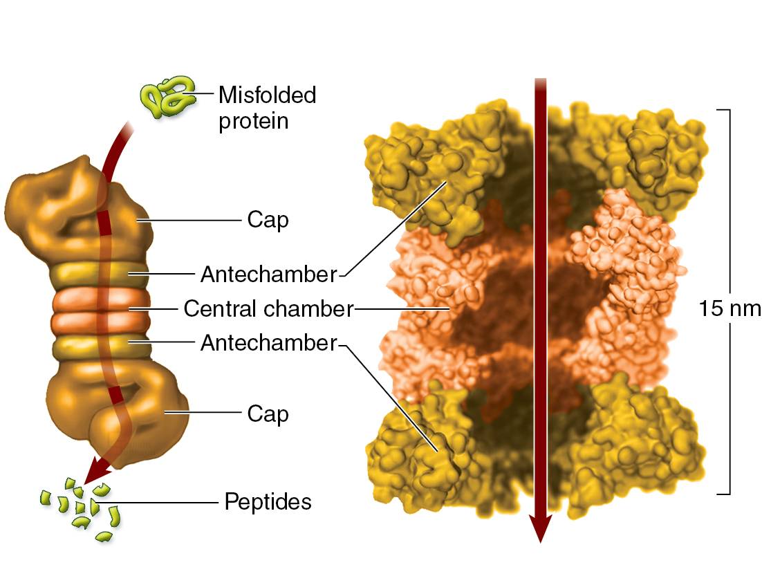
  • Proteasomes — These are protein-destroying organelles made up of protein subunits in a cylindrical shape.

  • They destroy abnormal or misfolded proteins one at a time, unlike lysosomes that destroy groups of proteins.

  • Proteins that need to be destroyed are tagged with ubiquitin, which signals the proteasome to degrade the protein into peptides (small chains of amino acids).

  • Proper functioning of proteasomes helps prevent diseases, such as Parkinson’s disease, where proteasomes fail to destroy defective proteins.
Peroxisomes

  • Peroxisomes — small vesicles containing enzymes. They are similar to lysosomes but specialize in detoxification.

  • They break down toxic substances like hydrogen peroxide (H₂O₂), using enzymes like catalase.

  • Peroxisomes contain the enzymes peroxidase and catalase, which are important in detoxification reactions involving hydrogen peroxide (H2 O2 ).

  • They are abundant in kidney and liver cells where detoxification is important.

Mitochondria

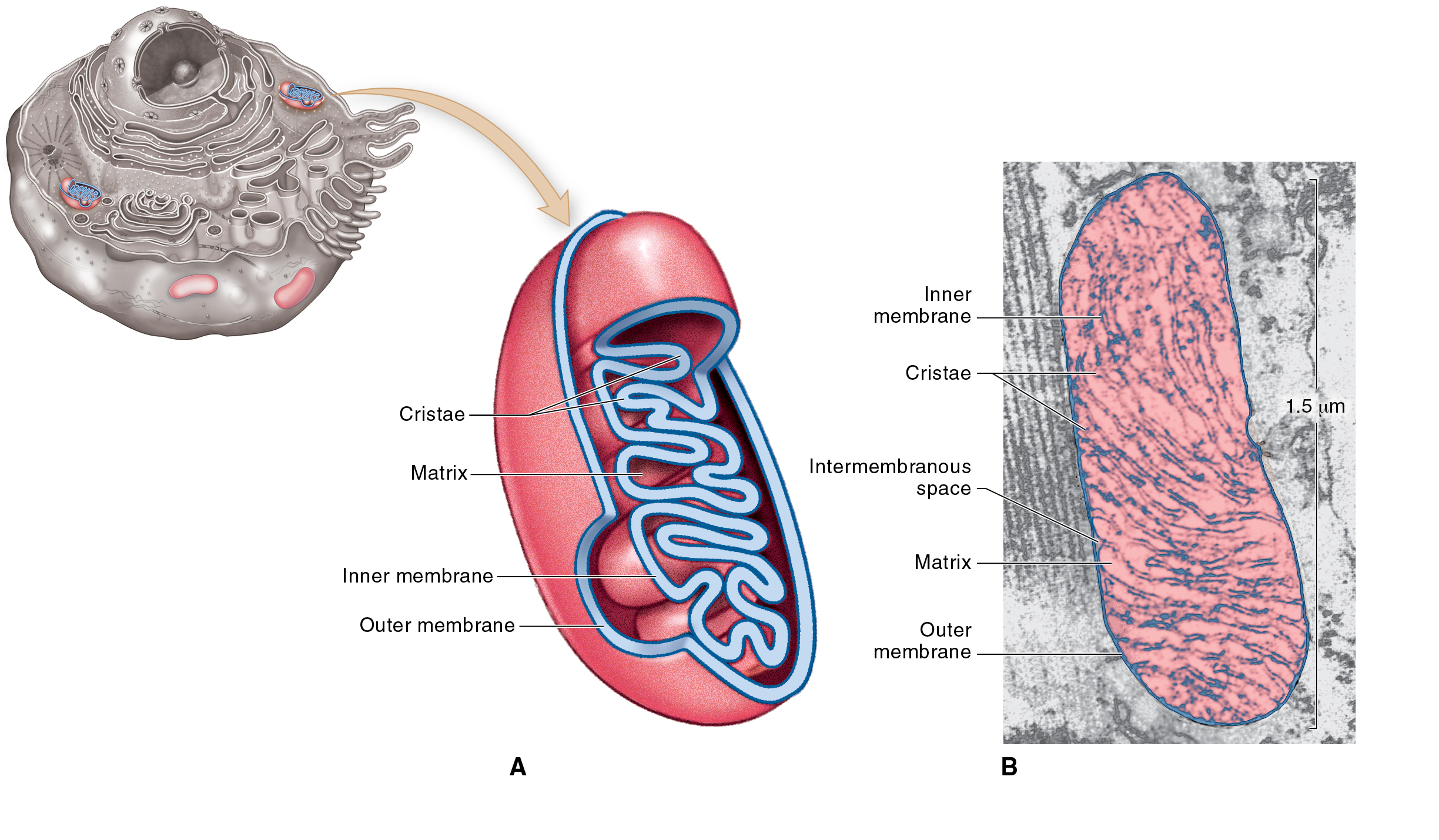
  • Mitochondria — small, sausage-shaped organelles known as the cell’s “power plants”.

  • They have two membranes—the outer membrane and the folded inner membrane, which forms cristae. The folds increase the surface area for chemical reactions.

  • Mitochondria generate ATP (adenosine triphosphate), the cell’s main energy source, through the breakdown of food molecules.

  • Cells with high energy needs (like liver cells) have many mitochondria, while less active cells (like sperm cells) have fewer.

  • Each mitochondrion has its own DNA and can replicate independently, which is why it is sometimes thought to have evolved from ancient bacteria.

Nucleus

  • Nucleus — one of the largest structures in the cell, usually found in the centre. Most cells have a single, spherical nucleus, but the shape and number of nuclei can vary between different types of cells.

  • Nucleoplasm — a viscous liquid that fills the nucleus, just like the cytosol of the cell.

  • The nucleus is surrounded by a nuclear envelope, which consists of two membranes with nuclear pores.

    • These pores regulate the movement of molecules in and out of the nucleus.

    • The nuclear envelope is continuous with the endoplasmic reticulum (ER), linking it to the cell's internal transport system.

    • Vaults — tiny, barrel-shaped organelles that may also also assist with transport of molecules to and from the nucleus.

  • Nuclear Pore Complex (NPC) — These are intricate structures that act as gatekeepers, allowing specific molecules to pass into and out of the nucleus while preventing unwanted material from entering.
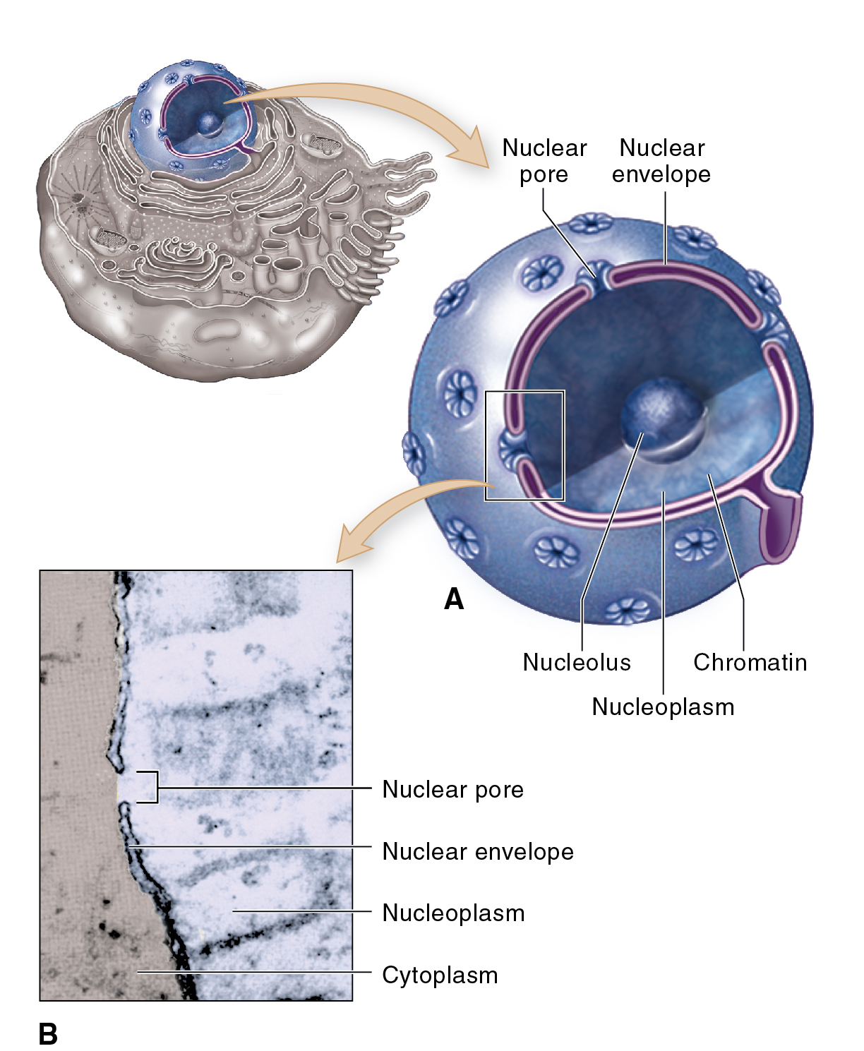
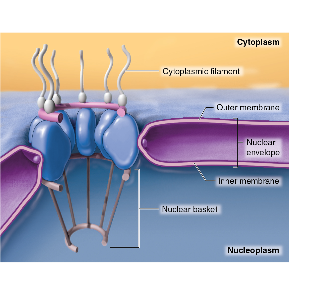
Nucleus<br />Nuclear Pore Complex (NPC)
  • Inside the nucleus, DNA molecules are present in the form of chromatin—tiny threads that are tangled and sprinkled with granules; this makes up the chromosomes.

    • Chromatin is named from the Greek word for "colour" because it stains easily.

    • In non-dividing cells, the chromatin strands occupy specific areas and move around within the nucleus.

  • When a cell begins to divide, chromatin becomes tightly coiled to form chromosomes.

    • Human cells (except sex cells) have 46 chromosomes, each made up of one DNA molecule and associated proteins.

  • The DNA in the nucleus carries the genetic code for making RNA, enzymes, and other proteins.

    • This genetic code controls both the structure and function of the cell, and the DNA is passed on during cell division, playing a crucial role in heredity.

  • Nucleolus — a small non-membranous body that stains densely, which is found inside the nucleus.

    • It is made primarily of RNA (ribonucleic acid), not DNA.

    • The nucleolus synthesizes ribosomal RNA (rRNA) and combines it with proteins to form ribosomal subunits, which later join together to create ribosomes (the protein factories of the cell).

    • Cells that produce large amounts of protein, like those in the pancreas, have larger nucleoli.


Cytoskeleton

  • Cytoskeleton — the internal supporting framework of a cell, comparable to the skeletal system in humans.

    • Consists of rigid, rod-liek fibres that provide support and allow movement.

    • It is involved in moving parts of the cell.

    • It allows for both internal (movement of organelles and vesicles) and external (whole cell movement) processes.

    • During cell division, the cytoskeleton helps in chromosome separation by forming the spindle.

    • The cytoskeleton can respond to signals and environmental changes by reorganising itself, allowing the cell to adapt.
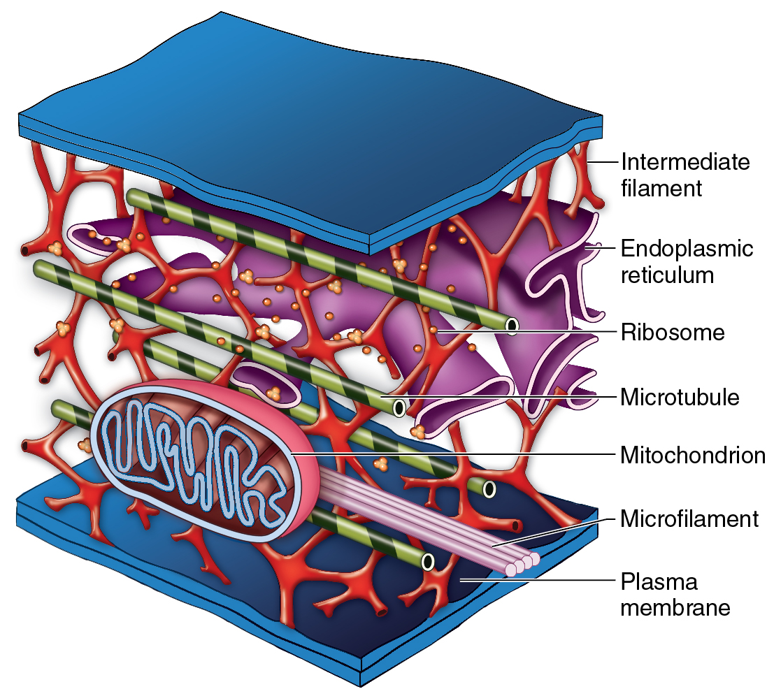
Cell Fibres

  • Cell fibres — these are long, thin structures found in cells that provide support, structure, and movement.

Types of Cell Fibres

  • Microfilaments — the smallest fibres, made of thin, twisted strands of protein.

    • They function as cellular muscles, enabling cells to contract and shorten.

    • This process is especially important in muscle cells, where bundles of microfilaments work together to generate force.

  • Intermediate Filaments — These fibres are thicker and stronger than microfilaments.

    • They are like the tendons and ligaments of a cell, providing structural support and resisting tension.

    • For example, the outer layer of skin cells contains many intermediate filaments, making them tough and resilient.

  • Microtubules — The thickest fibres, which are hollow tubes made of protein subunits.

    • They act as the engines of the cell, responsible for moving things around inside the cell and helping with processes like chromosome separation during cell division.

Centrosome

  • Centrosome — a region near the nucleus that coordinates the assembly and breakdown of microtubules.

    • It is also known as the microtubule-organising centre (MTOC).

  • Centrioles — two cylindrical structure found in the centrosome, which help in cell division by forming a spindle of microtubules that pulls chromosomes apart.

  • Pericentriolar material (PCM) — a cloudlike mass of material surrounding the centrioles, it is active in starting the growth of new microtubules.

  • Aster — a formation of microtubules radiating outward from the centrioles.
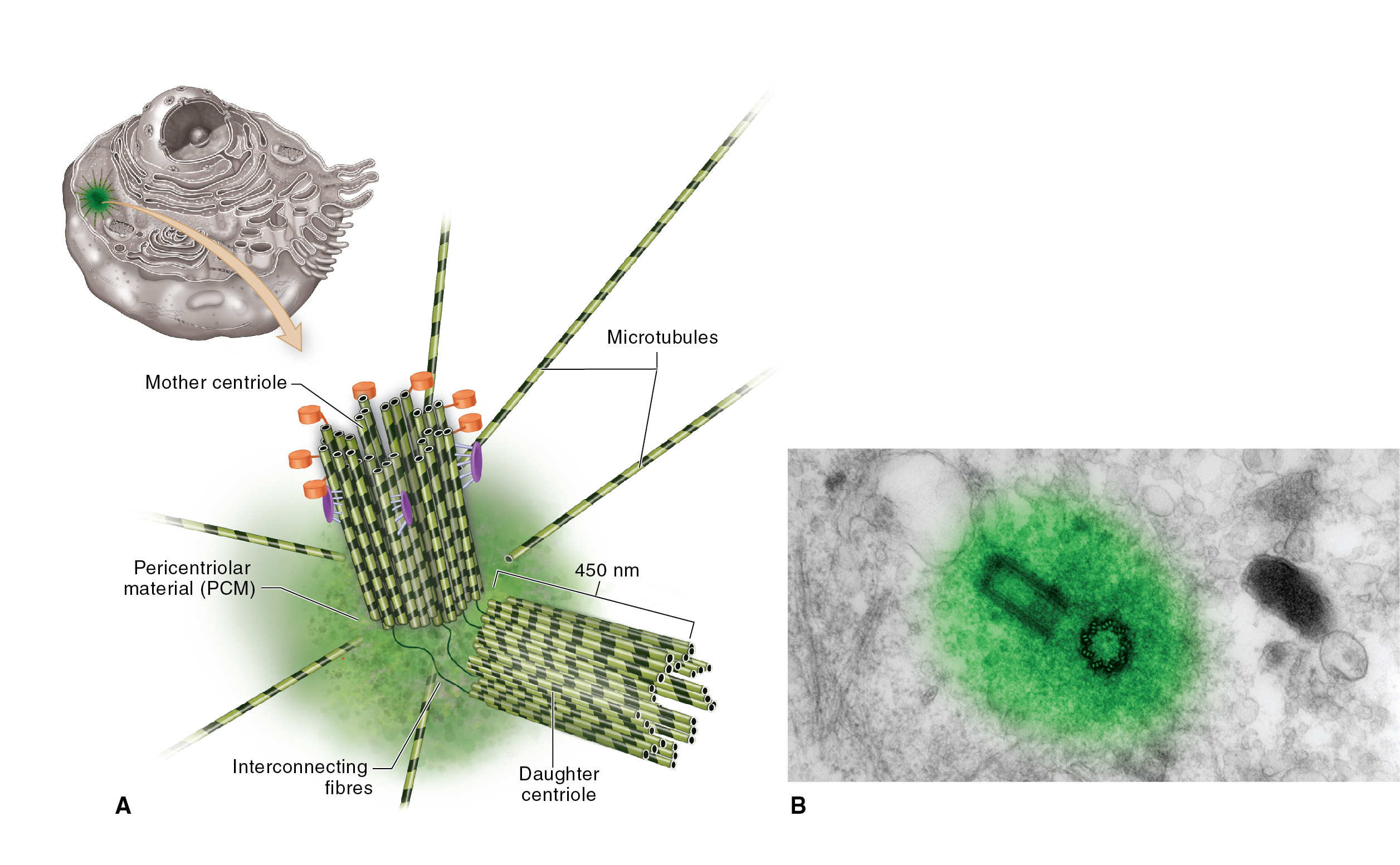
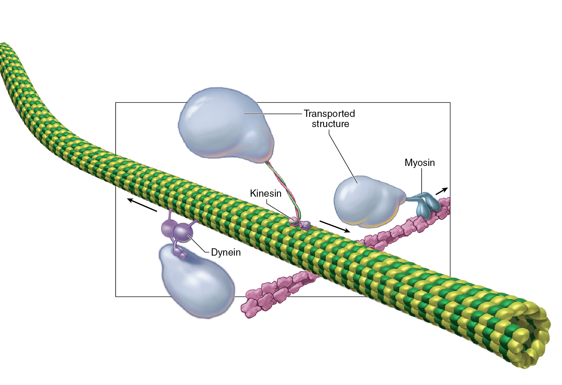
Molecular Motors

  • Molecular motors These are proteins that move vesicles, organelles, and other structures along the microtubules and microfilaments of the cytoskeleton.

  • These tiny motors transport materials within the cell like trains on a track and are also responsible for cell movement.

  • It also allows the cell’s framework to move with force, extending and contracting to create movements of the cell.
Cell Extensions

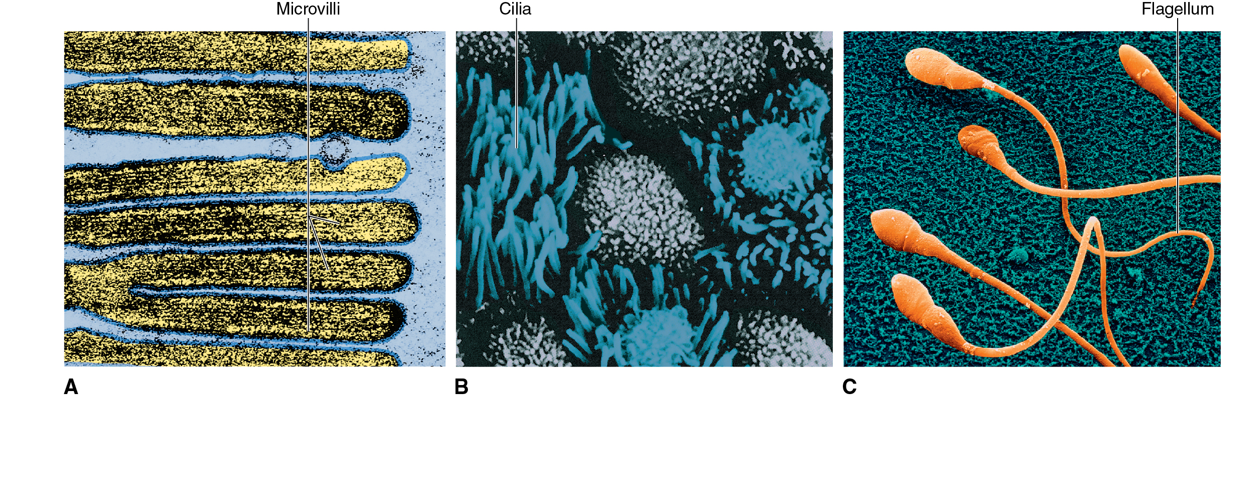
  • Microvilli — Tiny, finger-like projections found on cells where absorption is key (e.g., in the intestines).

    • They increase surface area and help with absorption.

    • Inside each microvillus is a bundle of microfilaments that provides support and mobility.

  • Cilia — Short, hair-like extensions that move substances (e.g., mucus) across the surface of cells.

    • They move in a coordinated, wave-like manner and are found in areas such as the respiratory tract.

  • FlagellaSingle, long extensions found on sperm cells.

    • It move in a wave-like motion, allowing the sperm to "swim."

Cell Connections

Extracellular Matrix Connections

  • Integrins — These are a group of integral membrane proteins that help cells attach to the extracellular material, or matrix, surrounding them.

  • Some integrins span the plasma membrane and link the cytoskeleton inside the cell to the extracellular fibres, anchoring the cell in place.

Direct Cell-to-Cell Connections

  • Cells can also form direct connections with each other, facilitated by proteins like integrins, selectins, cadherins, and immunoglobulins.

    • These connections are essential for holding cells together and sometimes enable direct communication between them.

  • Desmosomes — these act like small “spot welds” that hold adjacent cells tightly together, similar to how Velcro works.

    • The fibres on the surface of each of this interlock with each other, forming a strong bond.

    • Spot desmosomes are located at specific points between cells (e.g., in skin cells).

    • Belt desmosomes (also called adhesive belts or zona adherens) form a belt-like structure that encircles the cell, providing even more connection strength.

  • Gap Junctions — these form when membrane channels of neighbouring cells connect, creating “tunnels” between them.

    • This allows molecules, ions, and electrical signals to pass directly from one cell to another.

    • These are crucial in heart muscle cells, enabling electrical impulses to travel seamlessly across multiple cells, allowing coordinated heartbeats.

  • Tight Junctions — these occur when cells are tightly bound together near their apical surfaces (the top part of the cell).

    • The plasma membranes of adjacent cells fuse together, forming a tight seal.

    • These junctions prevent the passage of substances between cells, ensuring that molecules must pass through controlled channels in the cell membrane.

    • These are essential in areas like the intestines, where the body needs to regulate what gets absorbed into the bloodstream.


Cellular Disease

  • Type 2 Diabetes — form of diabetes mellitus (DM), often produced by a cellular response to obesity that triggers a reduction in the number of functioning membrane receptors for the hormone insulin.

  • Duchenne muscular dystrophy (DMD) — a severe inherited condition, results from “leaky” membranes in muscle cells.

    • Dystrophin — a protein that normally helps connect a muscle cell’s cytoskeleton to the plasma membrane and to the surrounding extracellular matrix, is missing.

    • Muscle contractions pull at the weakened connections to the plasma membrane and rip holes that allow calcium ions (Ca ++ ) to enter the cell. This flood of Ca ++ triggers chemical reactions that destroy the muscle, causing life-threatening paralysis.

  • Parkinson disease (PD)

    • Mitochondrial dysfunction reduces the cell’s energy production, affecting muscle control areas in the brain.

    • It involves the breakdown of microtubules in the cytoskeleton, contributing to movement difficulties.

  • Alzheimer's Disease (AD)

    • Dysfunction in proteasomes leads to the buildup of abnormal proteins, forming plaques in the brain.

    • These plaques are characteristic of AD and contribute to cognitive decline.

MA

5: Cell Structure

Functional Anatomy of Cells

  • Almost all human cells are microscopic in size.

  • Their diameters range from 7.5 micrometres to about 150μm.

Units of Size

UNIT

SYMBOL

EQUAL TO

USED TO MEASURE

Centimetre

cm

1/100 metre

Objects visible to the eye

Millimetre

mm

1/1,000 metre (1/10 cm)

Very large cells; groups of cells

Micrometre (micron)

μm

1/1,000,000 metre (1/1000 mm)

Most cells; large organelles

Nanometre

nm

1/1,000,000,000 metre (1/1,000 μm)

Small organelles; large biomolecules

Cell Types

TYPES

FEATURES

FUNCTIONS

Nerve Cells

  • Surface that is sensitive to stimuli

  • Long extensions

  • Detect changes in internal or external environment

  • Transmit nerve impulses from one part of the body to another

Muscle Cells

  • Elongated, threadlike

  • Contain tiny fibres that slide together forcefully

  • Contract (shorten) to allow movement of body parts

Red Blood Cells

  • Contain haemoglobin, a red pigment that attracts, then releases, oxygen

  • Transport oxygen in the bloodstream (from lungs to other parts of the body)

Gland Cells

  • Contain sacs that release a secretion to the outside of the cell

  • Release substances such as hormones, enzymes, mucus, and sweat

Immune Cells

  • Some have outer membranes able to engulf other cells

  • Some have systems that manufacture antibodies

  • Some are able to destroy other cells

  • Recognise and destroy “nonself” cells such as cancer cells and invading bacteria

Cells

Major Cell Structures and Their Functions

CELL STRUCTURE

DESCRIPTION

FUNCTIONS

Membranous

Plasma membrane

Phospholipid bilayer reinforced with cholesterol and embedded with proteins and other organic molecules

Serves as the boundary of the cell, maintains its integrity; protein molecules embedded in plasma membrane perform various functions— they serve as markers that identify cells of each individual, as receptor molecules for certain hormones and other molecules, and as transport mechanisms

Endoplasmic reticulum (ER)

Network of canals and sacs extending from the nuclear envelope; may have ribosomes attached

Ribosomes attached to rough ER synthesise polypeptides that enter rough ER for folding and finishing, then move on to smooth ER; ER synthesises IMPs and membrane lipids incorporated in cell membranes, steroid hormones, detoxification enzymes, glycogen-regulating enzymes, and carbohydrates used to form glycoproteins—also removes and stores Ca ++ from the cell’s interior.

Rough ER: produce proteins in the cells.
Smooth ER: produce lipids and also detoxify toxins in the body

Golgi apparatus

Stack of flattened sacs (cisternae) surrounded by vesicles

Synthesises carbohydrate, combines it with protein, and packages the product as globules of glycoprotein.

transport, sorting, and modification of both protein and lipids.

Vesicles

Tiny membranous bags

Temporarily contain molecules for transport or later use

move molecules, secrete substances, digest materials, or regulate the pressure in the cell

Lysosomes

Tiny membranous bags containing enzymes

Digestive enzymes break down defective cell parts (autophagy) and ingested particles; a cell’s “digestive system”; some lysosomes are involved in membrane repair or secretion

Peroxisomes

Tiny membranous bags containing enzymes

Enzymes detoxify harmful substances in the cell.

They break down molecules through oxidation; they are not part of the endomembrane system.

Mitochondria

Tiny membranous capsule surrounding an inner, highly folded membrane embedded with enzymes; has small, ringlike chromosome (DNA)

Catabolism; adenosine triphosphate (ATP) synthesis; a cell’s “power plants”

Nucleus

A usually central, spherical double-membrane container of chromatin (DNA); has large pores

Houses the genetic code, which in turn dictates protein synthesis, thereby playing an essential role in other cell activities, namely, cell transport, metabolism, and growth

Nonmembranous

Ribosomes

Small particles assembled from two tiny subunits of rRNA and protein

Site of protein synthesis; a cell’s “protein factories”

Proteasomes

Hollow protein cylinders with embedded enzymes

Destroys misfolded or otherwise abnormal proteins manufactured by the cell; a “quality control” mechanism for protein synthesis

Cytoskeleton

Network of interconnecting flexible filaments, stiff tubules, and molecular motors within the cell

Supporting framework of the cell and its organelles; functions in cell movement (using molecular motors); forms cell extensions (microvilli, cilia, flagella)

Centrosome

Region of cytoskeleton that includes two cylindrical groupings of microtubules called centrioles

Acts as the microtubule-organizing centre (MTOC) of the cell; centrioles assist in forming and organizing microtubules

Microvilli

Short, fingerlike extensions of plasma membrane; supported internally by microfilaments

Tiny, fingerlike extensions that increase a cell’s absorptive surface area

Cilia and flagella

Moderate (cilia) to long (flagella) hairlike extensions of plasma membrane; supported internally by cylindrical formation of microtubules, sometimes with attached molecular motors

Cilia move substances over the cell surface or detect changes outside the cell; flagella propel sperm cells

Nucleolus

Dense area of chromatin and related molecules within nucleus

Site of formation of ribosome subunits

Cell Structures

  • Main Cell Structure:

    • Plasma Membrane

    • Cytoplasm

    • Organelles

    • Nucleus

  • Each cell is surrounded by a plasma membrane that separates it from the environment.

  • Cytoplasm — gel-like substance inside the cell; it is made up of various organelles and molecules suspended in cytosol.

  • Cytosol — also known as the intracellular fluid; the fluid present in the cell and is a constituent of the cytoplasm.

Cytoplasm

Cell Membranes

  • Membranous organelles — sacs and canals made of the same type of membrane material as the plasma membrane.

    • This material is a very thin sheet (about 7.5 nm thick), made of lipid, protein, and other molecules.

Membrane Structure

  • Fluid Mosaic Model — concept of cell membranes, like the times in an art mosaic, the different molecules that make up a cell membrane are arranged in a sheet.

    • It shows that the molecules of a cell membrane are bound tightly enough to form a continuous sheet but loosely enough that the molecules can slip past one another.

Plasma Membrane
  • Chemical attractions — the forces that hold a cell membrane together.

  • The primary structure of a cell membrane is a double layer of phospholipid molecules.

    • Because their heads are hydrophilic and the tails are hydrophobic, these molecules naturally arrange themselves into bilayers in water.

    • Phospholipid bilayers appear wherever phospholipid molecules are scattered among the water molecules.

  • Cholesterol — a steroid lipid that mixes with phospholipid molecules to form a blend of lipids that stays just fluid enough to function properly at body temperature.

    • Without this, the cell membranes would break far too easily.

  • Phospholipids and cholesterol serve as a ‘fence’ that fits together to form a barrier.

    • Water and water-soluble things can’t easily pass through because most of the molecules are hydrophobic.

  • Different parts of the membrane can be stiffer or more flexible, depending on what the cell needs. Many of these are packed more densely with proteins, while others have fewer proteins.

  • Some membrane lipids combine with carbohydrates to form glycolipids.

    • Some membrane lipids combine with protein to form lipoproteins.

  • Lipid rafts — stiff groupings of membrane molecules that travel together around the membrane.

    • It helps organize things in the membrane and can even help the cell take substances.

  • Caveolae — tiny pits of the plasma membrane like tiny caves.

    • They form from lipid rafts and protein molecules that pinch in and move inside the cell.

    • It can capture extracellular material and shuttle it inside the cell.

    • Some of these may have CD36 cholesterol receptors that attract low-density lipoproteins.

    • When LDLs attach to these receptors, the caveola closes and brings the LDL inside the cell. The LDLs then build up behind the lining of the blood vessel, making it narrower and blocking blood flow. This can lead to serious problems like stroke and heart disease.

Membrane Function

  • Integral Membrane Proteins (IMPs) — protein integrated into the structure of the membrane itself.

Membrane Function

Description

Transport

IMPs act as transporters, forming gates that allow specific water-soluble molecules to pass through the membrane. Cells can control when these gates open or close

Identification

Some IMPs, in the form of glycoproteins, act as markers to help the immune system identify "self" from "nonself" cells, aiding in immune responses and compatibility

Signalling

IMPs function as receptors, reacting to hormones or chemicals to trigger metabolic changes through signal transduction, playing a vital role in cellular communication

  • Signal Transduction — the process which cells translate the signal received by a membrane receptor into a specific chemical change in the cell.

Connection

IMPs connect the cell membrane to other cells, membranes, or to internal/external structures like the cytoskeleton or extracellular matrix, supporting tissue formation

Functional Anatomy of Cell Membranes

ANATOMY

STRUCTURE

FUNCTION

Sheet (bilayer) of phospholipids stabilised by cholesterol

Maintains boundary (integrity) of a cell or membranous organelle

Integral membrane proteins that act as channels or carriers of molecules

Controlled transport of water-soluble molecules from one compartment to another

Receptor molecules that trigger metabolic changes in membrane (or on other side of membrane)

Sensitivity to hormones and other regulatory chemicals; involved in signal transduction

Enzyme molecules that catalyze specific chemical reactions

Regulation of metabolic reactions

Integral membrane proteins that bind to molecules outside the cell

Form connections between one cell and another

Integral membrane proteins that bind to support structures

Support and maintain the shape of a cell or membranous organelle; participate in cell movement; bind to fibres of the extracellular matrix (ECM)

Glycoproteins or proteins in the membrane that act as markers

Recognition of cells or organelles


Cytoplasm and organelles

Cytoplasm

  • Early scientists viewed cytoplasm as a uniform fluid. Advances in microscopy revealed its complexity, including organelles previously considered inclusions due to their undetected roles.

  • Components:

    • Cytosol

    • Organelles

Organelles

  • Membranous Organelles — Structures made of cell membranes (sacs or canals).

  • Nonmembranous Organelles — Structures made of microscopic filaments or other particles, not enclosed by membranes.

Endoplasmic Reticulum

  • Endoplasmic Reticulum — A network within the cytoplasm, consisting of membranous canals and sacs.

    • It is found throughout the cytoplasm and is involved in various cellular functions.

  • Types of ER

    • Rough Endoplasmic Reticulum (RER)

      • It is composed of flattened sacs with ribosomes on their surface, giving it a "rough" appearance.

      • Synthesizes proteins as new polypeptides enter the RER's lumen (internal cavity).

      • Helps fold these proteins with the assistance of chaperone molecules.

      • Produces phospholipids that contribute to the ER's membrane.

    • Smooth Endoplasmic Reticulum (SER)

      • It is more tubular in shape and lacks ribosomes, giving it a "smooth" appearance.

      • Continues chemical processing initiated in the RER.

      • Synthesizes lipids, carbohydrates, and steroid hormones.

      • Contains enzymes that detoxify harmful substances (like drugs) and regulate glycogen breakdown into glucose.

      • Synthesizes most phospholipids and cholesterol for cell membranes.

      • Stores calcium ions (Ca²⁺) to maintain low internal concentrations, which is important for various cellular functions.

ER

Ribosomes

  • Ribosomes — nonmembranous structures essential for protein synthesis, composed of two subunits (large and small).

  • The cell’s protein factories.

  • Found attached to the rough ER and free in the cytoplasm.

  • They translate the genetic code (from mRNA) to synthesize proteins.

  • They assemble amino acids into polypeptide chains as directed by mRNA, which contains the "recipe" for proteins.

  • Polyribosomes — groups of ribosomes that work together on the same mRNA strand, appearing like strings of beads under an electron microscope.

  • The two subunits temporarily join when mRNA is present and separate once the protein synthesis is complete.

  • Types of RNA in Ribosomes

    • Ribosomal RNA (rRNA) — Forms the core of ribosomes.

    • Messenger RNA (mRNA) — Carries genetic instructions from DNA for protein synthesis.

    • Transfer RNA (tRNA) — Transfers specific amino acids to the ribosome based on the mRNA sequence.

Ribosome

Golgi Apparatus

  • Golgi apparatus — a membranous organelle made of small sacs called cisternae. It's located near the nucleus and is also known as the Golgi complex.

  • It processes, modifies, and packages proteins and glycoproteins (proteins with attached carbohydrates) for export or use in the cell. The proteins come from the endoplasmic reticulum (ER) and pass through the Golgi apparatus for final modification before they leave the cell or become part of the plasma membrane.

  • Proteins are packaged in small vesicles from the ER, moved through the Golgi apparatus, and eventually secreted out of the cell through exocytosis.

    • Exocytosis — the process of vesicles fusing with the plasma membrane and releasing contents.

Golgi Apparatus

Lysosomes

  • Lysosomes — membranous vesicles that pinch off from the Golgi apparatus and contain digestive enzymes.

  • They break down unnecessary proteins and other cellular waste, a process called autophagy (self-eating).

  • It also help destroy bacteria or other harmful particles, earning them the nickname "digestive bags" or "suicide bags."

  • Lysosomes help recycle old organelles or proteins by breaking them down into basic components, which the cell can reuse.

Proteasomes

  • Proteasomes — These are protein-destroying organelles made up of protein subunits in a cylindrical shape.

  • They destroy abnormal or misfolded proteins one at a time, unlike lysosomes that destroy groups of proteins.

  • Proteins that need to be destroyed are tagged with ubiquitin, which signals the proteasome to degrade the protein into peptides (small chains of amino acids).

  • Proper functioning of proteasomes helps prevent diseases, such as Parkinson’s disease, where proteasomes fail to destroy defective proteins.

Peroxisomes

  • Peroxisomes — small vesicles containing enzymes. They are similar to lysosomes but specialize in detoxification.

  • They break down toxic substances like hydrogen peroxide (H₂O₂), using enzymes like catalase.

  • Peroxisomes contain the enzymes peroxidase and catalase, which are important in detoxification reactions involving hydrogen peroxide (H2 O2 ).

  • They are abundant in kidney and liver cells where detoxification is important.

Mitochondria

  • Mitochondria — small, sausage-shaped organelles known as the cell’s “power plants”.

  • They have two membranes—the outer membrane and the folded inner membrane, which forms cristae. The folds increase the surface area for chemical reactions.

  • Mitochondria generate ATP (adenosine triphosphate), the cell’s main energy source, through the breakdown of food molecules.

  • Cells with high energy needs (like liver cells) have many mitochondria, while less active cells (like sperm cells) have fewer.

  • Each mitochondrion has its own DNA and can replicate independently, which is why it is sometimes thought to have evolved from ancient bacteria.


Nucleus

  • Nucleus — one of the largest structures in the cell, usually found in the centre. Most cells have a single, spherical nucleus, but the shape and number of nuclei can vary between different types of cells.

  • Nucleoplasm — a viscous liquid that fills the nucleus, just like the cytosol of the cell.

  • The nucleus is surrounded by a nuclear envelope, which consists of two membranes with nuclear pores.

    • These pores regulate the movement of molecules in and out of the nucleus.

    • The nuclear envelope is continuous with the endoplasmic reticulum (ER), linking it to the cell's internal transport system.

    • Vaults — tiny, barrel-shaped organelles that may also also assist with transport of molecules to and from the nucleus.

  • Nuclear Pore Complex (NPC) — These are intricate structures that act as gatekeepers, allowing specific molecules to pass into and out of the nucleus while preventing unwanted material from entering.

Nucleus<br />Nuclear Pore Complex (NPC)
  • Inside the nucleus, DNA molecules are present in the form of chromatin—tiny threads that are tangled and sprinkled with granules; this makes up the chromosomes.

    • Chromatin is named from the Greek word for "colour" because it stains easily.

    • In non-dividing cells, the chromatin strands occupy specific areas and move around within the nucleus.

  • When a cell begins to divide, chromatin becomes tightly coiled to form chromosomes.

    • Human cells (except sex cells) have 46 chromosomes, each made up of one DNA molecule and associated proteins.

  • The DNA in the nucleus carries the genetic code for making RNA, enzymes, and other proteins.

    • This genetic code controls both the structure and function of the cell, and the DNA is passed on during cell division, playing a crucial role in heredity.

  • Nucleolus — a small non-membranous body that stains densely, which is found inside the nucleus.

    • It is made primarily of RNA (ribonucleic acid), not DNA.

    • The nucleolus synthesizes ribosomal RNA (rRNA) and combines it with proteins to form ribosomal subunits, which later join together to create ribosomes (the protein factories of the cell).

    • Cells that produce large amounts of protein, like those in the pancreas, have larger nucleoli.


Cytoskeleton

  • Cytoskeleton — the internal supporting framework of a cell, comparable to the skeletal system in humans.

    • Consists of rigid, rod-liek fibres that provide support and allow movement.

    • It is involved in moving parts of the cell.

    • It allows for both internal (movement of organelles and vesicles) and external (whole cell movement) processes.

    • During cell division, the cytoskeleton helps in chromosome separation by forming the spindle.

    • The cytoskeleton can respond to signals and environmental changes by reorganising itself, allowing the cell to adapt.

Cell Fibres

  • Cell fibres — these are long, thin structures found in cells that provide support, structure, and movement.

Types of Cell Fibres

  • Microfilaments — the smallest fibres, made of thin, twisted strands of protein.

    • They function as cellular muscles, enabling cells to contract and shorten.

    • This process is especially important in muscle cells, where bundles of microfilaments work together to generate force.

  • Intermediate Filaments — These fibres are thicker and stronger than microfilaments.

    • They are like the tendons and ligaments of a cell, providing structural support and resisting tension.

    • For example, the outer layer of skin cells contains many intermediate filaments, making them tough and resilient.

  • Microtubules — The thickest fibres, which are hollow tubes made of protein subunits.

    • They act as the engines of the cell, responsible for moving things around inside the cell and helping with processes like chromosome separation during cell division.

Centrosome

  • Centrosome — a region near the nucleus that coordinates the assembly and breakdown of microtubules.

    • It is also known as the microtubule-organising centre (MTOC).

  • Centrioles — two cylindrical structure found in the centrosome, which help in cell division by forming a spindle of microtubules that pulls chromosomes apart.

  • Pericentriolar material (PCM) — a cloudlike mass of material surrounding the centrioles, it is active in starting the growth of new microtubules.

  • Aster — a formation of microtubules radiating outward from the centrioles.

Molecular Motors

  • Molecular motors These are proteins that move vesicles, organelles, and other structures along the microtubules and microfilaments of the cytoskeleton.

  • These tiny motors transport materials within the cell like trains on a track and are also responsible for cell movement.

  • It also allows the cell’s framework to move with force, extending and contracting to create movements of the cell.

Cell Extensions

  • Microvilli — Tiny, finger-like projections found on cells where absorption is key (e.g., in the intestines).

    • They increase surface area and help with absorption.

    • Inside each microvillus is a bundle of microfilaments that provides support and mobility.

  • Cilia — Short, hair-like extensions that move substances (e.g., mucus) across the surface of cells.

    • They move in a coordinated, wave-like manner and are found in areas such as the respiratory tract.

  • FlagellaSingle, long extensions found on sperm cells.

    • It move in a wave-like motion, allowing the sperm to "swim."


Cell Connections

Extracellular Matrix Connections

  • Integrins — These are a group of integral membrane proteins that help cells attach to the extracellular material, or matrix, surrounding them.

  • Some integrins span the plasma membrane and link the cytoskeleton inside the cell to the extracellular fibres, anchoring the cell in place.

Direct Cell-to-Cell Connections

  • Cells can also form direct connections with each other, facilitated by proteins like integrins, selectins, cadherins, and immunoglobulins.

    • These connections are essential for holding cells together and sometimes enable direct communication between them.

  • Desmosomes — these act like small “spot welds” that hold adjacent cells tightly together, similar to how Velcro works.

    • The fibres on the surface of each of this interlock with each other, forming a strong bond.

    • Spot desmosomes are located at specific points between cells (e.g., in skin cells).

    • Belt desmosomes (also called adhesive belts or zona adherens) form a belt-like structure that encircles the cell, providing even more connection strength.

  • Gap Junctions — these form when membrane channels of neighbouring cells connect, creating “tunnels” between them.

    • This allows molecules, ions, and electrical signals to pass directly from one cell to another.

    • These are crucial in heart muscle cells, enabling electrical impulses to travel seamlessly across multiple cells, allowing coordinated heartbeats.

  • Tight Junctions — these occur when cells are tightly bound together near their apical surfaces (the top part of the cell).

    • The plasma membranes of adjacent cells fuse together, forming a tight seal.

    • These junctions prevent the passage of substances between cells, ensuring that molecules must pass through controlled channels in the cell membrane.

    • These are essential in areas like the intestines, where the body needs to regulate what gets absorbed into the bloodstream.


Cellular Disease

  • Type 2 Diabetes — form of diabetes mellitus (DM), often produced by a cellular response to obesity that triggers a reduction in the number of functioning membrane receptors for the hormone insulin.

  • Duchenne muscular dystrophy (DMD) — a severe inherited condition, results from “leaky” membranes in muscle cells.

    • Dystrophin — a protein that normally helps connect a muscle cell’s cytoskeleton to the plasma membrane and to the surrounding extracellular matrix, is missing.

    • Muscle contractions pull at the weakened connections to the plasma membrane and rip holes that allow calcium ions (Ca ++ ) to enter the cell. This flood of Ca ++ triggers chemical reactions that destroy the muscle, causing life-threatening paralysis.

  • Parkinson disease (PD)

    • Mitochondrial dysfunction reduces the cell’s energy production, affecting muscle control areas in the brain.

    • It involves the breakdown of microtubules in the cytoskeleton, contributing to movement difficulties.

  • Alzheimer's Disease (AD)

    • Dysfunction in proteasomes leads to the buildup of abnormal proteins, forming plaques in the brain.

    • These plaques are characteristic of AD and contribute to cognitive decline.

robot