Cell Biology: Microscopy (Practical)
Using a light microscope to observe, draw and label cells in an onion skin
In this experiment you will prepare a microscope slide to show the cells and their contents in an onion leaf. You will use an optical microscope to observe, draw and measure the cells in the onion skin. You will also need to identify structures within the cells.
METHOD
A small piece of onion
A knife or scalpel
A white tile
Forceps
A microscope slide
A coverslip
A microscope
Iodine solution in a dropping bottle.
INSTRUCTIONS
Microscope Prep
1) Use a dropping pipette to put one drop of water onto a microscope slide.
2) Separate one of the thin layers of the onion.
3) Peel off a thin layer of epidermal tissue from the inner surface.
4) Use forceps to put this thin layer on to the drop of water that you have placed on the microscope slide.
5) Make sure that the layer of onion cells is flat on the slide.
6) Put two drops of iodine solution onto the onion tissue.
7) Carefully lower a coverslip onto the slide. Do this by placing one edge of the coverslip on the slide and then using a mounted needle to lower the other edge onto the slide.
8) Use a piece of filter paper to soak up any liquid from around the edge of the coverslip.
9) Put the slide on the microscope stage.
Using the Microscope
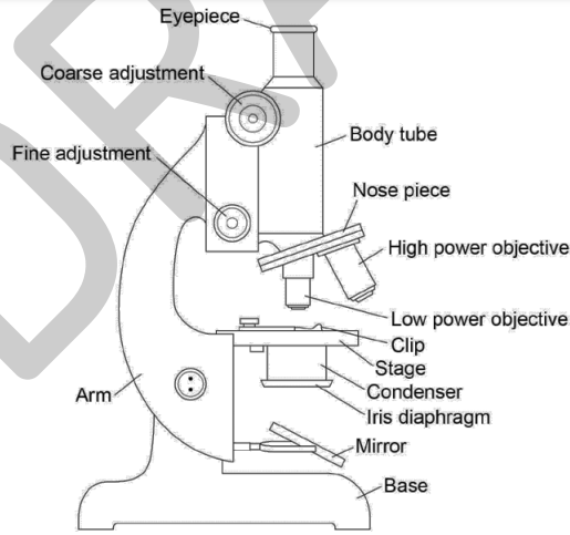
1) Turn the nosepiece to the lowest power objective lens.
2) Looking from the side (not through the eyepiece) turn the coarse adjustment knob so that the end of the objective lens is almost touching the slide.
3) Now looking through the eyepiece, turn the coarse adjustment knob in the direction to increase the distance between the objective lens and the slide. Do this until the cells come into focus.
4) Now rotate the nosepiece to use a higher power objective lens.
5) Slightly rotate the fine adjustment knob to bring the cells into a clear focus and use the low power objective (×40 magnification) to look at the cells.
6) When you have found some cells, switch to a higher power (×100 or × 400 magnification).
7) Make a clear, labelled drawing of some of these cells. Make sure that you draw and label any component parts of the cell.
Use an eyepiece graticule to measure the length of one of the epidermal cells that you have drawn. Remember to include the units.
9) Now measure the same cell in your drawing.
10) Calculate the magnification of your drawing, using the formula:
magnification = length of drawing of cell ÷ actual length of cell
11) Write the magnification underneath your drawing.
60 Copyright © 2015 AQA and its licensors. All rights reserved.