Chapter 1 and 2 of Neurology
Chapter 2: Neurons and Glia
History of neuroscience
Where is the seat of intelligence
Ancient Egyptians= proposed that the heart was the seat of intelligence
Aristotle= The role of the brain is to cool the blood
1600s= Gall= phrenology= behavioral traits could be inferred from bumps on the skull reflecting brain enlargements
1861= Dr. Paul Broca= studied patients unable to speak but they could understand language. They had a legion in the same part of the brain (now coined as the Broca area) which is important for language
1891= Dr. Carl Wernickle= studied patients that could speak but could not understand language
It had legions but just in a different part of the brain
Now: some functions are localized but others aren’t
How does information pass through nerves and move muscles?
Galen: discovers the brain’s fluid filled ventricles. Proposes that the nervous system operates via hydraulics
Descartes= agreed with Galen’s sentiment
Mid-1700s= Electricity is discovered→ Luigi Galbani demonstrated this is how neves and muscles work via electricity.
Debate: Is the nervous system a continuous “reticulum” or cells?
Late 1800s/Early 1900s
Golgi= argues that nervous system is a continuous reticulum like the circulatory system
Ramon y Cajal= argues that it’s made of cells that communicate via contact
Cajal= used silver nitrate stain (Golgi stain) Golgi developed to prove he was right and Golgi was wrong
Settled in 1950s with the help of the electron microscope
Neuron doctrine= the fact that the nervous system is a selection of cells called neurons
Nerve cell communication
Sparks= Information travels down n erve cells with electricity. This is how neurons communicate (how the gap closed)
(Correct theory) Soups= Information travels down nerve cells with electricity. But the connection is crossed via chemicals
Dominant framework in neuroscience since 1960s
Molecules → neurons & circuits → brain function and dysfunction
Genes >> proteins >> neurons>> circuits (brain activity) >> mind and behavior
E.g. Huntington Disease
Genetic mutation >> toxic proteins >> neurons die >> motor mood and cognitive impairment
Problems at the molecular level leads to problems at highest level
Dominant framework
genes → proteins→ neurons → circuit → brain activity→ mind and behavior
Genes: code for proteins that are used to form and operate neurons. They then form a network of circuits to communicate info which is called brain activity, which dictate mind/behavior
Neurons and Glia
Histology
Brain cells→ neurons and glia are very small
Size of brain cells 10-50 micrometers
Cannot be seen by the naked eye
Compound microscope (17th century) allowed the field of cellular neuroscience to begin
Brain tissue is similar to jello and cannot be sliced into sections unless hardened
Usually harden with formaldehyde and sliced with microme
It is uniform under a microscope
Nissil stain= primarily stains neurons not glia
Stains the cell body
I.e. not the hest stain does not show complete cytoarelinetine which is the arrangement of cells
Camillo Golgi
Golgi Stain 1873
Silver chromate solution
Two parts of neurons
Cell body= soma
Neurites= axons and dendrite
Santiago Ramon y Cajal
Used the Golgi stain 1888 and a light microscope to see and draw neurons and neural circuits
The neuron doctrine
Neuron is the basic functional unit of the nervous system
Cell theory applies to neurons
Contact, not continuity
Cell theory= the belief that the system is made of a collection of cells than a continuous reticulum
Cajal and the Neuron Doctrine
Neurons are discrete cells
Connected but not continuous
Synapse is 20-50 mm
Difference between axons and dendrites
Axons are the way a neuron sends outgoing signal
Dendrites are where a neuron receives a signal
Specificity to the connections among neurons
Unidirectional information flow
Basics of a neurons
Neurons sense changes in the environment, communicate them to other neurons and command the body’s response to the stimulus
Properties of neurons
Morphologically heterogeneous in size and shape each has a unique size/shape
Conduct bioelectric signals across long distances with no toss of signal strength
Possess specific connections with other nerve cells and with muscles and glands
Specific neurons are connected to specific efforts the connections aren’t arbitrary
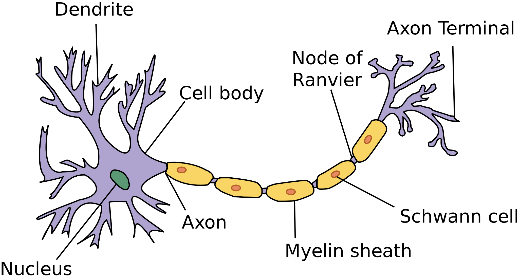
Protoypical neurons
Three parts
Soma: cell body; contains nucleus and other organelles
Performs most metabolic functions
Dendrites- projections from the soma that information
Axon; extension that sends information- via electrical signals from the cell body to the terminal buttons (transmits away)
Usually has one acon it may branch to form axon collaterals
Soma and its contents
Nucleus: contains DNA, which is transcribed into mRNA and translated into a protein
Chromosomes>> DNA
Gene expression: DNA → transcription→ mRNA
RNA processing (alternative splicing 1 gene > multiple proteins)
mRNA exists nucleus via pores in nuclear envelope for translation into protein
Nucleus Structure
Roughly spherical with double membrane nuclear envelope with folded inner membrane
Reinforced with nuclear pores for exchange of materials e.g. nucleotides, mRNA etc
Pores are 0.1 um. Contains all genetic material as DNA condemned into chromatin threads, which contains genes that each code for specific proteins
Ribosomes and Rough Endoplasmic reticulum (ER)
Ribosomes: proteins translation: mRNA >> protein
Bound ribosomes >> Rough ER
Aka. Nissl bodies: membrane with attached ribosomes (bound ribosomes)
Translated proteins inserted into plasma membrane or are exocytosed (ion channels)
Also they are packet in lysosomes
Lots in neurons
Free ribosomes
Translates proteins that are released into the cytosol
Smooth ER and Golgi Apparatus
Smooth ER: no ribosomes
Folds proteins giving them 3D structure
Regulates internal Ca2+ concentration
Golgi apparatus
“Post translational” modification of proteins
“Post office” sorts proteins destinations
Mitochondria
Site of cellular respiration
“Powerhouse”
Kreb’s cycle yield ATP, the cell’s energy source
Neurons require large amounts of ATP
Mitochondria are especially abundant where energy needs are greatest
Concentrated at synapses- neurotransmitter synthesis
Rough ER structure
Network of tubular membranes enclosing fluid filled sacs called eisternae each
1-1.5 um
Contains receptors to which ribosomes attach
Ribosome structure
Made of rRNA 2 subunit (1 small 1 big) contains peptidyl site, amino deysite and E site
Structure of smooth ER
More tubular than RER. modifies and transports lipid and helps 3’ folding of proteins
It regulates Ca 2+ in muscle cells → sarcoplasmic reticulum
Structure of Golgi apparatus
Stacks of flattened fluid filled sacs called cisternae. CONVEX: forming face where vesicles containing materials to modify enter. CONCAVE: maturing face, when vesicles with modified material huf off
Structure of mitochondria
1 um thick to 10 um in length
Double membrane envelope
Inner membrane folded cristae which increases SA for phosphorylation; impermeable to ions to set up electrochemical gradient for ATP synthesis
Contains circular DNA for binary fission not enclosed in a nuclear membrane, free in the matrix (bachynoid fluid), not associated with histones, 70s ribsomes for protein synthesis
Neural membrane structure
selectively permeable phospholipid bilayer controls exchange of materials between cell and environment
Contains protein channels carriers and pumps (K+/Na+)
Many of which are voltage gated which help to set of action and resting potential for transmitting impulses.
Cytoskeletal Proteins in Neurons
Type | Relative size | Function |
|---|---|---|
Microfilaments (actin) | Small (5 mm) | Dynamic involved in growth and movement Enriched in spin → dendrite |
Neurofilaments (intermediate filament) | Medium (10 mm) | Controls thickness of neurites (axons and dendrites) Enriched in axons Rope like |
Microtubules | Large (20 mm) | Composed of tubulin Runs down the length of nerites to give basic shape Acts as a “railroad” for transport |
The Axons (sends info)
No rough ER or free ribosomes
Membrane proteins are quite different
Axon hillock (beginning) where axons tanners away from soma
Axon proper (middle)
Collaterals (branches) = increase surface area
Axon terminal (end)
“Terminal bouton”
“Nerve terminal” (synaptic bulb)
Used for communication
Axon collaterals that branch and then return to communicate with original neurons are called cecurrent axon collaterals
Axon terminal
Presynaptic side
Presence of synaptic vesicles (w/ neurotransmitter (NT))
Large number of mitochondria (high demand)
Synaptic bulb heavily coated in proteins
When a neuron makes contact with another cell it is called innervation
The Synapse (gap between neurons)
Two sides
Presynaptic membrane
Filled with NT vesicles
Postsynaptic membrane
NT receptors inserted into membrane
Synaptic cleft= the space between
NT is released into here
Synaptic transmission= transfer of info at the synapse
Electrical to chemical to electrical: transfer of info at the synapse
Axoplasmic Transport
No protein translation in axon→ all protein is made in the soma
Axoplasmic transport: proteins made in the soma must be transported to the axon terminals
Uses the cytoskeleton
MT form the track along which packets of proteins travel by the action of motor proteins (kinesin & dynein)
Anterograde→ away froom the soma
Soma to axon terminal
Uses kinesin
Retrograde→ back to soma
Axon terminal to soma
Uses dynein
Dendrites (receive information)
“Antennae” of neurons
Dendritic “tree” aborization
collective= dendritic tree
1 branch= dendrite
Covered with neurotransmitter receptors
High ribosome content
Protein synthesis
Some covered with spines
Like leaves on a tree, increase surface
Dendritic spines
Described by Cajal in 1888 “brisitiling thorns or short spines”
“Leaves on the trees”
Contain neurotransmitter receptors
Form, enlarge, shrink, and retract throughout the animal’s lifespan
Dynamic in nature
Branching from original branch dendrite in the tree especially
Dendritic spines are induced in development to ensure neurons are connected to correct neurons
Axons | Dendrites |
|---|---|
Longer Thinner Uniform in diameter Fewer branches Presynaptic | Shorter Thicker Tapered Highly branched Postsynaptic |
There are some exceptions to this
Axons can be connected to other axons
Classifying neurons
Based on the number of neurites (axons and dendrites) on soma
Based on connections
Motor neurons
Direct output to muscles
Primary sensory neurons→ they have connections with receptors
Convert physical stimuli (like light or sound or pressure) into electrical signals
Interneurons
Connections only with other neurons in brain spinal cord
Do not make connections outside of PNS and CNS
Based on dendritic or somatic morphology
Stellate cells → can be spiny or aspinous
Pyramidal cells→ always spiny
Based on axonal length
Golgi type 1: long axons → projection neuron→ axon extend from 1 side of brain to other
Pyramidal cells
Golgi type 2: short axons→ axon doesn’t extend beyond
Stellate cells
Based on neurotransmitter type
Ex: glutamatergic: releases glutamate at synapses
Ex: dopaminergic: releases dopamine at synapses
Ex: cholinergic: release acetylcholine at synapse
MB: Every neuron will make for unique proteins or neurotransmitters for their corresponding receptors
Glia: The Glue that holds it altogether
Contribute to brain function in a supporting role. They act as insulators, nourish and support neighboring neurons and within spaces to limits neurite growth
Glia
Outnumber neurons 10 to 1
Support neuronal functions
Formation of synapses and development
Neuronal plasticity
Protection against neurodegeneration and toxicity
Response to i injury
Neurotransmission
Types of glia
Astrocytes
Oligodendrocytes
Ependymal cells
Microglia
Astrocytes
Most numerous glia in the brain
Envelop synapses
Neurotransmitter reuptake
Influence neurite growth (radial glia) → subtying glia
Myelinating glia
Produce myelin sheath that insulates axons
Schwann cells (peripheral NS)
One cell myelinates a single axon
Oligodendrocytes (central NS)
One cell can myelinate several axons
(probably due to density of neurons in CNS)
Lactating at Nodes of Ranvier
Allows propagation of action potential across the axon for faster transmission
Microglia
“Immune system of the brain”
Respond to insult or injury in the CNS
Microglial activation and motility following brain tissue injury
They act as phagocytes or repair the brain
Ependymal cells
Line the brain’s ventricular system
Produce cerebrospinal fluid CSF