Fungal Practical

Image

Description

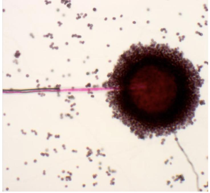
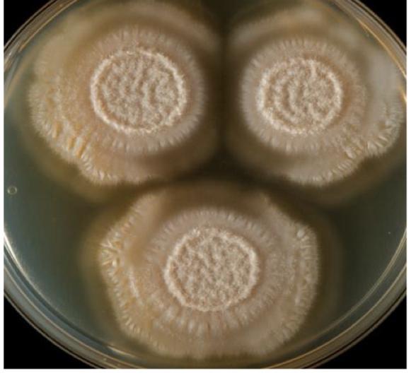
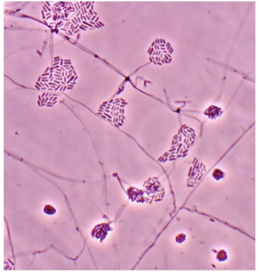
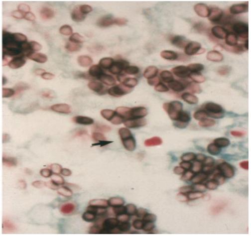
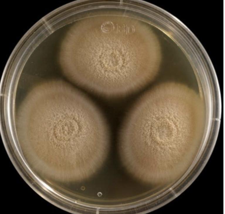
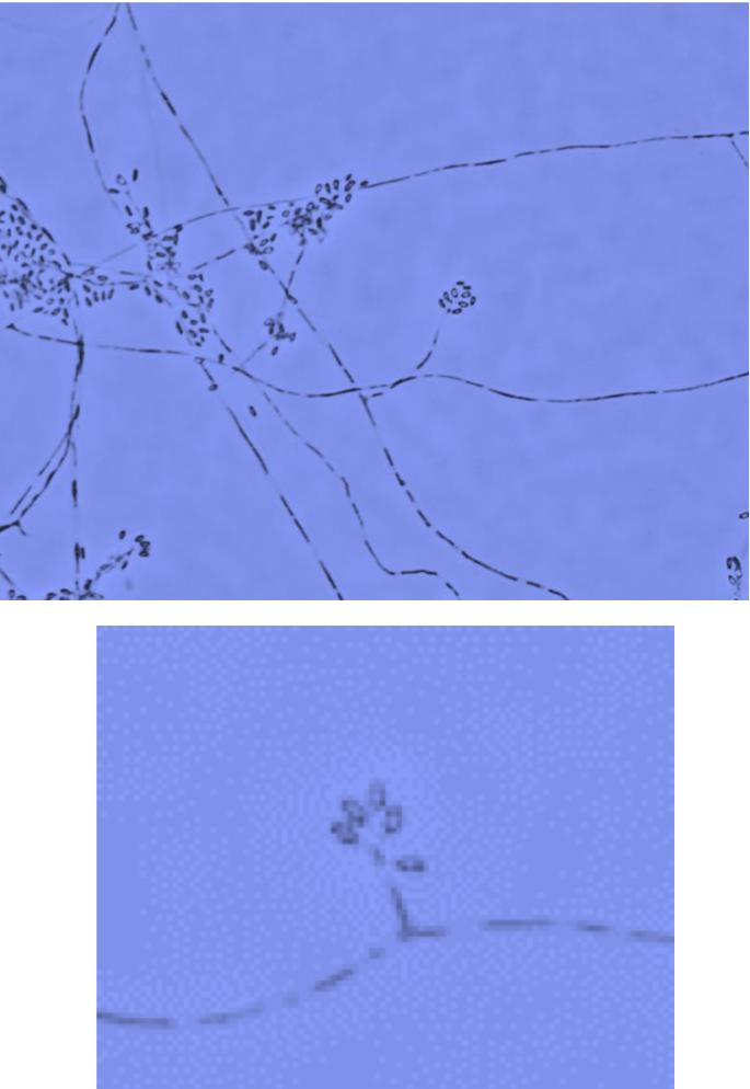
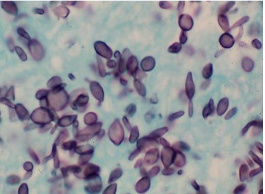
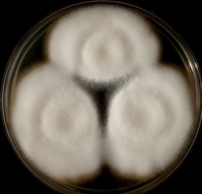
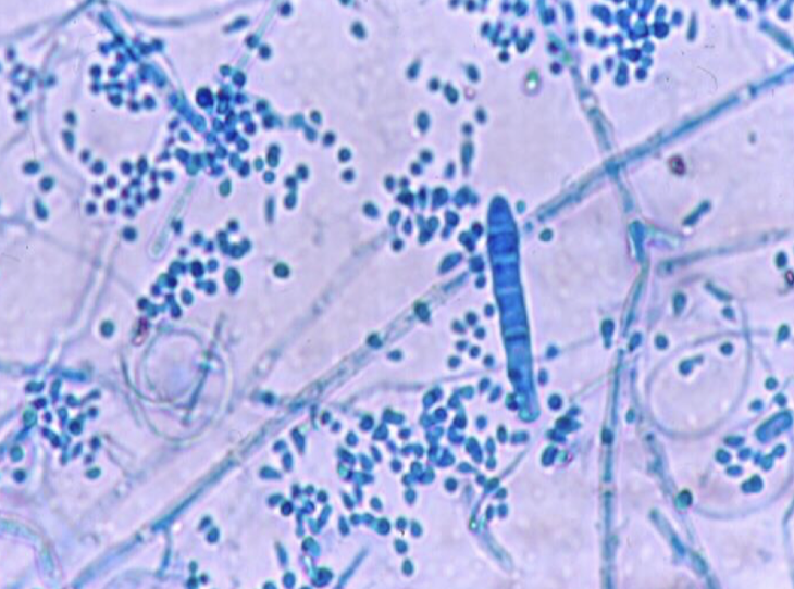
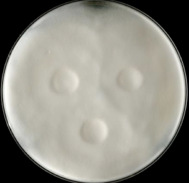
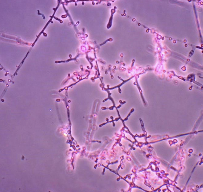
Fonsecaea Pedrosoi

  • Tropical areas of world

  • Cause Chromoblastomycosis

Scedosporium spp.

  • “Mycetoma belt”

  • North Africa and Asia

  • Around equator (warm tropics)

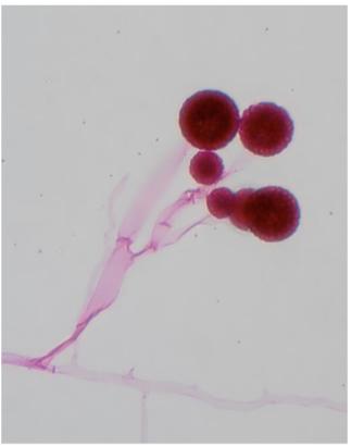
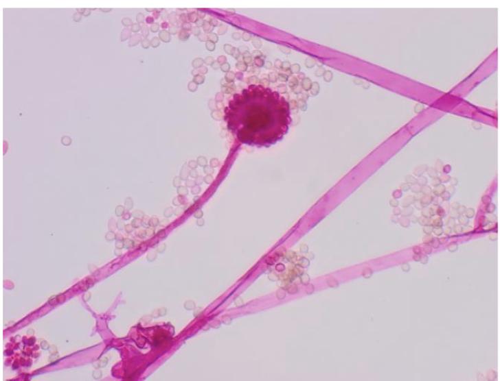
Rhizopus

  • Mucormycosis

  • Hyaline mold

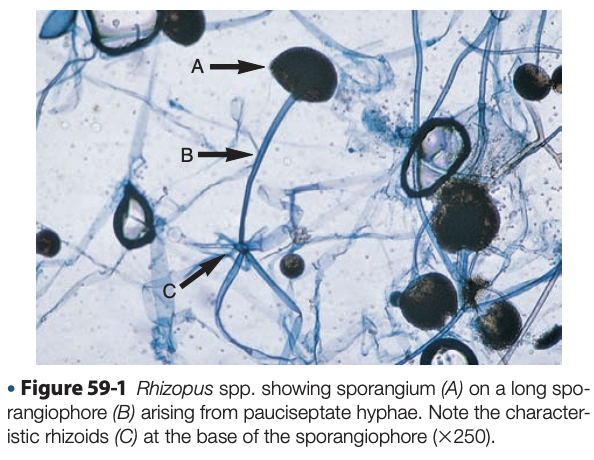
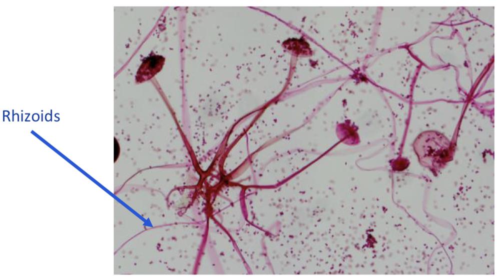
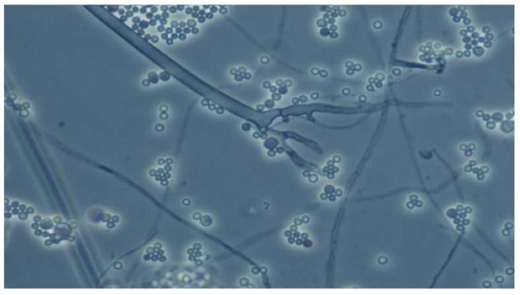
  • Unbranched sporangiophores with rhizoids

  • Recovered from respiratory secretions, skin, nose, brain, stool, wounds, ear, lung

  • Seen in rhinocerebral infection, pulmonary infection, mycotic keratitis

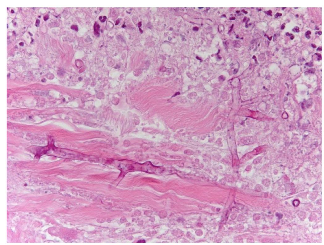
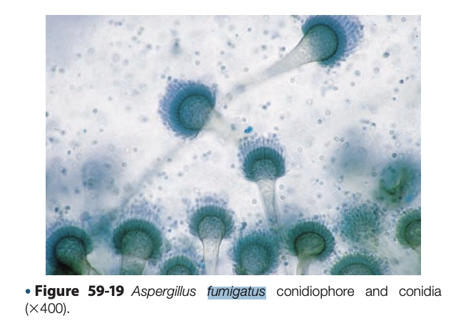
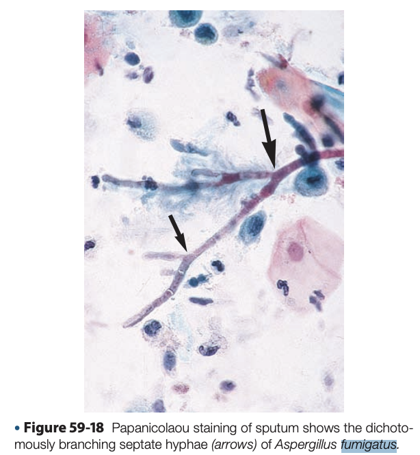
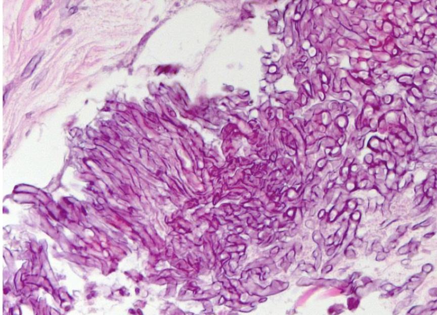
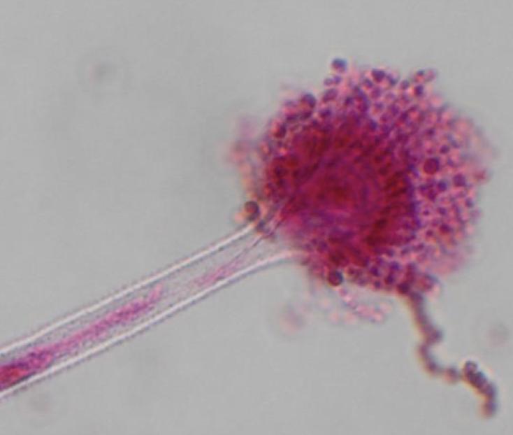
Aspergillus Fumigatus

  • Hyalohyphomycosis

  • Allergic bronchopulmonary infection, invasive pulmonary infection, skin and nail infections

  • Most commonly recovered spp from immunocompromised pts, common to see in the lab

  • Rapidly growing

  • Septate hyphae, short or long conidiophores with “foot cell” at base

  • 45 degrees C

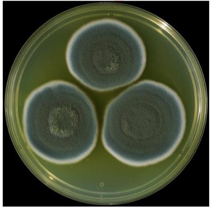
  • Blue green to greeny gray

Cladophialophora bantiana

  • Phaeohyphomycosis

  • Melanin pigment in cell wall

  • Can cause serious Brian infection in healthy patient extremely rare (HIS WORDS)

  • Brain abscess

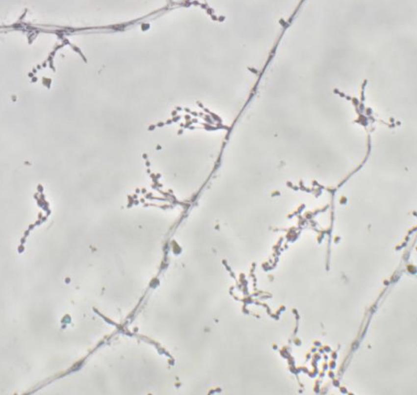
  • Long, poorly branched conidial chains

  • MAX growth temp 42-43C

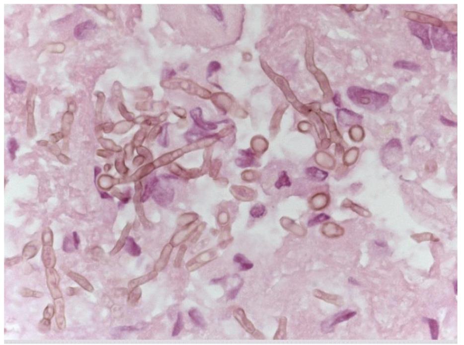
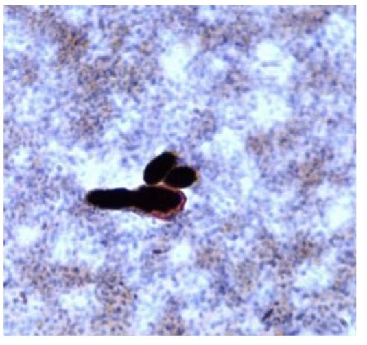
Fonsacaea spp.

  • presence of nodular, wart lesions

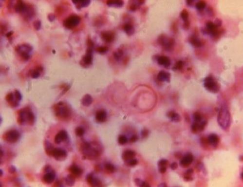
  • Presence of sclerotic bodies in tissue

  • Not forming hypee

  • From stepping on rock/ Stepping on stick

Coccidioides spp.

  • Dimorphic mold

  • Coccidioidomycosis

  • Lungs, skin, meninges

  • Rapid growth

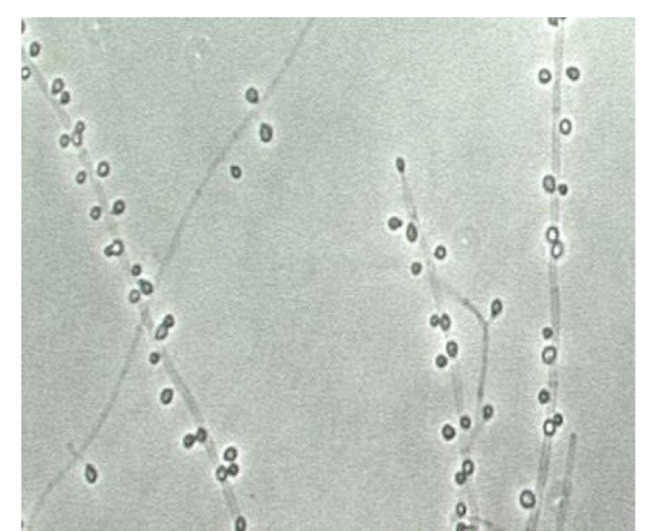
Trichophyton spp.

  • no chaining or branching

  • - only one spore per location

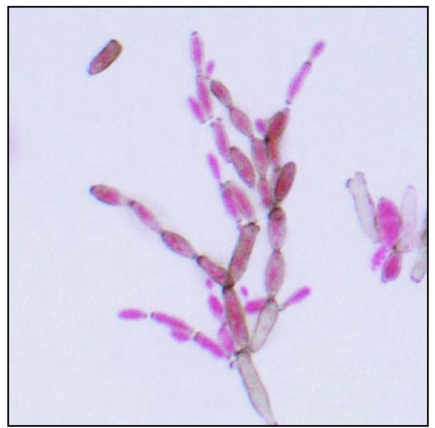
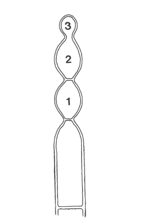
Acropetal chaining with branches

Youngest cell is at the tip of the chain

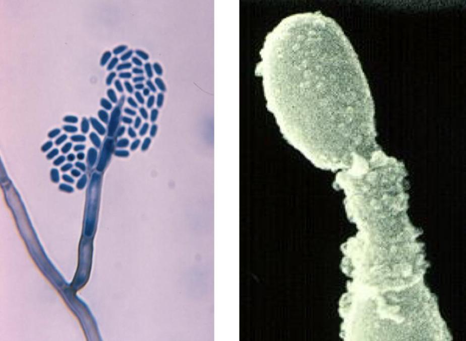
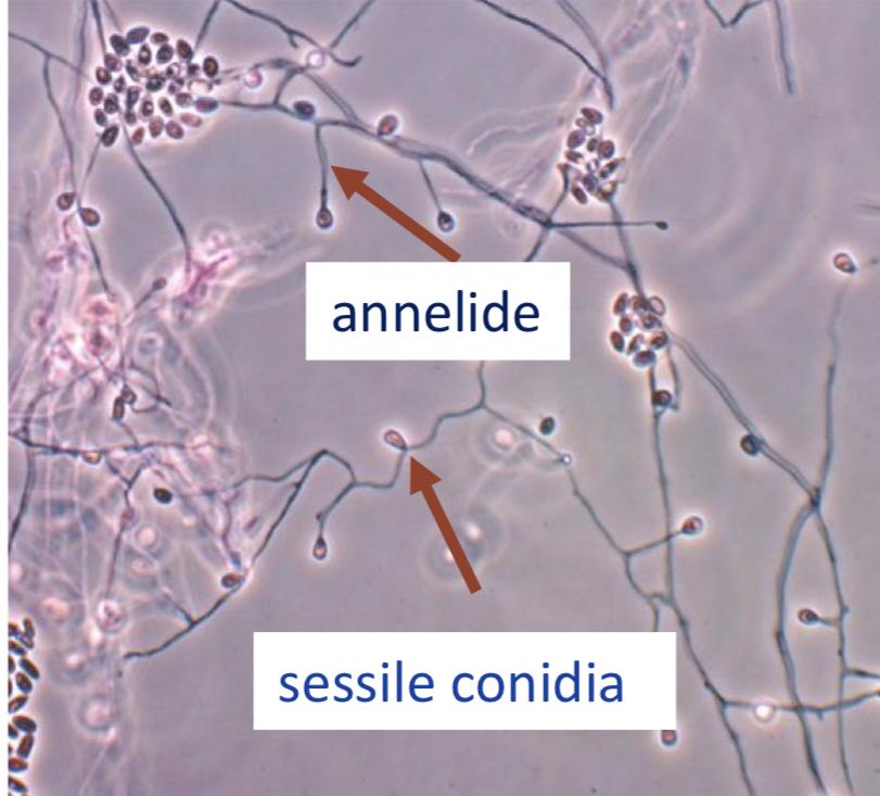
Conidia in wet masses

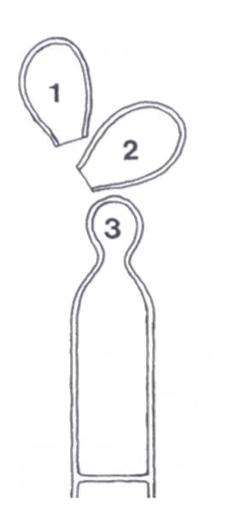
annelides

Phialides

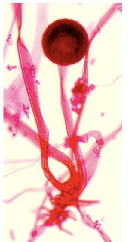
Rhizopus Oryzae

  • Make sporangia in a sporangium instead of conidia

  • have sporrangiophores

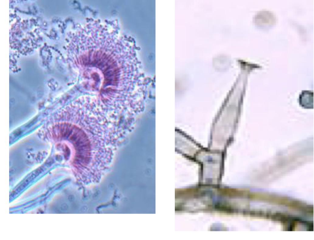
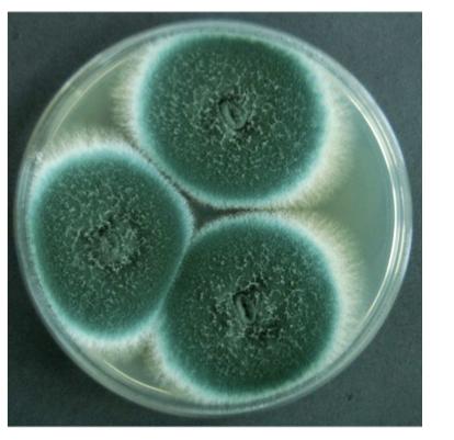
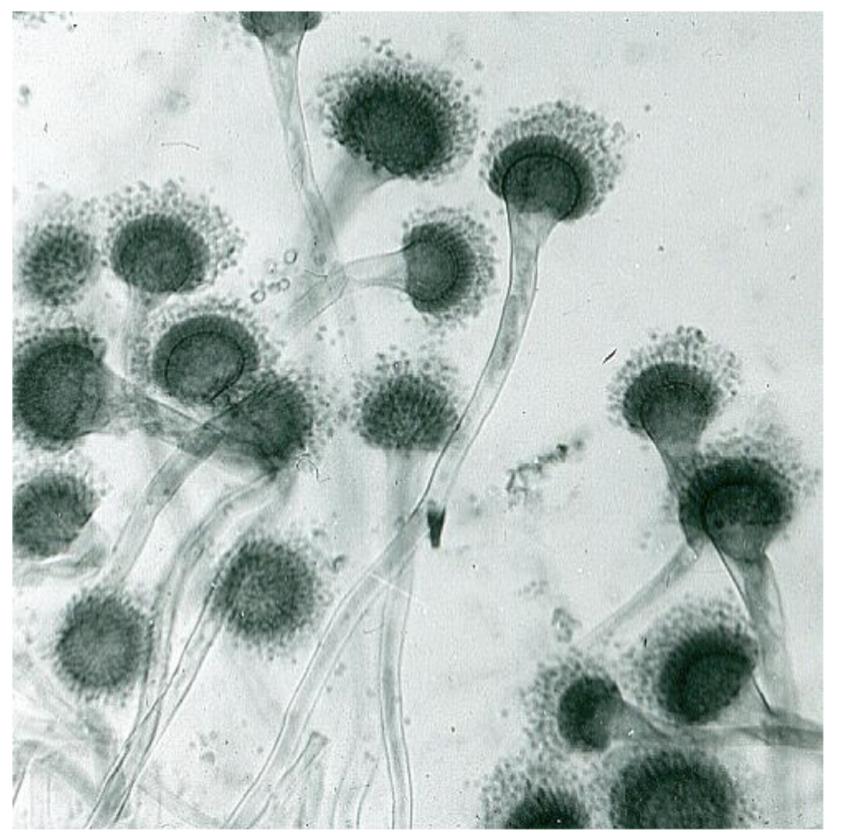
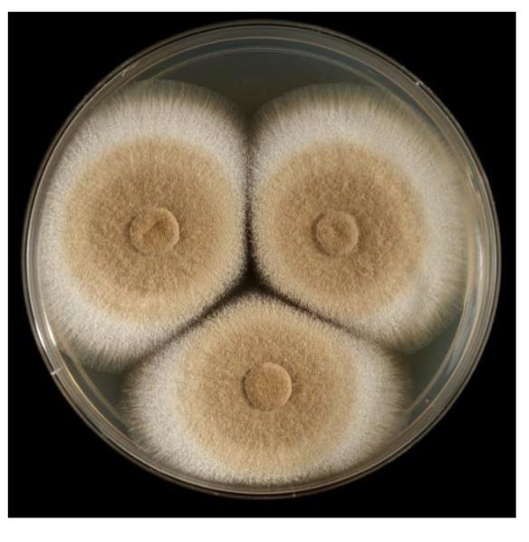
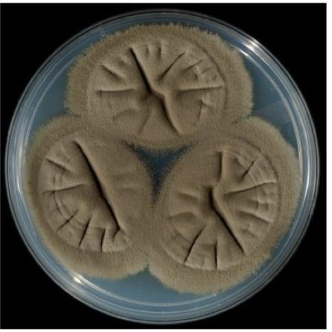
Aspergillus fumigatus

  • Bluish-green with a plate yellow reverse

  • Monoseriate, Phialides columnar, conidia in short chains

  • Grows at 45 C

  • Seen EVERYWHERE

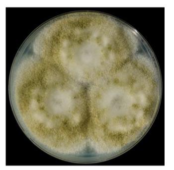
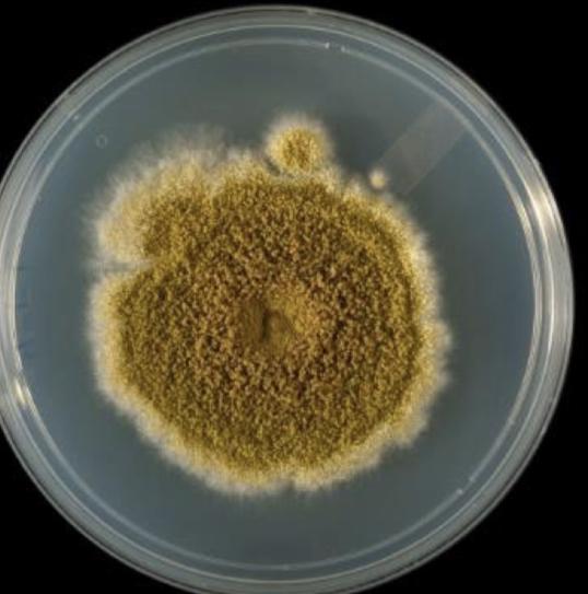
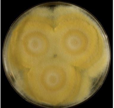
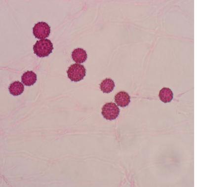
Aspergillus Flavus (yellow)

  • yellow-green to lime green with a cream reverse

  • conical heads radiate to columnar

  • Stipe walls roughened/ spiky edges

  • Largely biseriate but can be monoseriate

Aspergillus terreus

  • Cinnamon-brown colonies

  • Cream to deep brown reverse

  • Conidia heads columnar and covering upper half of vesicle

  • Biseriate with verylong chains of conidia

  • Amphotericin B resistant

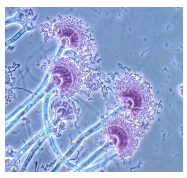
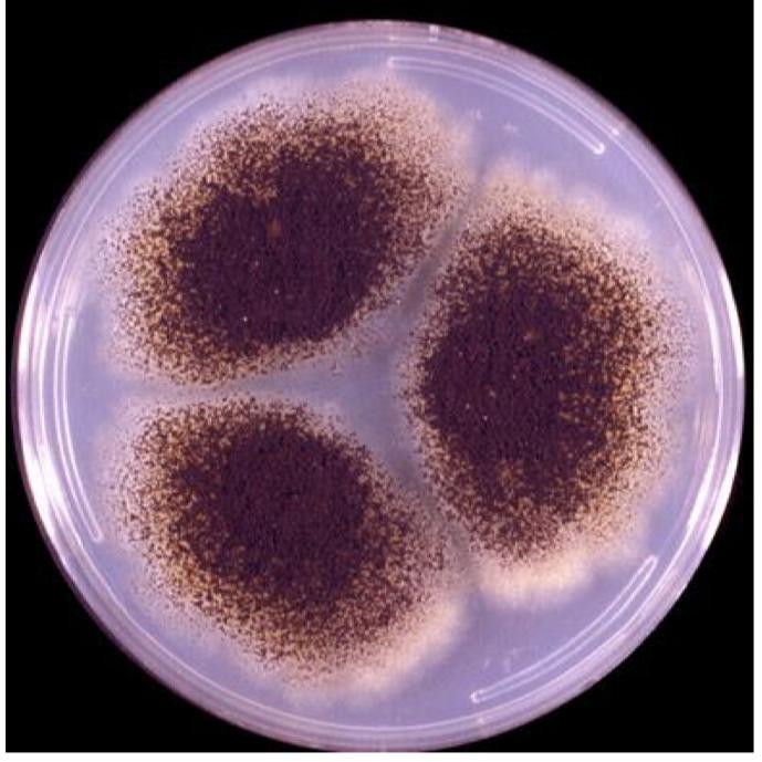
Aspergillus niger

  • White mycelia become black with age due to pigmented conidia

  • Colonies are large, microscopic conidial head is also large (viewed on 10X vs 40X)

  • Reverse is pale yellow

  • Conidia heads radiate- black conidia cover entire vesicle

  • Biseriate

  • Common in children ear infections

Sarocladium spp., (Acremonium)

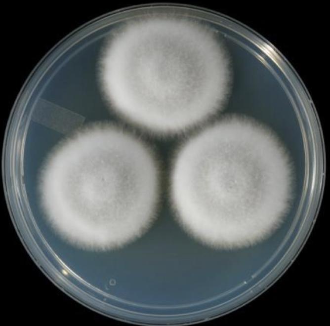
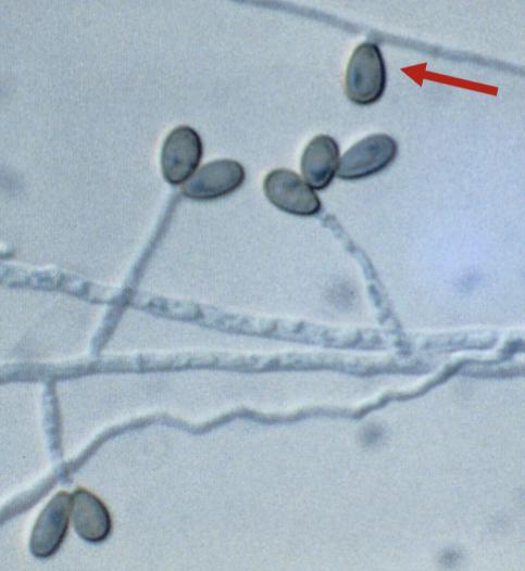
  • Conidia in wet masses

    • Single-celled

    • Cylindrical

  • Colonies are waxy

  • Pale-orange to brown

  • Will grow in blood culture (one of the only few that will)

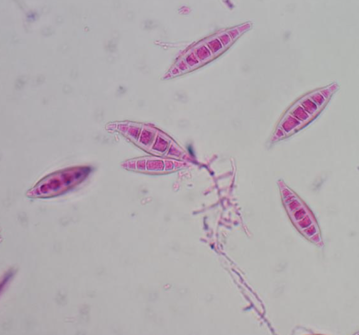
Fusarium spp.

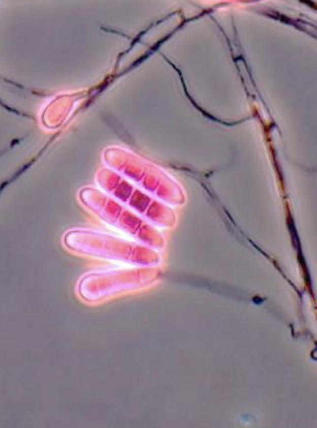
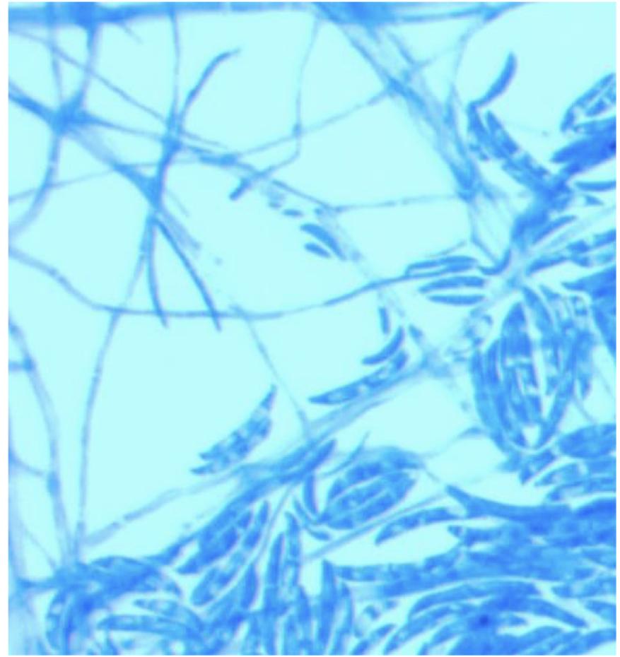
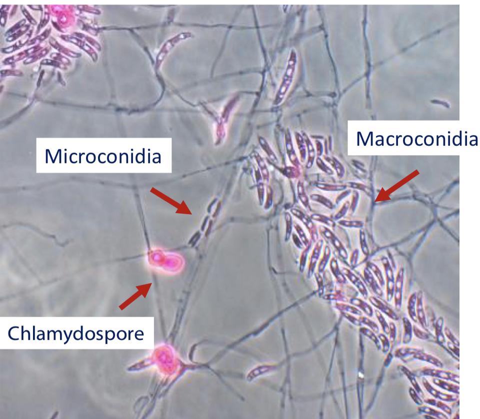
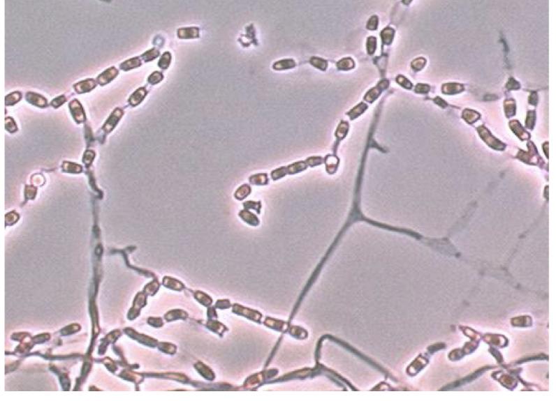
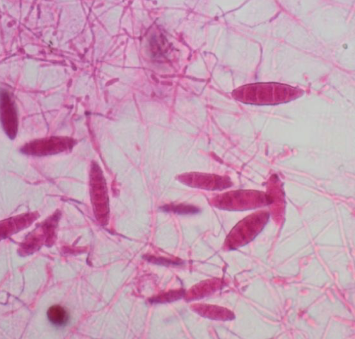
  • Macro and micro conidia

    • Multiseptate, curved like a canoe or a banana

  • Chlamydospores can be present

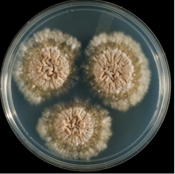
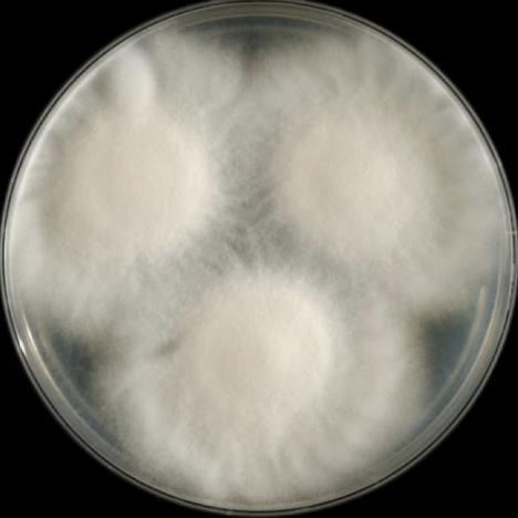
Fusarium solani

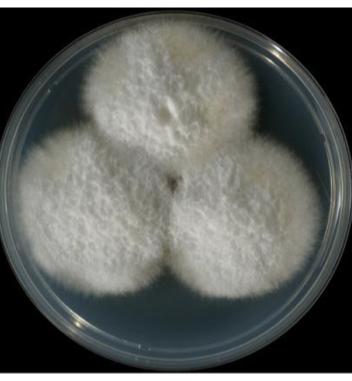
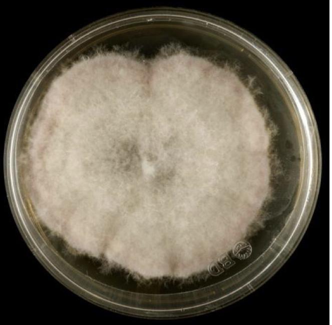
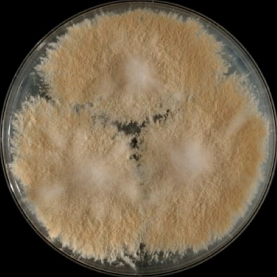
  • white to cream cottony with a white reverse

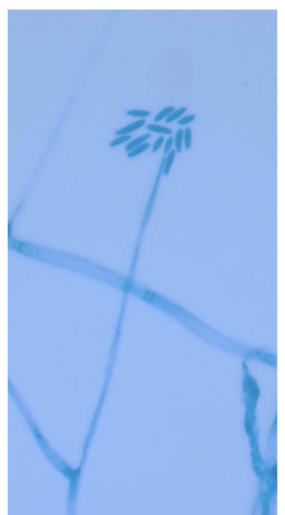
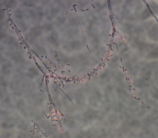
  • Long conidiophores that may contain septa

Fusarium oxysporum

  • Wooly white colonies becoming lavender to purple

    • Pretty pink and purple

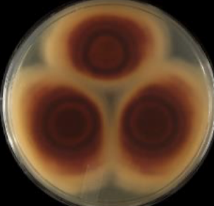
  • Reverse is white to dark purple

  • Short conidiophores without septa

  • Microconidia, macroconidia and chlamydospores

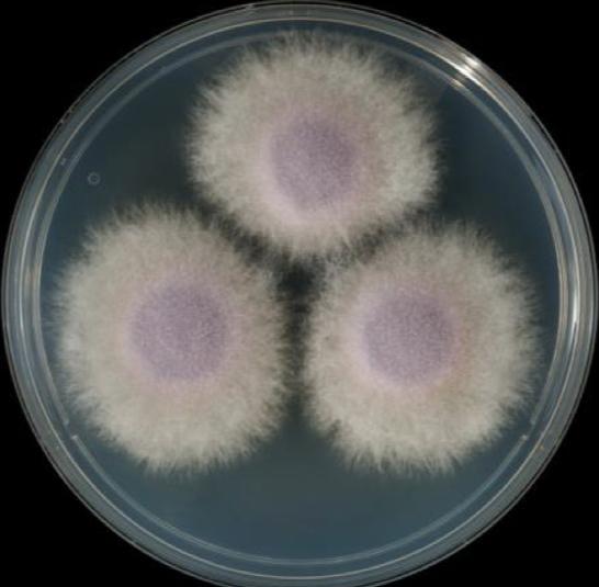
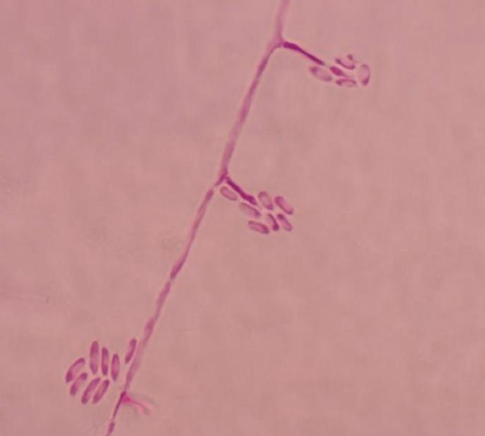
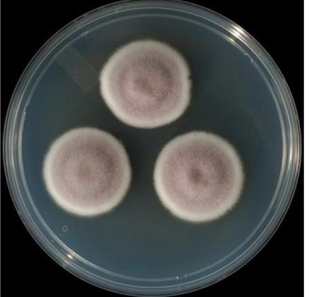
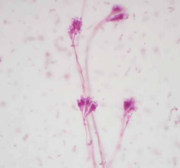
Purpureocillium lilacinum

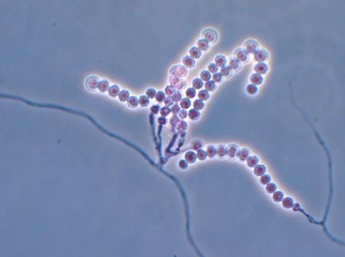
  • Enteroblastic conidia in basipetal chains

  • Colonies are powdery

  • Cream turning violet with age

  • Conidiophores are tapering phialides - similar to a bowling pin

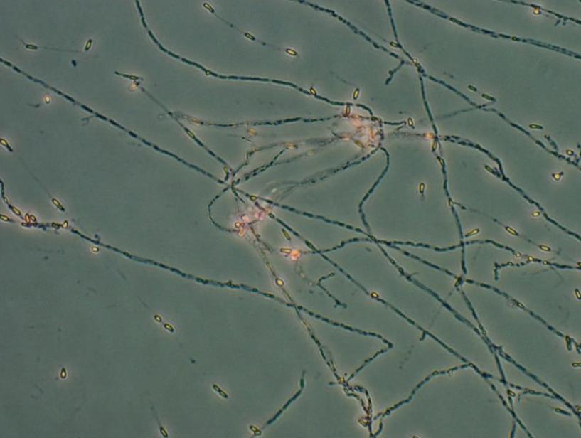
Scedosporium Apiospermum

  • will grow at 42C but NOT 45C

  • Colonies cottony to wooly

  • Conidiophores can be long or short, producing oval to globose conidia which become thick-walled and brown after release

  • Long annelids

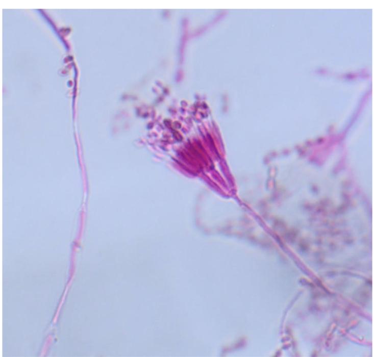
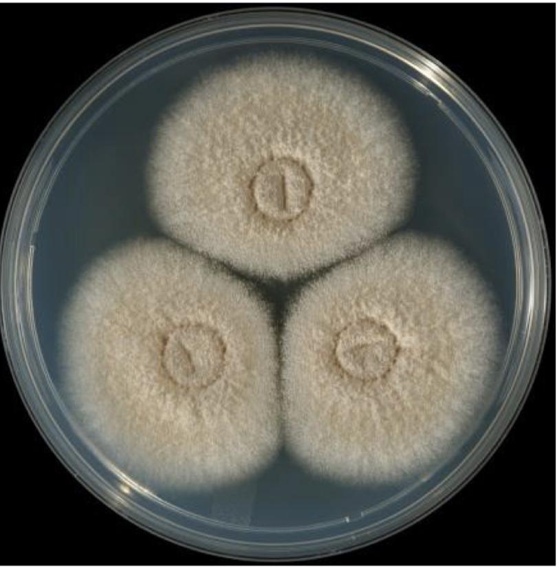
Penicillium species

  • green to slate-gray

  • Colonies velvety

  • Will not grow at 30C

  • Rarely cause disease

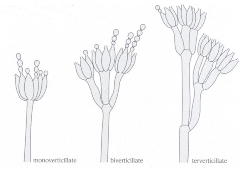
  • Can have 1-3 rows of phialides

Scopulariopsis Brevicaulis

  • Pinkish- brown colonies

  • Conidia are rough-walled, pyriform with truncated bases

  • Broom-like

Thermally dimorphic fungi

Histoplasma Capsulatum mold form

  • Macroscopic and microscopic pretty much exclusively

  • Has tubercules

  • Growth in culture (4-5 weeks)

  • Urinary antigen test, serology, giemsa-stained BAL

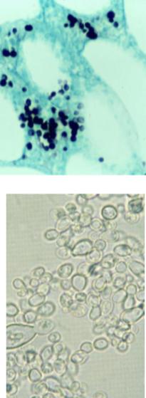
Histoplasma Capsulatum yeast form

Sepedonium spp.

  • Can look similar to Histoplasma but only has macroconidia, NO microconidia

  • Cream to amber colonies, “wooly”

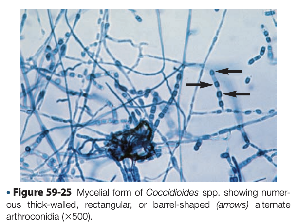
Coccidioides immitis

  • Suede-like, gray-white with a tan-brown reverse

  • Grows on cycloheximide

  • Dx: growth in culture, serology

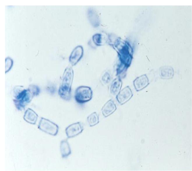
  • Barrel-shaped arthroconidia

  • Disjunctorcells that break apart cells and release spores

  • Lab-acquired infections

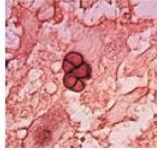
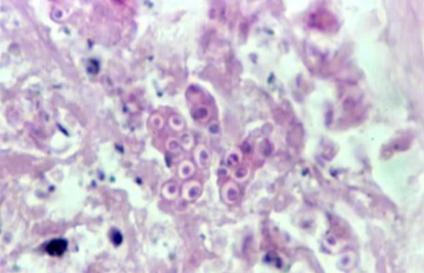
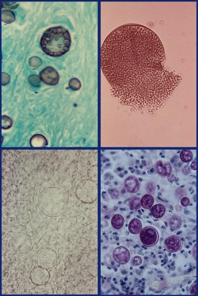
Coccidioides immitis spherules

  • Filled with endospores that can be released upon spherule bursting

Malbranchea spp

  • Look-alike to Coccidiodes arthroconidia

  • Very tiny when compared to Coccidioides

Blastomyces dermatitidis mold

  • Short stem with small conidia on end, resemble lollipops

  • Culture waxy to whitish or cream, turning brown with age

  • Dx: culture growth, urinary antigen test, serology, thermal conversion

  • *usually diagnosed by distinct yeast form (has to be grown at 37 degrees C

Blastomyces dermatitidis yeast form

  • Grows at 37 degrees C on BHI agar

Talaromyces marneffei

  • Penicillium-like

  • Philades mono- or biverticillate

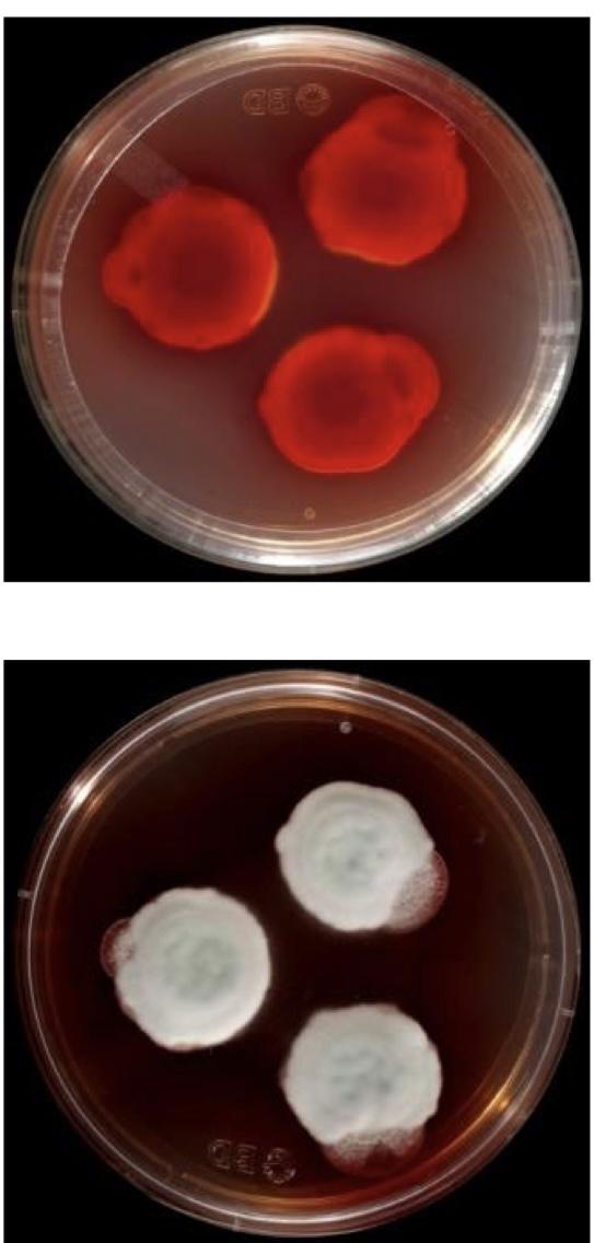
  • Red pigment that secretes into the media (reverse side)

  • Pt is usually already sick when infection w/ this, does not usually affect healthy person outside of Asia

Talaromyces marneffei yeast phase

  • Binary fission

  • Yeast on BHI agar at 37 degrees C

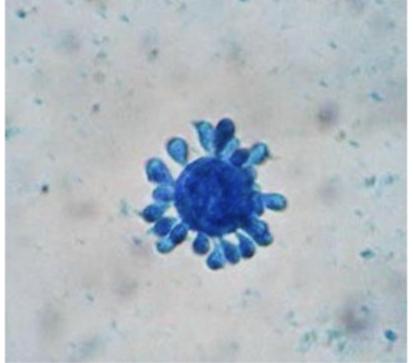
Paracoccidioides brasiliensis

  • Hyphae on short stalks

  • Smooth-walled conidia that resemble lollipops

  • Similar to Blastomyces

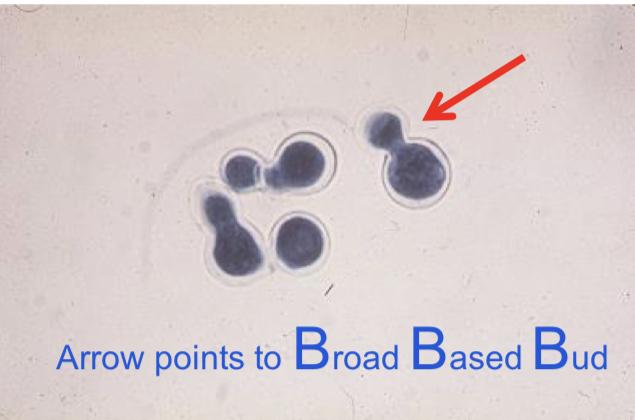
  • Large yeast with multiple buds- “mariner’s wheel”

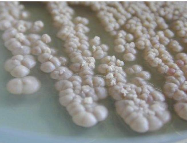
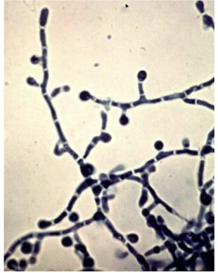
Sporothrix schenckii mold

  • Colonies are waxy to cottony white to cream, turning dark with age (one of two waxy)

  • “Daisy petal”

Sporothrix schenckii yeast form

  • Pleomorphic with some cigar-shaped cells when budding

  • “Rose-grower’s disease” due to certain rose treatment products (sphagnomoss) causing this infection

  • Can have similar lymphogenic affects like nocardia

Mucormycetes

Mucor circinelloides

  • Will not grow above 37 degrees C

  • No rhizoids

  • Colonies initially white but become grayish-brown

  • Branched sporangiophores

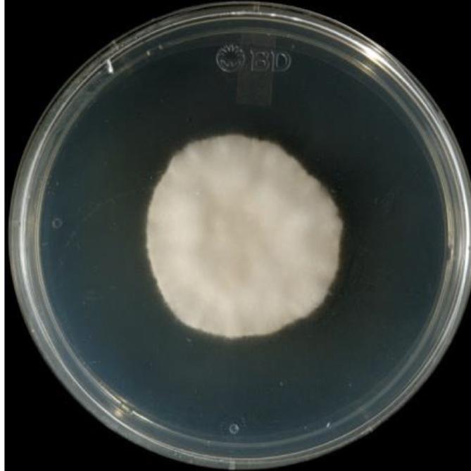
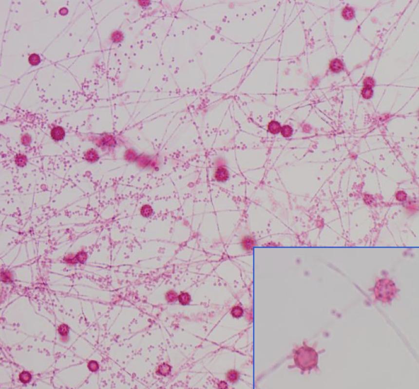
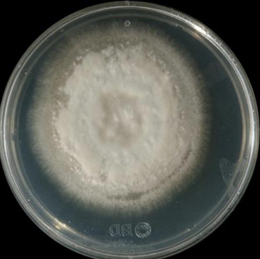
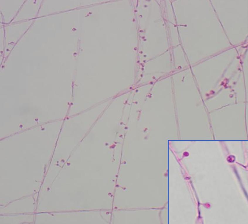
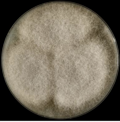
Rhizopus oryzae (arizus)

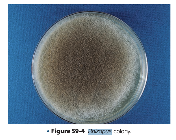
  • Colonies white, turning gray with age

  • Growth at 40 degrees C but not 45 degrees C

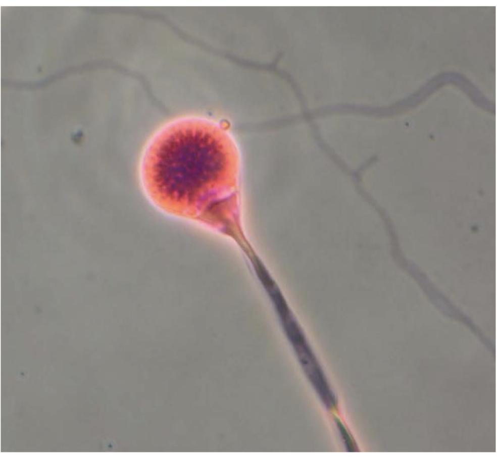
  • Sporangiophores single and unbranched

  • Rhizoids

  • Columella seen when ruptured

Lichtheimia corymbifer

  • Growths better at 35 degrees C than at 25 (likes higher temps- 48-52)

  • Sporangiophores are branched

  • “Champagne glass” apophysis in second image

  • Rhizoids present

Cunninghamella bertholletiae

  • Sporangiophores end in a swollen vesicle with one-celled sporangiophores covering surface

  • Colonies white to tannish gray

  • Growth up to 45 degrees C

Dematiaceous/Dark molds (black on the back)

Exophiala dermatitidis

  • Mold but also known as a “black yeast”

  • Growth at 40-42 degrees C

  • Colony brown, velvety to waxy, often with the brown pigment diffusing into the media

  • Phaeohyphomycosis

  • Chromoblastomycosis

  • Keratitis

  • Long phialides with mic of yeast and conidia growing along

Cladosporium species

  • Colonies are slow-growing, olive to brown, dark brown reverse

  • Short, highly branched drains of conidia from a poorly differentiated conidiophore

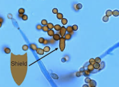
  • “Shield” cells often present after conidia break off

  • Very common environmental mode- it’s everywhere, not pathogenic

Cladophialophora bantiana

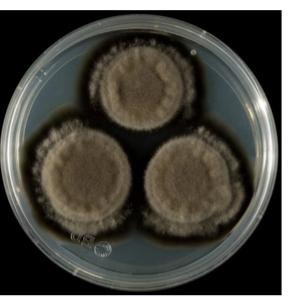
  • Colony wooly to velvety, olivaceous black with black reverse

  • Grows at 40-42

  • Conidiophores often cannot be distinguished from hyphae

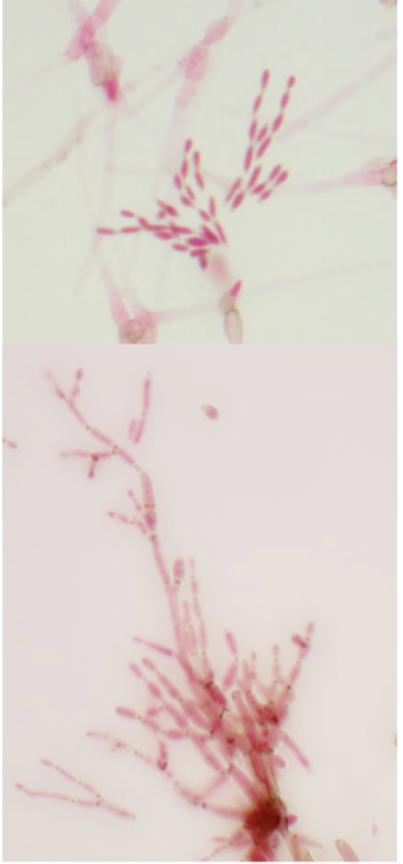
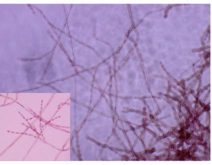
  • Conidia in long, sparsely-branched chains that remain intact, conidia are lemon-shaped

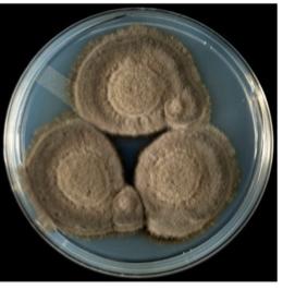
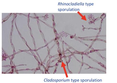
Fonsecaea pedrosoi

  • Colony grows very slowly, velvety to cottony, oliver to brown-black, black reverse

  • Unique bc it can produce 4 types of conidiogenesis but most isolates display 2-3 types

  • Chromoblastomycosis in south america

Holoblastic dematiaceous fungi with multi celled conidia

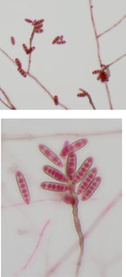
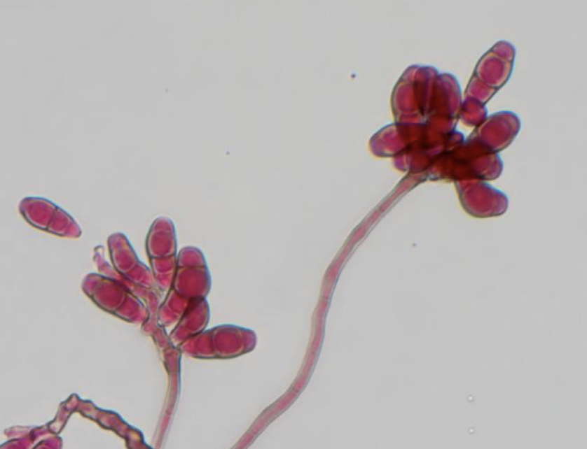
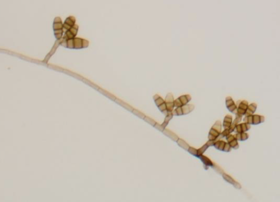
Curvularia spicifera

  • Conidia are ellipsoidal, straight or curved and divided into compartments by distosepta (false septa)

  • Conidia contain 3-6 cells

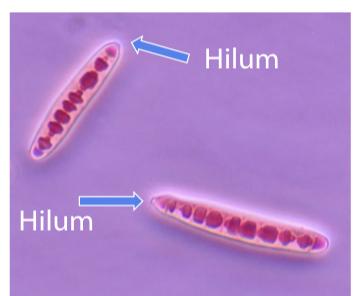
  • Hilum has slight protrusion

  • Phaeohyphomycosis and eye infections

  • Causes fungal sinusitis

Curvularia lunata

  • Pear-shaped or curved and divided into compartments

  • Can resemble a croissant - cent cell larger than other cells

  • Conidia contain 4-5 cells

Curvularia geniculata

  • Geni means knee-like

  • Sympodial conidiogenesis

  • Croissant

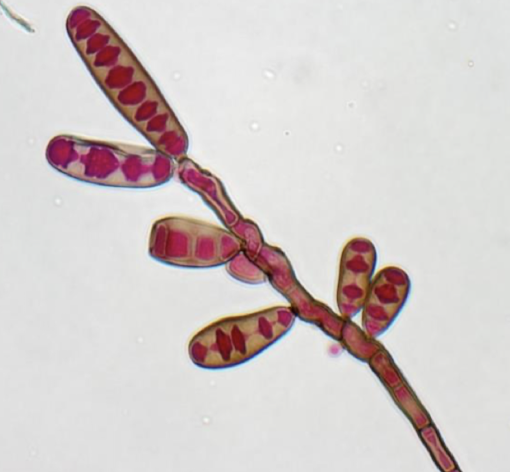
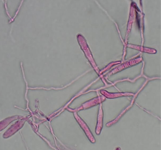
Exserohilum rostratum

  • No dark line between cells- false septa

  • Condia are ellipsoidal, straight or curved and divided into compartments

  • Cells on ends are darker than other cells

  • Conidia contain 7-12 cells

  • Hilum is prominent - fungal meningitis

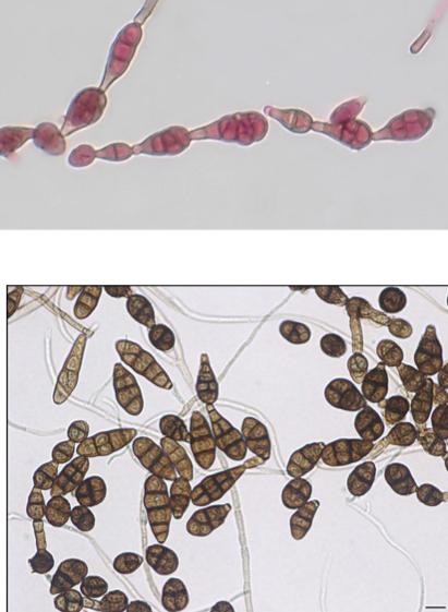
Alternaria sp.

  • Common plant pathogen

  • Conidia in unbranched chains, are flat, brown, and have a “beaked” apex

  • Septations may be both transverse and longitudinal

  • Phaeohyphomycosis, fungal sinusitis

  • “Alternating alternaria” big ol’ turkey leg

Dermatophytes

Grow on nonliving outer layer of epidermis: the stratum corneum- skin hair nails

Epidermophyton floccosum

  • Colonies are velvety to powdery, pale yellow to mustard yellow - colonies highly variable

  • “Beaver tail”

  • Athlete’s foot - use of common shower or gym facilities

  • Macroconidia only

Microsporum canis

  • Skin hair infection NOT nails

  • Macro- and microconidia - spindle-shaped, often with a slightly bent terminal knob

Microsporum gypseum

  • Macro and microconidia

  • Not bent at the tip, ends are rounded instead of point when compared to to canis

  • Zoonotic

  • Does not fluoresce under UV light

Thricophyton rubrum

  • Rubrum- red

  • Club-shaped microconidia

  • Referred to as “birds on a wire”

  • Urease negative

  • V contagious, outbreaks in daycare settings

Trichophyton mentagrophytes

  • Cylindrical walls with thin, smooth walls 3-8 cells

  • Clusters of conidia

  • Spiral hyphae

Trichophyton tonsurans

  • Microconidia are variable in morphology, can be club-shaped, cylindrical, balloon-shaped

  • Abundant chlamydospores

  • Fairy lights

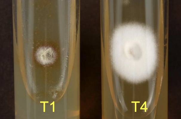
Trichophyton agars (T-media)

  • 7 T-medias, T1 and T4 most commonly used

  • Contains vitamin-free casamino acids + individual supplements

  • MALDI-TOF more commonly used now