knowt logo

2 Anatomy of the Nervous System

Anatomy of the Nervous System

  • Neuroanatomy: Study of the nervous system

    • Refers to the study of the various parts of the nervous system and their respective function(s).

    • The nervous system consists of many substructures, each comprised of many neurons.
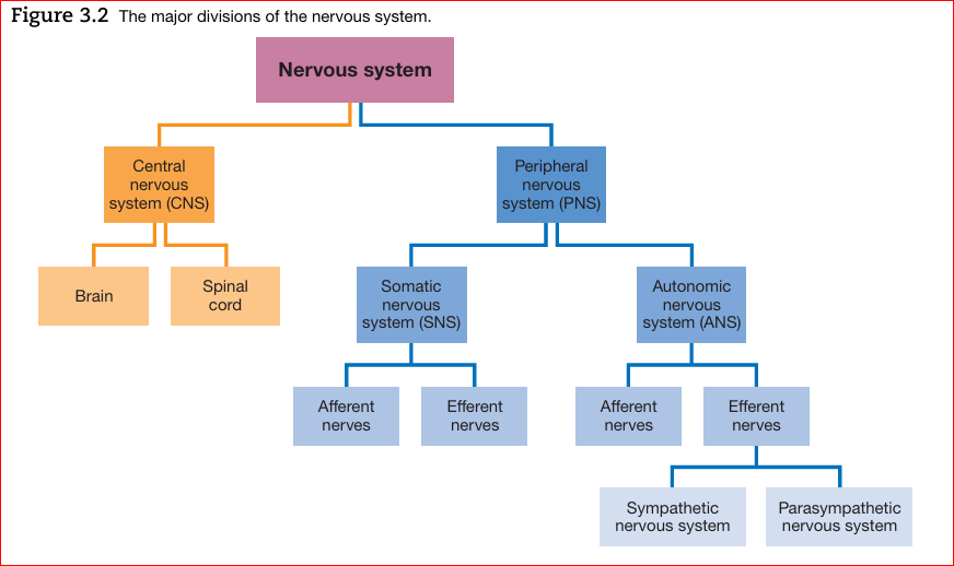
The Nervous system has two divisions:

  • Central nervous system (CNS) is the division of the nervous system located within the skull and spine

  • Peripheral nervous system (PNS) is the division located outside the skull and spine.


Central nervous system (CNS)

Two divisions:

  1. the brain is the part of the CNS located in the skull

  2. the spinal cord is the part located in the spine.

Peripheral nervous system (PNS)

Two divisions:

  1. Somatic nervous system (SNS) is the part of the PNS that interacts with the external environment. It is composed of:

    • Afferent nerves - carry sensory signals from the skin, skeletal muscles, joints, eyes, ears, and so on, to the central nervous system.

    • Efferent nerves that carry motor signals from the central nervous system to the skeletal muscles.

  2. Autonomic nervous system (ANS) is the part of the peripheral nervous system that regulates the body’s internal environment.

    • Sympathetic vs Parasympathetic:

      • Sympathetic: Fight or flight response. Act in threatening situations. Sympathetic changes are indicative of psychological arousal

      • Parasympathetic: Rest and digest. Act to conserve energy. Parasympathetic changes are indicative of psychological relaxation.

    • Has two kinds of efferent nerves:

      1. Sympathetic nerves - from the CNS in the lumbar (small of the back) and thoracic (chest area) regions of the spinal cord.
        - synapse on second-stage neurons at a substantial distance from their target organs

      2. Parasympathetic nerves - 2 sides = Cranium (Cranial nerves) and sacral (lower back) region of the spinal cord = Craniosacral nerves

        - synapse near their target organs

        *Both are two-stage: sympathetic and parasympathetic neurons project from the CNS and go only part of the way to the target organs before they synapse on other neurons (second-stage neurons) that carry the signals the rest of the way.

        *Both operate in contrasting fashion from one another in certain organs whilst in other organs they work in sync. (Pupil dilation = sympathetic; Pupil Constriction = parasympathetic) vs (Sexual arousal = sympathetic NS vs climax = parasympathetic NS)

        *Sweat glands, Piloerection (goose bumps), cutaneous blood vessel, adrenal medulla (cortisol), are ONLY innervated by the sympathetic nervous system.

        *Generally, we develop from parasympathetic dominance to sympathetic dominance in life

Sympathetic Nerves: Hypertension

  • Generally speaking, colder weather produces higher blood pressure because the cold narrows blood vessels ergo the heart has a harder time to send blood out (effect is reduced when medicated).

  • Humidity coupled with heat on the other hand increases blood pressure because the heart has a harder time to supply more blood to the skin which is the effect of the environment. This happens because the body is attempting to radiate heat (cool down).


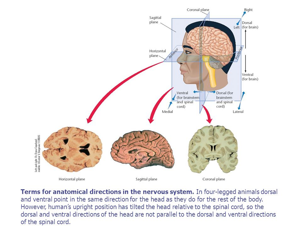
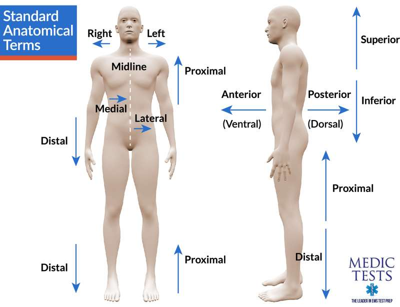
Anatomical Terms Referring to Directions

  • Directional Terms:

    • Dorsal: Toward the back. Away from the ventral (stomach) side.
      Ex. Top Brain

    • Ventral: Toward the stomach. Away from the dorsal (back) side.

    • Anterior: Toward the front.

    • Posterior: Toward the rear end.

    • Superior: Above another part.

    • Inferior: Below another part.

    • Lateral: Toward the side.

    • Medial: Toward the midline.

    • Proximal: Close to the point of origin.

    • Distal: Distant from the point of origin.
      *Far but still connected to a part

    • Ipsilateral: Same side of the body.

    • Contralateral: Opposite side of the body.

  • Planes:

    • Coronal: Front view.

    • Sagittal: Side view.

    • Horizontal: Top view.

Brain

Protective Layers

  • Layers of Protection:

    1. Scalp - first protective layer

    2. Periosteum - provides a waxy protective substance

    3. Skull - bone protective cavity for the brain

    4. Dura Mater - outermost meninx

    5. Arachnoid Space - Made because of the blood vessels but doesn’t contain blood vessels.

    6. Pia Mater - last protective layer

    7. Brain
      *Foramen magnum - the opening in the base of the skull that connects the spinal cord to the brain.


Meninges, Ventricles, and CSF

  • Meninges: Three protective membranes inside the skull.

    1. Dura Mater - means “Hard Mother”. Outer meninx.

      • Tough and outermost layer of protection inside the skull. Contains CSF to form a sac-like protection for the brain

      • Virtually connected to the skull UNLESS there is bleeding.

    2. Arachnoid Space (spider-web-like membrane) - network of connective tissue and is avascular

      • Contains the Subarachnoid space which contains many large blood vessels and cerebrospinal fluid

    3. Pia Mater (pious mother)- the delicate and innermost meninx which adheres to the surface of the CNS.


  • Cerebrospinal Fluid (CSF):

    • Supports and cushions the brain.

    • Fills the subarachnoid space, central canal of the spinal cord, and the cerebral ventricles of the brain.

    • Produced by the choroid plexus - networks of capillaries, or small blood vessels that protrude into the ventricles from the pia mater

  • Central canal is a small central channel that runs the length of the spinal cord

  • Cerebral Ventricles: Pathways for CSF flow. The four large internal chambers of the brain: the two lateral ventricles, the third ventricle, and the fourth ventricle.

    • The subarachnoid space, central canal, and cerebral ventricles are interconnected by a series of openings and thus form a single reservoir.

  • Cerebral Aqueduct / Aqueduct of Sylvius – Connects Third Ventricle and Diencephelon and the Fourth Ventricle

  • excess CSF is continuously absorbed from the subarachnoid space into large blood-filled spaces, or dural sinuses which run through the dura mater and drain into the large jugular veins of the neck;

  • Hydrocephalus (water head): The resulting buildup of fluid in the ventricles causes the walls of the ventricles, and thus the entire brain, to expand.

    • treated by draining the excess fluid from the ventricles and trying to remove the obstruction.


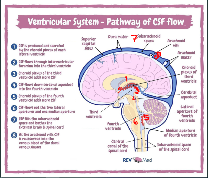
Flow of CSF

  1. CSF secreted by choroid plexus in lateral ventricles.

  2. CSF Flows through interventricular foramina into the third ventricle.

  3. Additional CSF added in the third ventricle.

  4. Flows down the cerebral aqueduct to the fourth ventricle.

  5. Choroid plexus of 4th ventricle adds more CSF.

  6. CSF Flowsout the two lateral apertures and one median aperture.

  7. Flows out into the subarachnoid space.

  8. Reabsorbed into venous blood of the dural venous sinuses at arachnoid villi.

Blood-Brain Barrier

  • Function: Barrier consisting of cerebral blood vessels which inhibit the passage of harmful substances from entering the brain

  • a consequence of the special structure of cerebral blood vessels.


Blood Circulation in the Brain

  • Circle of Willis: Named after Thomas Willis, is a circle like structure that supplies blood in the brain and the surrounding areas.

  • Located near the Interpeduncular Fossa at the base of the Brain

  • Creates redundancy for collateral circulation in the cerebral circulation. If one part of the circle becomes blocked or narrowed (stenosed), or one of the arteries supplying the circle is blocked or narrowed, blood flow from other vessels can often preserve the cerebral perfusion well enough to avoid the symptoms of ischemia (loss of blood or oxygen thus resulting in death of tissue)


Triangle of Death

  • The section of your face from the bridge of your nose to the corners of your mouth is sometimes known as the “danger triangle of the face,” or even the “triangle of death.” And it's one place where you should never pop a pimple, as it can lead to an infection in your brain.


Cells of the Nervous System

  • Neurons – specialized cells for reception, conduction, and transmission of electrochemical signals
    *There are about 200Bn Neurons contained in the CNS

  • Neuron cell membrane is composed of a lipid bilayer, or two layers of fat molecules.

    • Some membrane proteins are channel proteins, through which certain molecules can pass; others are signal proteins, which transfer a signal to the inside of the neuron when particular molecules bind to them on the outside of the membrane

      • Classes of Neurons:

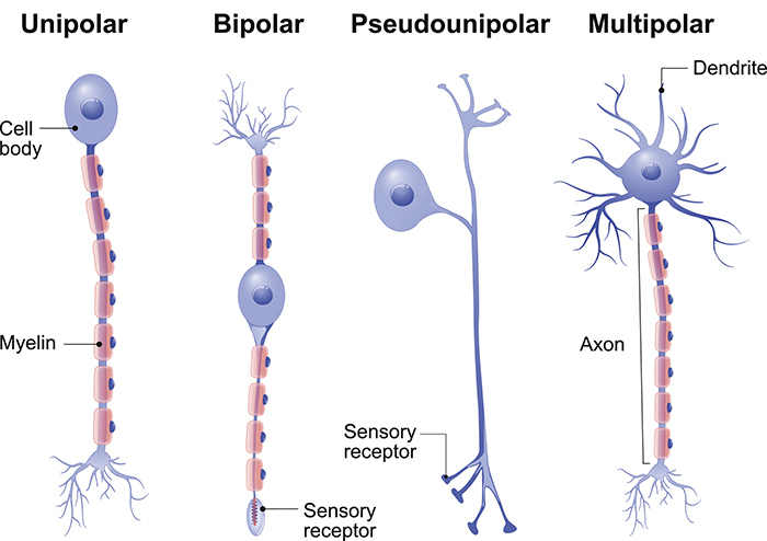
        1. Multipolar - A neuron with more than two processes extending from its cell body

        2. Unipolar - with one process extending from its cell body

        3. Bipolar - with two processes extending from its cell body

        4. Interneurons - with a short axon or no axon at all
      Two kinds of gross neural structures in the nervous system:

      1. Those composed primarily of cell bodies:

        • In the CNS, clusters of cell bodies are called nuclei

        • In the PNS, they are called ganglia

      2. Those composed primarily of axons:

        • In the CNS, bundles of axons are called tracts

        • In the PNS, they are called nerves

  • Glial Cells: Surround neurons and provide support and insulation.

    • Classes of Glia:

      1. Oligodendrocytes - Cells with extensions that wrap around axons thus myelinating them.

        • provides several myelin segments, often on more than one axon

      2. Schwann Cells - second class of glia. Functions similarly to Oligodendrocytes except that it can guide axonal regeneration and is found in the PNS.

        • constitutes one myelin segment

      3. Microglia - smaller than other glial cells—thus their name. They respond to injury or disease by multiplying, engulfing cellular debris or even entire cells which then triggers inflammatory responses.

      4. Astrocytes - largest glial cells. Plays a role in the passage of some chemicals from blood into the CNS, Structures neurons, send and receive signals from neurons and other glial cells to control the establishment and maintenance of synapses between neurons, modulate neural activity, maintain function of axons, and participate in glial circuits.

        • Their extensions cover the outer surface of blood vessels and also make contact with neuron. They have the ability to contract or relax blood vessels based on the blood flow demands of particular brain regions.

  • Myelin - a fatty insulating substance

    • Myelin sheaths - increase the speed and efficiency of axonal conduction

  • Peripheral vs Central Neurons: Peripheral neurons are more susceptible to injury.


Motor vs Sensory Neurons

  • Motor Neurons: Outward to skeletal muscles.

    • Motor neurons also branch out to the autonomic nervous system (target: Glands. Ex. Salivation) and controls functions

  • Sensory Neurons: Toward the CNS to give environmental information and by target organs.

Spinal Nerves
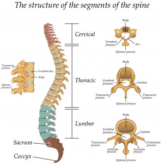
  • Total: 62 nerves (31 pairs).

  • Divisions:

    1. Cervical - 8 (C1-C8)

    2. Thoracic - 12 (T1-T12)

    3. Lumbar - 5 (L1-L5)

    4. Sacral - 5 (S1-S5)

    5. Coccygeal

SPINAL CORD

  • The part of the CNS found within the spinal column and communicates with the sense organs and muscles below the level of the head.

  • Spinal cord comprises two different areas:

    1. Gray matter: Unmyelinated.

    2. White matter: Myelinated.

  • The two dorsal arms of the spinal gray matter are called the dorsal horns, and the two ventral arms are called the ventral horns.

  • 31 pairs of spinal nerves = 62 spinal nerves

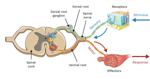
    • Pairs of spinal nerves are attached to the spinal cord— Each of these 62 spinal nerves divides as it nears the cord, and its axons are joined to the cord via one of two roots: the dorsal root or the ventral root.

      1. DORSAL ROOT AXONS = SENSORY (DORSAL ROOT GANGLIA)

        • All dorsal root axons, whether somatic or autonomic, are sensory (afferent) unipolar neurons with their cell bodies grouped together just outside the cord to form the dorsal root ganglia.

      2. VENTRAL ROOT AXONS = MOTOR (VENTRAL ROOT GANGLIA)

        • All ventral root neurons are motor (efferent) multipolar neurons with their cell bodies in the ventral horns.

        • Those that are part of the somatic nervous system project to skeletal muscles; those that are part of the autonomic nervous system project to ganglia, where they synapse on neurons that in turn project to internal organs (heart, stomach, liver, etc.).

  • The Bell-Magendie law states the entering dorsal roots carry sensory information and the exiting ventral roots carry motor information.
Basic Functions of the Central Nervous System

  • Functions:

    • Homeostasis

    • Voluntary Movement

    • Perception

    • Abstraction

Lobes of the Brain

  • Lobes:

    • Frontal Lobe - voluntary movement, expressive language and for managing higher level executive functions.

    • Parietal Lobe - sensory perception and integration, including the management of taste, hearing, sight, touch, and smell.

    • Occipital Lobe - visual processing

    • Temporal Lobe - processing auditory information and with the encoding of memory.

Cranial Nerves

  • Cranial Nerves Overview:

    • CN I: Olfactory (smell)

    • CN II: Optic (vision)

    • CN III: Oculomotor (eye muscles)

    • CN IV: Trochlear (superior oblique muscle)

    • CN V: Trigeminal (face sensation)

    • CN VI: Abducens (lateral rectus, eye abduction)

    • CN VII: Facial (facial expression)

    • CN VIII: Vestibulocochlear (hearing)

    • CN IX: Glossopharyngeal (tongue and pharynx)

    • CN X: Vagus (heart, lungs, GIT)
      *Longest cranial nerves which contain motor and sensory fibers traveling to and from the gut.

    • CN XI: Accessory (sternocleidomastoid)

    • CN XII: Hypoglossal (tongue movement)


    *The cranial nerves include purely sensory nerves such as the olfactory nerves (I) and the optic nerves (II), but most contain both sensory and motor fibers.

NEURON
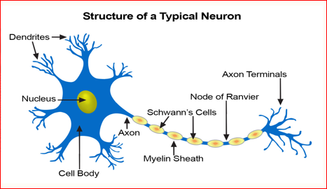
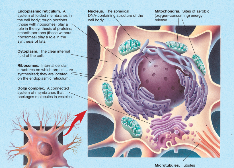
The major internal features of a neuron

  • Endoplasmic reticulum. A system of folded membranes in the cell body; rough portions (those with ribosomes) play a role in the synthesis of proteins; smooth portions (those without ribosomes) play a role in the synthesis of fats.

  • Cytoplasm. The clear internal fluid of the cell.

  • Ribosomes. Internal cellular structures on which proteins are synthesized; they are located on the endoplasmic reticulum.

  • Golgi complex. A connected system of membranes that packages molecules in vesicles.

  • Nucleus. The spherical DNA-containing structure of the cell body. Mitochondria. Sites of aerobic (oxygen-consuming) energy release

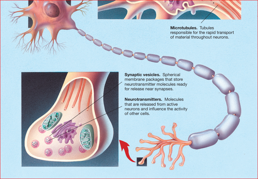
  • Microtubules. Tubules responsible for the rapid transport of material throughout neurons

  • Synaptic vesicles. Spherical membrane packages that store neurotransmitter molecules ready for release near synapses.

  • Neurotransmitters. Molecules that are released from active neurons and influence the activity of other cells.
The major external features of a neuron

  • Cell membrane. The semipermeable membrane that encloses the neuron.

  • Dendrites. The short processes emanating from the cell body, which receive most of the synaptic contacts from other neurons.

  • Axon hillock. The cone-shaped region at the junction between the axon and the cell body.

  • Myelin. The fatty insulation around many axons.

  • Axon. The long, narrow process that projects from the cell body

  • Nodes of Ranvier (pronounced “RAHN-vee-yay”). The gaps between sections of myelin

  • Cell body. The metabolic center of the neuron; also called the soma.

  • Buttons. The buttonlike endings of the axon branches, which release chemicals into synapses.

  • Synapses. The gaps between adjacent neurons across which chemical signals are transmitted

DG

2 Anatomy of the Nervous System

Anatomy of the Nervous System

  • Neuroanatomy: Study of the nervous system

    • Refers to the study of the various parts of the nervous system and their respective function(s).

    • The nervous system consists of many substructures, each comprised of many neurons.

The Nervous system has two divisions:

  • Central nervous system (CNS) is the division of the nervous system located within the skull and spine

  • Peripheral nervous system (PNS) is the division located outside the skull and spine.


Central nervous system (CNS)

Two divisions:

  1. the brain is the part of the CNS located in the skull

  2. the spinal cord is the part located in the spine.

Peripheral nervous system (PNS)

Two divisions:

  1. Somatic nervous system (SNS) is the part of the PNS that interacts with the external environment. It is composed of:

    • Afferent nerves - carry sensory signals from the skin, skeletal muscles, joints, eyes, ears, and so on, to the central nervous system.

    • Efferent nerves that carry motor signals from the central nervous system to the skeletal muscles.

  2. Autonomic nervous system (ANS) is the part of the peripheral nervous system that regulates the body’s internal environment.

    • Sympathetic vs Parasympathetic:

      • Sympathetic: Fight or flight response. Act in threatening situations. Sympathetic changes are indicative of psychological arousal

      • Parasympathetic: Rest and digest. Act to conserve energy. Parasympathetic changes are indicative of psychological relaxation.

    • Has two kinds of efferent nerves:

      1. Sympathetic nerves - from the CNS in the lumbar (small of the back) and thoracic (chest area) regions of the spinal cord.
        - synapse on second-stage neurons at a substantial distance from their target organs

      2. Parasympathetic nerves - 2 sides = Cranium (Cranial nerves) and sacral (lower back) region of the spinal cord = Craniosacral nerves

        - synapse near their target organs

        *Both are two-stage: sympathetic and parasympathetic neurons project from the CNS and go only part of the way to the target organs before they synapse on other neurons (second-stage neurons) that carry the signals the rest of the way.

        *Both operate in contrasting fashion from one another in certain organs whilst in other organs they work in sync. (Pupil dilation = sympathetic; Pupil Constriction = parasympathetic) vs (Sexual arousal = sympathetic NS vs climax = parasympathetic NS)

        *Sweat glands, Piloerection (goose bumps), cutaneous blood vessel, adrenal medulla (cortisol), are ONLY innervated by the sympathetic nervous system.

        *Generally, we develop from parasympathetic dominance to sympathetic dominance in life

Sympathetic Nerves: Hypertension

  • Generally speaking, colder weather produces higher blood pressure because the cold narrows blood vessels ergo the heart has a harder time to send blood out (effect is reduced when medicated).

  • Humidity coupled with heat on the other hand increases blood pressure because the heart has a harder time to supply more blood to the skin which is the effect of the environment. This happens because the body is attempting to radiate heat (cool down).


Anatomical Terms Referring to Directions

  • Directional Terms:

    • Dorsal: Toward the back. Away from the ventral (stomach) side.
      Ex. Top Brain

    • Ventral: Toward the stomach. Away from the dorsal (back) side.

    • Anterior: Toward the front.

    • Posterior: Toward the rear end.

    • Superior: Above another part.

    • Inferior: Below another part.

    • Lateral: Toward the side.

    • Medial: Toward the midline.

    • Proximal: Close to the point of origin.

    • Distal: Distant from the point of origin.
      *Far but still connected to a part

    • Ipsilateral: Same side of the body.

    • Contralateral: Opposite side of the body.

  • Planes:

    • Coronal: Front view.

    • Sagittal: Side view.

    • Horizontal: Top view.

Brain

Protective Layers

  • Layers of Protection:

    1. Scalp - first protective layer

    2. Periosteum - provides a waxy protective substance

    3. Skull - bone protective cavity for the brain

    4. Dura Mater - outermost meninx

    5. Arachnoid Space - Made because of the blood vessels but doesn’t contain blood vessels.

    6. Pia Mater - last protective layer

    7. Brain
      *Foramen magnum - the opening in the base of the skull that connects the spinal cord to the brain.


Meninges, Ventricles, and CSF

  • Meninges: Three protective membranes inside the skull.

    1. Dura Mater - means “Hard Mother”. Outer meninx.

      • Tough and outermost layer of protection inside the skull. Contains CSF to form a sac-like protection for the brain

      • Virtually connected to the skull UNLESS there is bleeding.

    2. Arachnoid Space (spider-web-like membrane) - network of connective tissue and is avascular

      • Contains the Subarachnoid space which contains many large blood vessels and cerebrospinal fluid

    3. Pia Mater (pious mother)- the delicate and innermost meninx which adheres to the surface of the CNS.


  • Cerebrospinal Fluid (CSF):

    • Supports and cushions the brain.

    • Fills the subarachnoid space, central canal of the spinal cord, and the cerebral ventricles of the brain.

    • Produced by the choroid plexus - networks of capillaries, or small blood vessels that protrude into the ventricles from the pia mater

  • Central canal is a small central channel that runs the length of the spinal cord

  • Cerebral Ventricles: Pathways for CSF flow. The four large internal chambers of the brain: the two lateral ventricles, the third ventricle, and the fourth ventricle.

    • The subarachnoid space, central canal, and cerebral ventricles are interconnected by a series of openings and thus form a single reservoir.

  • Cerebral Aqueduct / Aqueduct of Sylvius – Connects Third Ventricle and Diencephelon and the Fourth Ventricle

  • excess CSF is continuously absorbed from the subarachnoid space into large blood-filled spaces, or dural sinuses which run through the dura mater and drain into the large jugular veins of the neck;

  • Hydrocephalus (water head): The resulting buildup of fluid in the ventricles causes the walls of the ventricles, and thus the entire brain, to expand.

    • treated by draining the excess fluid from the ventricles and trying to remove the obstruction.


Flow of CSF

  1. CSF secreted by choroid plexus in lateral ventricles.

  2. CSF Flows through interventricular foramina into the third ventricle.

  3. Additional CSF added in the third ventricle.

  4. Flows down the cerebral aqueduct to the fourth ventricle.

  5. Choroid plexus of 4th ventricle adds more CSF.

  6. CSF Flowsout the two lateral apertures and one median aperture.

  7. Flows out into the subarachnoid space.

  8. Reabsorbed into venous blood of the dural venous sinuses at arachnoid villi.


Blood-Brain Barrier

  • Function: Barrier consisting of cerebral blood vessels which inhibit the passage of harmful substances from entering the brain

  • a consequence of the special structure of cerebral blood vessels.


Blood Circulation in the Brain

  • Circle of Willis: Named after Thomas Willis, is a circle like structure that supplies blood in the brain and the surrounding areas.

  • Located near the Interpeduncular Fossa at the base of the Brain

  • Creates redundancy for collateral circulation in the cerebral circulation. If one part of the circle becomes blocked or narrowed (stenosed), or one of the arteries supplying the circle is blocked or narrowed, blood flow from other vessels can often preserve the cerebral perfusion well enough to avoid the symptoms of ischemia (loss of blood or oxygen thus resulting in death of tissue)


Triangle of Death

  • The section of your face from the bridge of your nose to the corners of your mouth is sometimes known as the “danger triangle of the face,” or even the “triangle of death.” And it's one place where you should never pop a pimple, as it can lead to an infection in your brain.


Cells of the Nervous System

  • Neurons – specialized cells for reception, conduction, and transmission of electrochemical signals
    *There are about 200Bn Neurons contained in the CNS

  • Neuron cell membrane is composed of a lipid bilayer, or two layers of fat molecules.

    • Some membrane proteins are channel proteins, through which certain molecules can pass; others are signal proteins, which transfer a signal to the inside of the neuron when particular molecules bind to them on the outside of the membrane

      • Classes of Neurons:

        1. Multipolar - A neuron with more than two processes extending from its cell body

        2. Unipolar - with one process extending from its cell body

        3. Bipolar - with two processes extending from its cell body

        4. Interneurons - with a short axon or no axon at all

      Two kinds of gross neural structures in the nervous system:

      1. Those composed primarily of cell bodies:

        • In the CNS, clusters of cell bodies are called nuclei

        • In the PNS, they are called ganglia

      2. Those composed primarily of axons:

        • In the CNS, bundles of axons are called tracts

        • In the PNS, they are called nerves

  • Glial Cells: Surround neurons and provide support and insulation.

    • Classes of Glia:

      1. Oligodendrocytes - Cells with extensions that wrap around axons thus myelinating them.

        • provides several myelin segments, often on more than one axon

      2. Schwann Cells - second class of glia. Functions similarly to Oligodendrocytes except that it can guide axonal regeneration and is found in the PNS.

        • constitutes one myelin segment

      3. Microglia - smaller than other glial cells—thus their name. They respond to injury or disease by multiplying, engulfing cellular debris or even entire cells which then triggers inflammatory responses.

      4. Astrocytes - largest glial cells. Plays a role in the passage of some chemicals from blood into the CNS, Structures neurons, send and receive signals from neurons and other glial cells to control the establishment and maintenance of synapses between neurons, modulate neural activity, maintain function of axons, and participate in glial circuits.

        • Their extensions cover the outer surface of blood vessels and also make contact with neuron. They have the ability to contract or relax blood vessels based on the blood flow demands of particular brain regions.

  • Myelin - a fatty insulating substance

    • Myelin sheaths - increase the speed and efficiency of axonal conduction

  • Peripheral vs Central Neurons: Peripheral neurons are more susceptible to injury.


Motor vs Sensory Neurons

  • Motor Neurons: Outward to skeletal muscles.

    • Motor neurons also branch out to the autonomic nervous system (target: Glands. Ex. Salivation) and controls functions

  • Sensory Neurons: Toward the CNS to give environmental information and by target organs.

Spinal Nerves

  • Total: 62 nerves (31 pairs).

  • Divisions:

    1. Cervical - 8 (C1-C8)

    2. Thoracic - 12 (T1-T12)

    3. Lumbar - 5 (L1-L5)

    4. Sacral - 5 (S1-S5)

    5. Coccygeal

SPINAL CORD

  • The part of the CNS found within the spinal column and communicates with the sense organs and muscles below the level of the head.

  • Spinal cord comprises two different areas:

    1. Gray matter: Unmyelinated.

    2. White matter: Myelinated.

  • The two dorsal arms of the spinal gray matter are called the dorsal horns, and the two ventral arms are called the ventral horns.

  • 31 pairs of spinal nerves = 62 spinal nerves

    • Pairs of spinal nerves are attached to the spinal cord— Each of these 62 spinal nerves divides as it nears the cord, and its axons are joined to the cord via one of two roots: the dorsal root or the ventral root.

      1. DORSAL ROOT AXONS = SENSORY (DORSAL ROOT GANGLIA)

        • All dorsal root axons, whether somatic or autonomic, are sensory (afferent) unipolar neurons with their cell bodies grouped together just outside the cord to form the dorsal root ganglia.

      2. VENTRAL ROOT AXONS = MOTOR (VENTRAL ROOT GANGLIA)

        • All ventral root neurons are motor (efferent) multipolar neurons with their cell bodies in the ventral horns.

        • Those that are part of the somatic nervous system project to skeletal muscles; those that are part of the autonomic nervous system project to ganglia, where they synapse on neurons that in turn project to internal organs (heart, stomach, liver, etc.).

  • The Bell-Magendie law states the entering dorsal roots carry sensory information and the exiting ventral roots carry motor information.

Basic Functions of the Central Nervous System

  • Functions:

    • Homeostasis

    • Voluntary Movement

    • Perception

    • Abstraction

Lobes of the Brain

  • Lobes:

    • Frontal Lobe - voluntary movement, expressive language and for managing higher level executive functions.

    • Parietal Lobe - sensory perception and integration, including the management of taste, hearing, sight, touch, and smell.

    • Occipital Lobe - visual processing

    • Temporal Lobe - processing auditory information and with the encoding of memory.

Cranial Nerves

  • Cranial Nerves Overview:

    • CN I: Olfactory (smell)

    • CN II: Optic (vision)

    • CN III: Oculomotor (eye muscles)

    • CN IV: Trochlear (superior oblique muscle)

    • CN V: Trigeminal (face sensation)

    • CN VI: Abducens (lateral rectus, eye abduction)

    • CN VII: Facial (facial expression)

    • CN VIII: Vestibulocochlear (hearing)

    • CN IX: Glossopharyngeal (tongue and pharynx)

    • CN X: Vagus (heart, lungs, GIT)
      *Longest cranial nerves which contain motor and sensory fibers traveling to and from the gut.

    • CN XI: Accessory (sternocleidomastoid)

    • CN XII: Hypoglossal (tongue movement)


    *The cranial nerves include purely sensory nerves such as the olfactory nerves (I) and the optic nerves (II), but most contain both sensory and motor fibers.

NEURON

The major internal features of a neuron

  • Endoplasmic reticulum. A system of folded membranes in the cell body; rough portions (those with ribosomes) play a role in the synthesis of proteins; smooth portions (those without ribosomes) play a role in the synthesis of fats.

  • Cytoplasm. The clear internal fluid of the cell.

  • Ribosomes. Internal cellular structures on which proteins are synthesized; they are located on the endoplasmic reticulum.

  • Golgi complex. A connected system of membranes that packages molecules in vesicles.

  • Nucleus. The spherical DNA-containing structure of the cell body. Mitochondria. Sites of aerobic (oxygen-consuming) energy release

  • Microtubules. Tubules responsible for the rapid transport of material throughout neurons

  • Synaptic vesicles. Spherical membrane packages that store neurotransmitter molecules ready for release near synapses.

  • Neurotransmitters. Molecules that are released from active neurons and influence the activity of other cells.

The major external features of a neuron

  • Cell membrane. The semipermeable membrane that encloses the neuron.

  • Dendrites. The short processes emanating from the cell body, which receive most of the synaptic contacts from other neurons.

  • Axon hillock. The cone-shaped region at the junction between the axon and the cell body.

  • Myelin. The fatty insulation around many axons.

  • Axon. The long, narrow process that projects from the cell body

  • Nodes of Ranvier (pronounced “RAHN-vee-yay”). The gaps between sections of myelin

  • Cell body. The metabolic center of the neuron; also called the soma.

  • Buttons. The buttonlike endings of the axon branches, which release chemicals into synapses.

  • Synapses. The gaps between adjacent neurons across which chemical signals are transmitted

robot