Y8 Biology (help me please) (copy)
Cells - Objectives 1 - 8
Definition
smallest structural and functional unit of all living organisms
building blocks of life
Cell theory
all living things made up of cells
cells are basic building blocks of life
new cells are formed form the division of parent cells (mitosis)
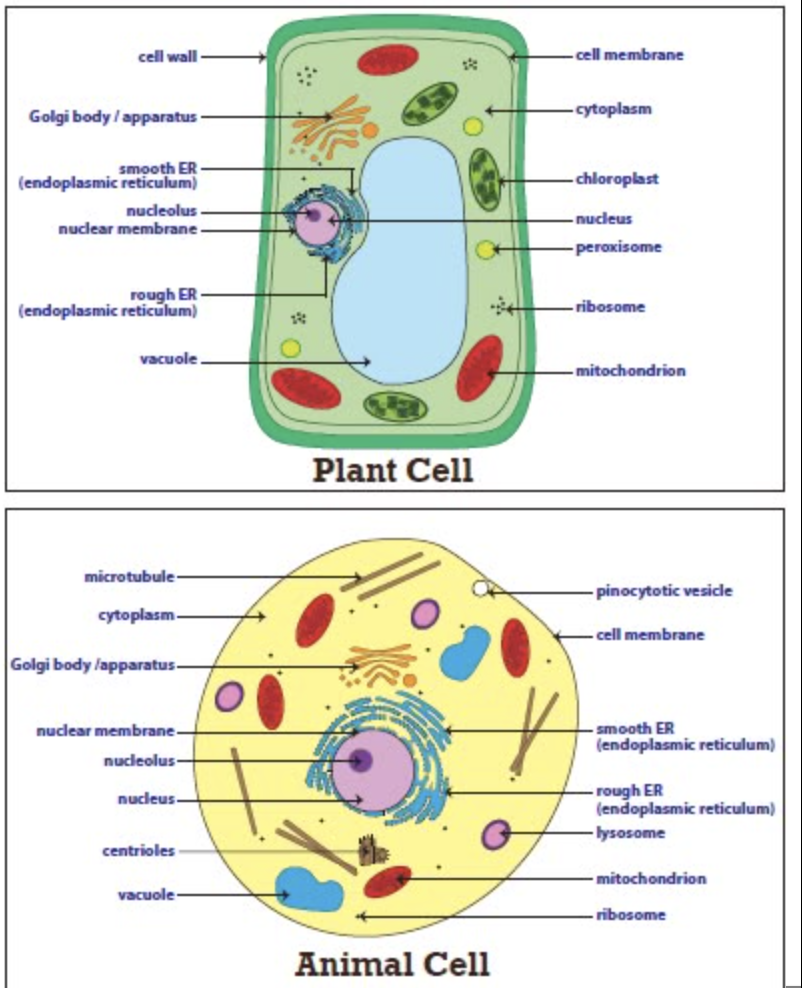
Structures
cell membrane
delicate membrane that surrounds the cell
regulates what enters and exits
cytoplasm
watery jelly-like substance
contains dissolved substances and organelles
fluid part is called cytosol
nucleus
membrane-bound sac that stores all the cell’s chromosomes/DNA
has pores
mitochondria
powerhouse of the cell
helps convert glucose into energy that the cell can use
ribosomes
builds protein for amino acids
can be free flowing or attached to the endoplasmic reticulum
endoplasmic reticulum (ER)
smooth ER - do not have ribosomes attached and builds lipids and carbohydrates
rough ER - has ribosomes attached and stores protein made by ribosomes
golgi apparatus
takes in sacs of raw material from ER and sends out sacs containing finished cell products
lysosome
only in animal cell
sacs filled with digestive enzymes
digests worn out cells
digests food/nutrients absorbed by cell
centrioles
pair of bundled tubes
organizes cell division
cytoskeleton
made out of microtubules
found throughout cytoplasm
gives shape to cells and moves organelles around inside
next 3 for plant cells only
cell wall
very strong, rigid, structure
made of cellulose
protects cells from rupturing
sits outside cell membrane
glued to other cells it is next to
chloroplasts
contains chlorophyll - the chemical that allows photosynthesis to occur
carries out photosynthesis
vacuole
huge, water-filled sac
permanent
keeps the cell pressurised
stores starch
animal cells also have vacuoles, but not permanent or big, and do not keep the cell pressurised
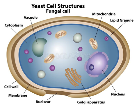
Fungal cells have all of the animal organelles and a cell wall made of chitin
Prokaryotes and Eukaryotes
Prokaryotes
organelles lack a membrane
ribosomes are the only organelle
genetic material floats in the cytoplasm
circular DNA
unicellular, smaller in size
large number of organisms
Eukaryotes
organelles covered by a membrane
multiple organelles - including ribosomes
have a nuclear membrane covering genetic material
linear DNA
can be unicellular or multicellular
cells larger in size
smaller number of organisms
Unicellular and Multicellular
Unicellular
simple and composed of a singular cell
i.e. yeast, amoeba, bacteria
Multicellular
complex and of multiple cells that carry out different functions
i.e. animals, plants, fungi
Microscopes - Objectives 9 - 14
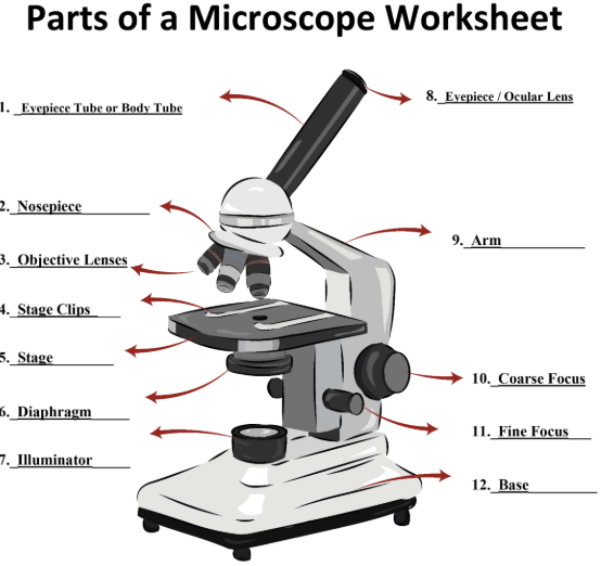
Main parts of a light miscroscope
body tube - used to look at the specimen and magnify the image for the second time
revolving nosepiece - can be rotated clockwise or counterclockwise to change the magnification
objective lens (4x, 10x, 40x) - magnifies the image for the first time
stage clips - holds the slide in place
diaphragm - controls the amount of light that reaches the specimen
light source - projects up through the slide to illuminate the sample
ocular lens (eyepiece) - used to look at the specimen
arm - supports the microscope when carried
stage - where the specimen is placed for viewing
course adjustment knob - moves stage up and down for focusing
fine adjustment knob - moves the stage slightly to sharpen the focus
base - supports and provides stability for the microscope
Magnification
As the field of view under the microscope increases, the magnification decreases
As the field of view under the microscope decreases, the microscope increases
to get the total magnification of a microscope, multiply the ocular lens magnification and the objective lens (i.e 10x ocular, 4x objective = 40x total)
1mm = 1000µm
magnification = image size/actual size, (actual size being the µm given on the scale bar and the image size being the scale bar measured in mm and multiplied to get the µm. i.e. if the scale bar said 600µm, you would measure the length of the bar with a ruler and convert that to µm, x, and perform the sum x/600 to get the magnification)
Advantages and disadvantages between a light and an electron microscope
Advantages | Disadvantages | |
Light |
|
|
Electron |
|
|
Mitosis - Objectives 15-18
Definition
Mitosis is the simple duplication and division of a cell and all of its parts, and occurs for growth and repair
It duplicates chromosomes and the 2 new cells (daughter cells) are identical
Stages - IPMAT
Interphase - normal state of cell. DNA duplicates here.
Prophase - When the cell gets the idea that it is time to divide, so it prepares by coiling the chromosomes, the mitotic spindle (what comes out the centrioles) begins to form, and the nuclear membrane begins to fade.
Metaphase - The DNA lines up along a central axis and the centrioles send out specialised tubules that connect to the DNA
Anaphase - When the separation begins. Half of the chromosomes are pulled to one side of the cell while half go the other way
Telophase - When the chromosomes uncoil, the nuclear membrane reappears and the mitotic spindle breaks down.
Telophase pt.2; Cytokinesis - when the cytoplasm divides

Digestive System - Objectives 19 - 26
Structural Units - general hierarchy, not specific to digestive system
cell - a structural and functional unit (i.e a nerve cell)
tissue - a group of cells with common structures/functions (i.e nervous tissue)
organs - group of tissues working together for a common task (i.e brain)
system - composed of several organs working together (i.e nervous system)
organism - an individual containing several organ systems (i.e human)
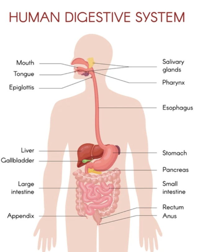
Major Structures and their functions
mouth
where food enters
coated in saliva which contains digestive enzymes that break down food (chemical digestion)
teeth used for chewing which breaks food into smaller pieces (mechanical digestion)
pharynx
a tube where food and air travel through
when food is being swallowed, a special structure called the epiglottis closes over the entrance airway (trachea) so that food doesn’t go into the lungs
connected to the oesophagus
oesophagus
a tube where food travels down after swallowing for digestion
food is moved down through peristalsis (wave-length muscular contractions)
stomach
both chemical (acid breaking down complex molecules and a new substance is formed) and mechanical digestion (breaking down food through movement) occur here
bag-like muscular structure that stores and churns food to break it down
contains enzymes that start to break down food and hydrochloric acid that kills bacteria and sterilises food
liver
makes bile for digestion of fats/lipids
processes the blood containing the nutrients absorbed from the small intestine
detoxifies potentially harmful chemicals in the blood
gallbladder
stores and concentrates the bile made by the liver
releases the bile into the small intestine
pancreas
produces enzymes for digestion
also produces insulin for controlling the absorption of glucose into cells
small intestine
final point of chemical digestion
large surface area so contents released from the stomach come into contact with the enzymes to break them down
villi, microvilli, and circular folds contribute in increasing the surface area
once the food has been digested, the villi and microvilli lining this tube absorb the nutrients and send them into the bloodstream
large intestine
vitamins and minerals are absorbed
waves or peristalsis move undigested food waste through this tube
waste becomes more solid as it moves through this tube due to the absorption of water from the waste back into the body
rectum
stores the waste (faeces) leftover after digestion
anus
controls the process of defecation and gets rid of waste
Key functions
ingestion - taking in food
digestion - breaking food down into molecules small enough to be absorbed
difference between chemical and mechanical digestion - chemical digestion uses enzymes to break down food into smaller, absorbable molecules and produce a new substance when doing this, while mechanical digestion breaks down food physically like chewing and churning.
absorption - absorbing the nutrients
egestion/defecation - eliminating undigested waste product
Nutrients in food
Type of Food Nutrient | Type of digestive enzyme | Where digestive enzyme is produced | Example |
Carbohydrates | Amylase |
| Bananas |
Proteins | Protease |
| Pineapple |
Lipids/fats | Lipase |
| Avocados |
Importance of enzymes
they break down important components in food so that they are easier to absorb
speed up chemical reactions in living organisms
without them, many processes would occur to slowly to sustain life
Comparing digestive systems
carnivores
short digestive tracts
sharp teeth
strong digestive enzyme
herbivores
long digestive tracts
flat teeth
omnivores
shorter digestive tracts than herbivores but longer than carnivores
range of digestive enzymes
range of sharp and flat teeth
Why do they have different digestive systems?
due to difference in diet
carnivores - sharp teeth to tear and hold down prey; since meat is easy to digest, they have strong enzymes to break it down and a short digestive tract to process food quickly
herbivores - flat teeth to grind fibrous plants; long digestive tracts to maximise nutrient absorption
omnivores - eat both meat and plants, so their digestive systems are in between carnivores and herbivores
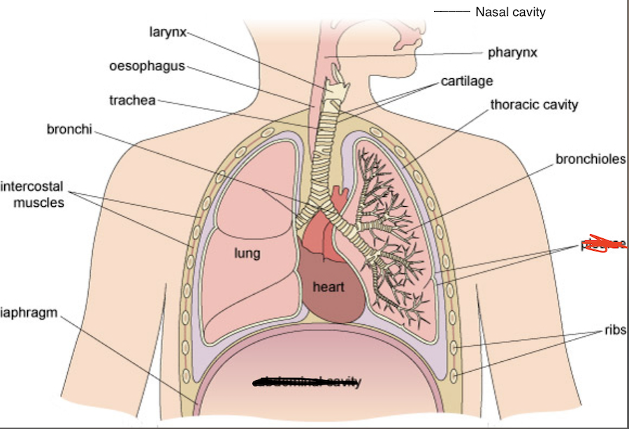
Respiratory System - Objectives 27 - 31
Major Structures and their functions
nasal cavity
the air enters through the nostrils
filtered by hairs
warmed, humidified
larynx
voice box
reinforced with cartilage to help protect airway
keeps fluid and food from entering the airway
trachea
windpipe
rings of cartilage that maintain the shape to prevent it from closing
cilia helps sweep particles and fluid from getting into the lungs
forks into the bronchi
bronchi
each bronchus leads into a lung and branches into smaller and smaller bronchioles
distributes air into bronchioles
bronchioles
during
alveoli
small, balloon like sac that is 200-500µm in diameter
found at the end of bronchioles in lungs
each alveolus plays an important role in letting oxygen and carbon-dioxide move into and from the blood stream during inhalation and exhalation
gas exchange occurs here - where oxygen moves from the lungs to the bloodstream, while carbon-dioxide simultaneously passes from the blood to the lungs
the rest are not where the air flows through, but parts that are still included in the respiratory system
lungs
where the two bronchi lead to, and where the bronchioles and alveoli are found
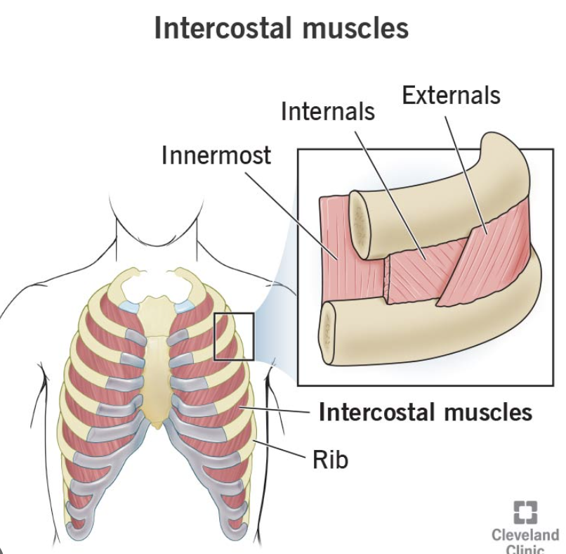
intercostal muscles
located in the space between the ribs
during inhalation, the external intercostal muscles contract and lift the ribs upward, expanding the chest cavity and creating negative pressure, which draws air into the lungs.
during exhalation the internal intercostal muscles contract, they pull the ribs downward, reducing the chest cavity and forcing air out of the lungs
ribs
protects the lungs
allows for expansion and contraction during breathing
sternum
breast bone
central bone in the ribs (therefore same function)
Inhalation and exhalation
When you breathe in:
intercostal muscles between the ribs contract, pulling the chest walls up and out
diaphragm muscle below the lungs contracts and flattens, increasing the size of the chest
lungs increase in size, so the pressure inside them falls. This causes air to rush in through the nose or mouth
When you breathe out:
intercostal muscles between the ribs relax so that the chest walls move in and down
the diaphragm muscle below the lungs relaxes and bulges up, reducing the size of the chest
the lungs decrease in size, so the pressure inside increased and the air is pushed up the trachea and out through the nose or mouth
Gas exchange
air rich in oxygen enters the lungs and reaches the alveoli
oxygen diffuses from the alveoli into the blood in the capillaries, driven by the partial pressure gradient
carbon dioxide diffuses from the blood in the capillaries into the alveoli, also driven by the partial pressure gradient
air rich in carbon dioxide is exhaled from the lungs.
oxygen-rich blood is transported to the tissues throughout the body, where oxygen is released to cells for cellular respiration.
carbon dioxide, a waste product of cellular respiration, is transported back to the lungs for exhalation
Comparing Fish and Animal respiratory system
Animal
have a mouth, a pharynx, lungs, a trachea, bronchi, bronchioles, diaphragm, alveoli
Fish
d support 1 or 2 filaments
spiracles that are an opening used to draw water into the gills for respiration
most-efficient system for exchanging water and carbon dioxide between blood and water
differences reflects the adaptations to the environment
Circulatory System - Objectives 32 - 38
all circulatory systems consist of a pump, carrier, fluid, and tubes/vessels
4 components of blood
plasma
makes up just over half of the blood
water based-liquid
white blood cells, red blood cells, platelets all in this solution
red blood cells (erythrocytes)
carry oxygen from the lungs to the rest of the body
contains haemoglobin that carries the oxygen and the carbon dioxide to and from the heart/lungs
white blood cells (leukocytes)
help fight infections and aid in the immune system
platelets (thrombocytes)
broken down parts of cells
help in blood clotting
form scabs
Types of circulatory systems
closed circulatory system
blood is always contained in the vessels and never directly makes contact with the body’s tissues
double circulatory system
blood pumps through the heart twice on every full circuit of the body and involves two distinct circuits - the pulmonary circulation and the systemic circulation
pulmonary circulation
pumps blood to the lungs from the heart, and then returns to the heart again
systemic circulation
pumps blood to the rest of the body and then back to the heart
Arteries, veins, and capillaries
arteries
carries oxygenated blood away from the heart and to the rest of the body
very strong and thick walls to cope with the high pressure of the blood flowing through them
thick walls stretch and then flex back into place because of the elastic fibres in the walls
capillaries
arteries divide into smaller and smaller vessels - smallest being the capillaries
very small and penetrate every part of the body
takes nutrients and oxygen to cells and takes waste products away
very thin walls so the substances can get in and out very quickly
veins
capillaries eventually join again and form veins
carries deoxygenated blood back to the heart
by the time blood gets to the veins, it is at a lower pressure than it was in the arteries
have thinner, less strong walls compared to the arteries
have valves to stop blood from flowing backwards
Heart
located in the centre of your chest in between the lungs
4 chambers - 2 atriums, 2 ventricles
Sexual and Asexual Reproduction - Objectives 41 - 49
reproduction is the biological process by which new individual organisms, or "offspring," are produced from their "parent" or parents, ensuring the continuity of species
Asexual and Sexual reproduction
Difference between asexual and sexual reproduction - asexual reproduction only requires one parent and produces a genetically identical offspring, whereas sexual reproduction requires 2 parents and produces a genetically unique offspring through the fusion of gametes
asexual reproduction examples
plants - vegetative propagation (when new plants grow from parts of the parent plant such as stems, leaves, or roots, without the use of runner, i.e. strawberries)
animals - parthenogenesis (when an unfertilised egg develops into a new individual without fertilisation by sperm, allowing females to reproduce without males, i.e. wasps)
Disadvantages and advantages
Disadvantages | Advantages | |
Sexual |
|
|
Asexual |
|
|
Reproductive system of a plant
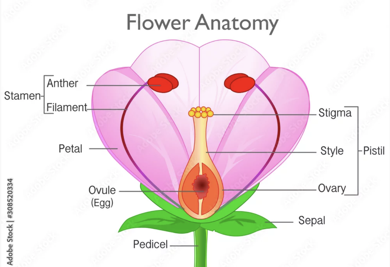
Structures:
petals
colourful and sweet
purpose is to attract pollinators
stalk/stem
holds up flower and gives support
also provides a highway for water and food to supply the plant with its needs
nectary
where nectary (sweet liquid) is produced by plants to attract pollinating animals
ovary
protects ovules
ovules
once they are fertilised they become seeds, and the ovary will become a fruit that protects the seed
receptacle
thickened part of a stem from which the flower grows
also where the flower attaches to the stalk
some cases it becomes a part of the fruit after fertilisation
style
stalk of the pistil that rises up from the ovary
stigma
part of the pistil that catches the pollen
sticky substance on the tip to catch
shaped different depending on the type of flower
pistil
female part of the flower
contains the ovary, ovules, stigma, style
stamen
male part of the flower
made of filament and anthers
also responsible for producing pollen
filament
stalk that holds up the anther
anther
located on the top of the filament and holds the pollen until the they mature
once the anthers mature, they burst, releasing the pollen
pollen
fertilising element of flowering plants
made of fine, powdery grains or spores
Pollination v Fertilisation
Pollination is the transfer of pollen from the male anther to the female stigma, while fertilisation is the fusion of male and female gametes (sperm and egg) to form a zygote, which eventually develops into a seed
Human reproductive system
Female

ovary
2 ovaries
primary female reproductive organs
produce and release ova
oviduct
2 uterine tubes that extend from the ovaries and lead to the uterus
the fimbriae at the ends of the tubes direct the ovum into tubes where ciliated epithelial cells and muscle contractions guide the ovum down to the uterus
clitoris
made up of erectile tissue, nerves an blood vessels
sensitive to touch and when stimulated becomes the ‘erect’ and engorged with blood
labia minora
two folds
lack fatty tissue and pubic hair
inner folds of skin that surround the clitoris and protect the urethral opening
labia majora
two folds
consist of fatty and fibrous tissue that protects the entrance to the vagina
many glands secrete oil keeping the inner surface moist
outer surface covered in pubic hair
vagina
canal that extends from the cervix and leads outside the body
lined with mucous membranes
receive the penis during intercourse
changes to become the birth canal during birth
cervix
neck of the uterus
protrudes into the vagina
where the semen is deposited during intercourse
uterus
composed of smooth muscle
lined with the endometrium
blastocyst implants here during pregnancy and as it grows the uterus expands
Male
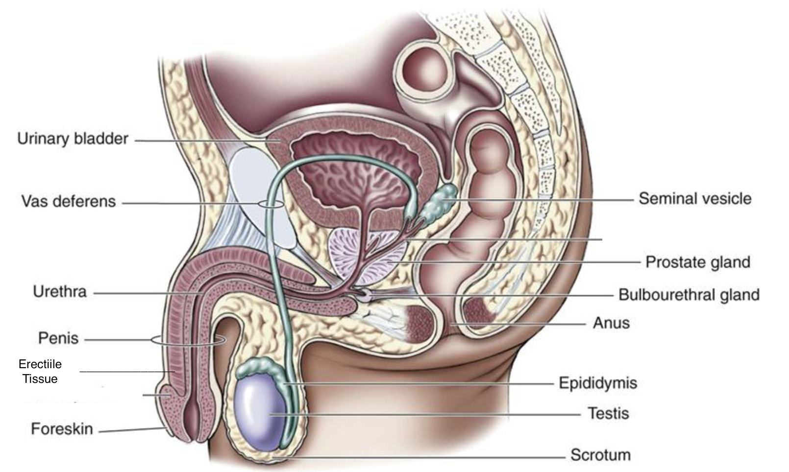
seminal vesicles
2 seminal vesicles
secrete fluid that makes up most of semen
thick fluid contains sugars to nourish the sperm
prostate gland
secretes fluid that is part of semen
thin, milky, alkaline fluid
bulbourethral glands
2 glands secrete a clear mucus just before the semen is ejaculated
acts as a lubricant
scrotum
skin covered pouch
holds the testes
internally divided into 2 pouches
muscles can move it away or towards the body
testes
2 primary male reproductive organs
produce sperm
epididymas
folded tube where sperm are stored and matured
5-6m long when stretched and sits behind the testes
erectile tissue
have a lot of spongy spaces filled with blood that cause the penis to erect
once erect the penis can be inserted into the vagina
urethra
tube that runs through the penis
can transport urine or semen out of the body
vas deferens
2 tubes
connects the epididymis of each testis to the urethra
carries sperm away from the testes to the urethra