G Protein - coupled Receptors
Summary Sheet:
G protein coupled receptors are made of single polypeptide chains.
The chains are folded and embedded into cell membranes.
The chains fold seven times so 7 proteins are present in the cell membrane from one receptor.
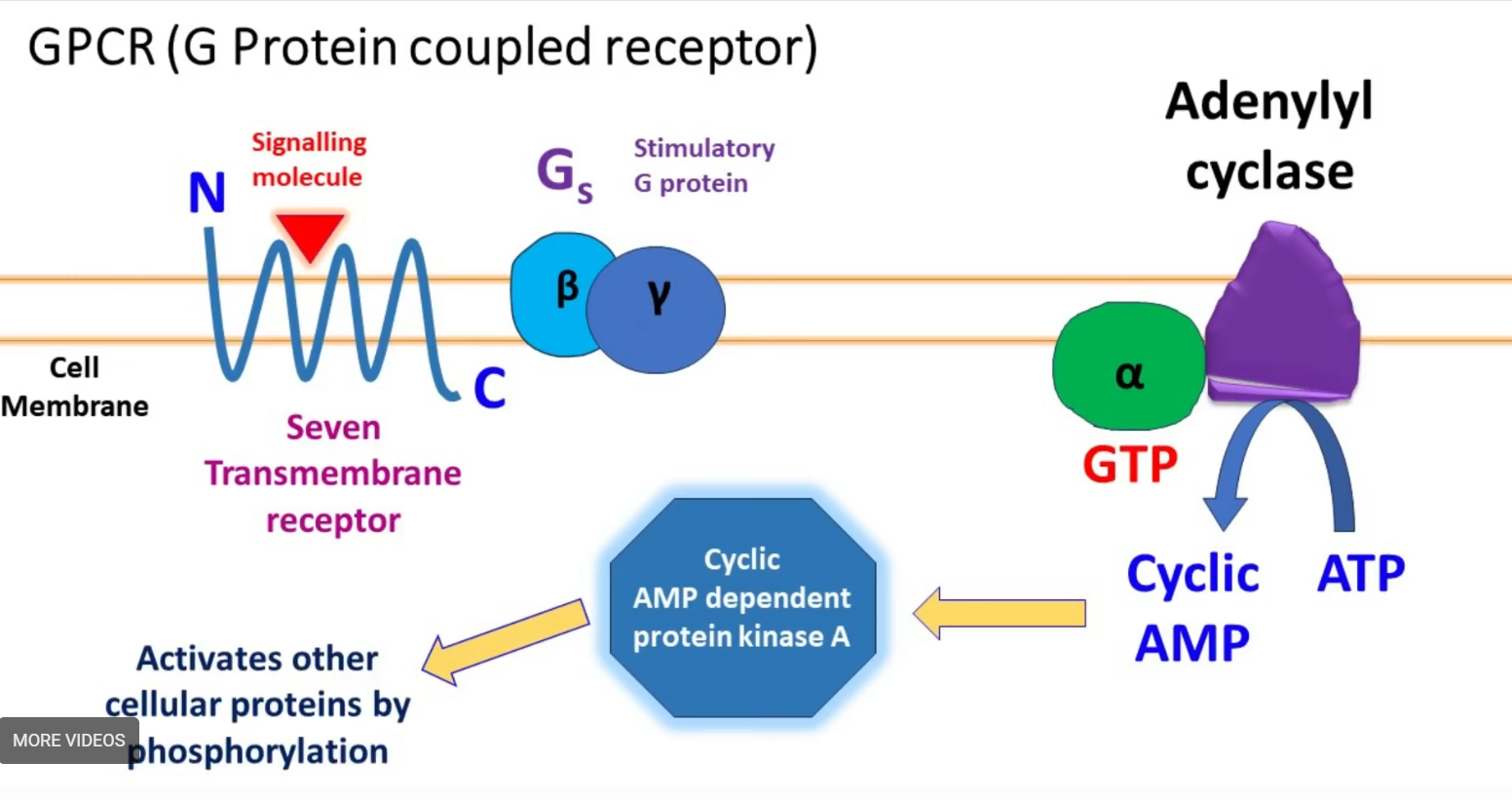
the N domain = extracellular
the C domain = intracellular
the C domain is attached to a Stimulatory G protein or Gs
the Gs protein has 3 domains
1) beta subunit
2) alpha subunit
3) gamma subunit
the alpha subunit is attached to GDP = inactive form of Gs protein
Steps
When a signalling molecule from the external environment binds to G protein-coupled receptor, the GDP attached to the alpha subunit is replaced with GTP = active form of Gs.
The GTP is hydrolysed to GTF allowing the alpha subunit to move and attach to Adenylyl cyclase enzyme.
The adenylyl cyclase enzyme catalyses the formation of cyclic AMP from ATP.
The cyclic AMP activates AMP-dependent protein kinase A.
The activated protein kinase A further activates other cellular proteins by phosphorylation.
GTP attached to alpha subunit gets hydrolysed to GDP.
GDP bound alpha subunit is inactive and returns to beta and gamma subunit which is still attached to G protein-coupled receptor.
G Protein coupled receptors
Gs - stimulates adenylate cyclase
Gi - inhibits adenylate cyclase
Gq - activates phospholipase C
Cyclic AMP (cAMP) acting as a second messenger
Adenylate cyclase converts ATP into cAMP
cAMP binds to activates other molecules like PKA
PKA (protein kinase) can phosphorylate other proteins, leading to various cellular responses,
Enzyme-linked receptors
cell surface receptors
have intracellular domains
2 types:
1) intracellular domain of receptor is an enzyme
2) intracellular domain interacts with an enzyme in order to catalyse a reaction
Kinase = enzyme that transfers phosphate groups to a protein or other target
Receptor Tyrosine Kinase (RTKs)
a class of enzyme-linked receptors found in humans and many other species
a receptor tyrosine kinase transfers phosphate groups specifically to the amino acid tyrosine
Receptor Tyrosine Kinase signalling
signalling molecules bind to the extracellular domains of two nearby RTKs.
ligand-binding induces the neighbouring RTKs to come together and dimerise.
the dimerisation activates the receptors’ intracellular tyrosine-kinase domain and the receptors attach phosphates to tyrosine in each other’s intracellular domain.
the phosphorylated tyrosine can transmit the signal to other molecules in the cell, initiating complex intracellular pathways.
Insulin Receptors
Insulin molecules bind to the alpha-subunits of the insulin receptor.
A conformational change occurs that activates the receptor’s tyrosine kinases
The activated tyrosine kinases add phosphate groups to specific tyrosine residues on themselves (autophosphorylation)
This activated receptor then phosphorylates other proteins, particularly insulin receptor substrates, which creates docking sites for more signalling molecules.
Signalling pathways are initiated, including the Ras/MAPK pathway and the PI3K/Akt pathway.
Cellular responses are triggered, such as moving the GLUT4 glucose transporter to the cell surface.
Intracellular Receptors
receptor proteins found on the inside of the cell, typically in the cytoplasm of nucleus
the ligands of intracellular receptors are typically small, hydrophobic molecules since they must be able to cross the plasma membrane to reach their receptors
Example:
the receptors for hydrophobic steroid hormones such as the sex hormones estradiol and testosterone
Nuclear Receptors (to trigger transcription of genes)
hormones like the steroid hormones are lipid soluble and can diffuse through the plasma membrane.
inside the cell they bind to their receptors, causing a conformational change.
the conformational change allows a dimer to form.
the dimer can then enter the nucleus
the dimer binds to recognition sites on DNA and triggers (or inhibits) transcription of specific genes