Human Osteology notes
Directional Terms
Superior (cranial) – top
Inferior (caudal) – bottom
Anterior (ventral) – front
Posterior (dorsal) – back
Medial – toward midline
Lateral – away from midline
Proximal – nearest to torso
Distal – furthest from the torso
Bone as living tissue: bone responds to stress, muscle action, disease, trauma, and nutritional status
When examining a bone, look for articulation facets (smooth surfaces where two bones meet) and for muscle/ligament attachments which are raised, roughened areas
Attachment size reflects muscle size: larger muscles need larger attachment areas
Terminology for surface features:
Line: small, slightly raised marking
Ridge: more protruding mark
Crest: prominent, protruding attachment area
Foramina (singular: foramen): holes in the bone
Skeleton division: cranial vs postcranial material
Cranial Components
Skull consists of cranium and mandible
Cranium includes face and calvarium; calvarium = brain case
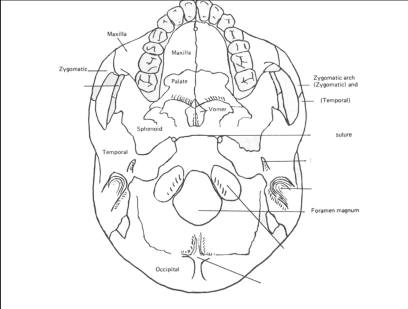
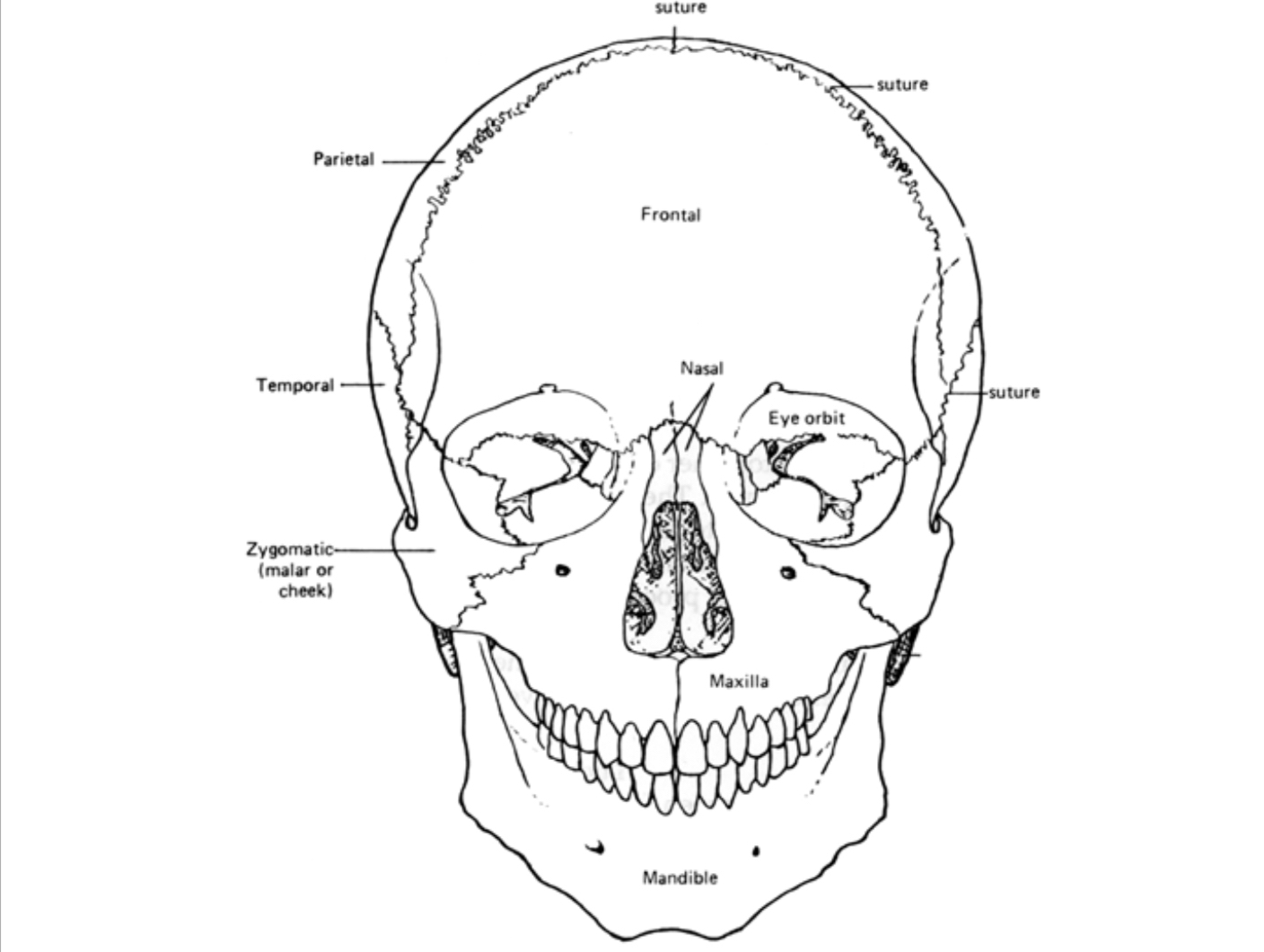
The skull is composed of a number of distinct bones; note major cranial bones and their articulations in your lab
Sutures: lines where cranial bones meet; major sutures include Coronal, Lambdoidal, Squamosal, and Sagittal
Sutures knit together (close) as maturity increases; degree of closure can indicate age (loose sutures in infants allow birth canal passage)
Mandible: lower jaw; formed by left and right dentary bones fused at the midline in humans (in many mammals they do not fuse, forming a flexible joint)
Teeth: incisors, canines, premolars, and molars visible in both maxilla (upper jaw) and mandible
Dental pattern shorthand (dental formula) describes tooth types per quadrant
Example: human dental pattern in one quadrant is
This means: 2 incisors, 1 canine, 2 premolars, 3 molars in each quadrant
Total tooth count = 32
Palate, incisors, canine, maxilla, zygomatic, etc. are illustrated in figures; major bones and sutures shown in Figures 2–5 in the source
Hyoid bone: a small U-shaped bone in the throat that floats (no articulation with other bones); origin for tongue and floor-of-mouth muscles; can move side to side when palpated in the throat
Postcranial Elements
Postcranial skeleton = axial skeleton + appendicular skeleton
Axial skeleton includes vertebral column + ribs + sternum; articulates with ribs via costal cartilage
Appendicular skeleton includes pectoral girdle, upper limbs, pelvic girdle, lower limbs
Axial Skeleton - Vertebral Column, Ribs and Sternum
Vertebral column features are shown in Figures 6–10; identify distinctive features of each vertebra
Cervical vertebrae (7): unique for having transverse foramina (foramen transversarium)
Atlas (C1) and Axis (C2) are the first two cervical vertebrae; Dens is a key feature on Axis
Thoracic vertebrae (12): have rib articulation facets (costal facets) for articulation with ribs
Lumbar vertebrae (5): distinctive S-curve; large size; short, squared spinous processes; lack of foramina or rib facets
Sacrum: typically 5 fused sacral vertebrae; articulates with the pelvis
Coccyx: 4–5 very small coccygeal vertebrae; represents the end of the backbone in humans; in other primates these elements would be larger and tail (caudal) vertebrae
Anterior view vs dorsal view show key features: vertebral foramen, spinous process, transverse process, rib articulation facets
Ribs
Humans have 12 ribs on each side
Ribs meet thoracic vertebrae dorsally and costal cartilage ventrally
Cartilaginous connections provide flexibility for breathing
Ribs 1–10 join the sternum via costal cartilage; Ribs 11–12 are floating ribs (do not join sternum)
First rib is relatively small and flat (superior–inferior dimension small)
Ribs 2–10 are flat front-to-back; superior surface rounded; inferior surface sharp
Sternum
Flat bone known as the breastbone
Three segments: manubrium (superior), body (middle), xiphoid process (inferior)
In adults these segments are usually fused, though sometimes unfused
Pectoral Girdle and Upper Limb
Scapula (shoulder blade): triangular, located dorsally on the thorax
Muscles envelop the blade except for the spine running along the dorsal aspect
Spine ends at the acromion process
Coracoid process projects anteriorly; glenoid fossa is the lateral surface that articulates with the humerus
Humeral head fits into the glenoid fossa to form the shoulder joint
Range of motion: humeral head rotates in the glenoid; full arm elevation is limited by shoulder joint; to raise arm above head, scapula rotates—example: touch top of head with right arm involves maximal humeral rotation plus counterclockwise scapular rotation to position the glenoid fossa upward
Clavicle (collarbone): S-shaped, lies between the scapula and sternum
Very strong ligaments hold it in place; rarely dislocates
More likely to fracture under extreme stress than to dislocate
Functions as a lateral brace for the shoulder
Upper limb bones (overview): humerus, radius, ulna, carpals, metacarpals, and phalanges
Humerus
Largest bone in the arm
Proximal head articulates with the glenoid fossa; distal end articulates with radius and ulna (forms part of the elbow and shoulder joints collectively with the scapula)
Deltoid tuberosity on the lateral midshaft where the deltoid muscle inserts
Distal end features: medial epicondyle, trochlea (articulates with ulna), capitulum (articulates with head of radius), and olecranon fossa (posterior; accommodates the olecranon of the ulna when the arm is extended)
The ulnar nerve runs medially near the medial epicondyle (funny bone sensation when struck)
Radius
Lateral bone of the forearm (thumb side) in anatomical position; the palm faces forward
Proximally articulates with the capitulum of the humerus and the radial notch of the ulna
Distally articulates with the carpal bones and the head of the ulna
Pronation: turning the hand over; radius crosses over the ulna so the dorsal surface of the hand faces forward
Supination: returning the palm to anatomical position; radius and ulna uncross
Ulna
Medial bone of the forearm; has a hook-like proximal end called the olecranon process (articulates with the humerus’ trochlea)
Lateral with the radial head via the radial notch
Distal end has the ulnar head; articulates with the radius at the ulnar notch
Function: flexion and extension of the forearm
Distal end features include a prominent styloid process
Carpals and the Hand
Carpals: eight wrist bones; each with a unique shape and name (not required to learn all names in this lab)
Metacarpals: five long bones in the palm; numbered 1–5 from the thumb (pollex to pinky)
Phalanges: finger bones
Pollex (thumb) has two phalanges (proximal and distal)
Digits 2–5 each have three phalanges (proximal, middle, distal)
Pelvis
Pelvic girdle consists of left and right innominate bones (coxal bones) that articulate with the sacrum
Each innominate appears as a single bone in the adult but is formed from three bones in juveniles: ilium (top blade), pubis (anterior), ischium (posterior-inferior)
In adults, these three bones fuse into a single innominate bone on each side
The pelvis is critical for anthropological work: key for age and sex estimation and for identifying locomotion (bipedalism) patterns in humans
Figure reference: pelvis with innominate, pubis, etc.
Lower Limb
Femur (thighbone): largest bone in the body
Femoral head articulates with the acetabulum (hip socket) forming a ball-and-socket joint; highly stable but relatively less flexible than the shoulder
Posterior shaft commonly shows a raised line called the linea aspera, origin site for hamstring muscles
Distal femur articulates with the tibia via the condyles; distal anterior surface articulates with the patella via the patellofemoral joint
Quadriceps muscle group forms the anterior thigh; tendon crosses the knee and inserts on the tibia; the patella enhances the leverage of the quadriceps
Patella
Kneecap, embedded within the quadriceps tendon; increases pull efficiency of the quadriceps across the knee
Tibia (shinbone)
Large, medial bone of the lower leg; articulates with the femur at the tibial plateau
Tibial tuberosity: insertion site for the quadriceps tendon; located below the plateau
Medial malleolus: prominent projection on the medial distal tibia; forms part of the ankle joint
Anterior crest: a ridge along the anterior surface of the shaft
Distal articulation with the fibula and with the tarsal bones (
ankle region)
Fibula
Slender bone on the lateral side of the lower leg
Articulates with the tibia proximally and with the tarsals distally
Lateral malleolus: distal end projection (outer ankle bump); not weight-bearing but stabilizes the ankle
Tarsals and Foot
Tarsals: seven ankle bones, including the calcaneus (heel bone)
Metatarsals: five long bones in the foot; numbered 1–5 from the big toe
Phalanges: toe bones; like the hand, the big toe (hallux) has two phalanges; digits 2–5 have three phalanges
Figure reference: Left foot diagram illustrating phalanges, metatarsals, and tarsals
Summary of Key Concepts and Implications
The skeleton is organized into cranial and postcranial components, with the skull housing the brain and sense organs, and the postcranial skeleton supporting movement and protection
Sutures in the skull serve both a functional and developmental role: flexibility for birth and age estimation through suture closure
The dental formula provides a compact summary of dentition and total tooth count; humans typically have 32 teeth with a per-quadrant pattern of
The vertebral column shows regional specialization: cervical with transverse foramina (specialization for vertebral arteries in the neck), thoracic with rib facets, lumbar with large bodies and short processes; sacrum and coccyx anchor the spine to the pelvis and tail region, respectively
The rib cage articulates with the spine via costal facets and with the sternum via costal cartilage, providing protection for thoracic organs and contributing to respiration dynamics
The pelvis is central for age and sex estimation and is essential to understanding habitual locomotion in humans
The shoulder complex demonstrates integrated mobility: the glenoid joint with the humerus is stabilized by the scapula’s rotation and the clavicle’s anchoring role
Pronation and supination describe forearm rotation driven by radius and ulna arrangement; the radius crossing over the ulna enables pronation, and uncrossing enables supination
The musculoskeletal system shows consistent relationships between bone morphology and function (e.g., deltoid tuberosity as a site of shoulder muscle attachment; linea aspera as a hamstring origin)
Practical lab insights include palpation landmarks (medial epicondyle, olecranon fossa, medial malleolus, etc.) and movements that reveal joint mechanics
The dental and skeletal references connected to real-world applications: anatomy of movement, age/sex estimation, and anthropological interpretation of locomotion and evolution
Directional Terms
Superior (cranial) – top
Inferior (caudal) – bottom
Anterior (ventral) – front
Posterior (dorsal) – back
Medial – toward midline
Lateral – away from midline
Proximal – nearest to torso
Distal – furthest from the torso
Bone responds to stress, muscle action, disease, trauma, and nutrition. Look for articulation facets (smooth surfaces) and raised muscle/ligament attachments (size reflects muscle size).
Terminology for surface features:
Line: small, slightly raised mark
Ridge: more protruding mark
Crest: prominent, protruding attachment area
Foramina (singular: foramen): holes in the bone
Skeleton division: cranial vs postcranial material
Cranial Components
Skull = cranium (face + brain case/calvarium) + mandible.
Cranial bones meet at sutures (Coronal, Lambdoidal, Squamosal, Sagittal), which close with age.
Mandible: lower jaw, fused dentary bones in humans.
Teeth: incisors, canines, premolars, molars in maxilla (upper jaw) and mandible.
Dental pattern shorthand: human formula is per quadrant (2 incisors, 1 canine, 2 premolars, 3 molars), totaling 32 teeth.
Hyoid bone: U-shaped, floats in throat, origin for tongue muscles.
Postcranial Elements
Postcranial skeleton = axial skeleton (vertebral column + ribs + sternum) + appendicular skeleton (pectoral/pelvic girdles + limbs).
Axial Skeleton - Vertebral Column, Ribs and Sternum
Vertebral column:
Cervical (7): unique with transverse foramina. Atlas (C1) and Axis (C2, with Dens).
Thoracic (12): have rib articulation facets.
Lumbar (5): large, short/squared spinous processes, no foramina/rib facets.
Sacrum (5 fused): articulates with pelvis.
Coccyx (4–5 small): end of backbone.
Ribs:
12 pairs. Meet thoracic vertebrae (dorsally) and costal cartilage/sternum (ventrally).
Ribs 1–10 join sternum; Ribs 11–12 are floating.
Sternum (breastbone): manubrium, body, xiphoid process; usually fused in adults.
Pectoral Girdle and Upper Limb
Scapula (shoulder blade): triangular; spine ends at acromion; coracoid process projects anteriorly; glenoid fossa articulates with humerus.
Clavicle (collarbone): S-shaped; strong brace for shoulder, often fractures under stress.
Upper limb bones: humerus, radius, ulna, carpals, metacarpals, phalanges.
Humerus: largest arm bone; head articulates with glenoid fossa; distal end articulates with radius/ulna (elbow joint).
Radius: lateral forearm bone (thumb side); crosses over ulna during pronation, uncrosses during supination.
Ulna: medial forearm bone; olecranon process forms part of elbow; ulnar nerve near medial epicondyle ("funny bone").
Carpals: 8 wrist bones.
Metacarpals: 5 palm bones (1-5 from thumb).
Phalanges: finger bones; thumb (pollex) has 2, digits 2-5 have 3.
Pelvis
Pelvic girdle: left and right innominate bones, articulate with sacrum.
Each innominate (adult) is fused from ilium, pubis, ischium (juvenile).
Critical for age/sex estimation and locomotion (bipedalism) patterns.
Lower Limb
Femur (thighbone): largest bone; head articulates with acetabulum (hip socket, stable but less flexible than shoulder); linea aspera on posterior shaft; distal end articulates with tibia (condyles) and patella.
Patella (kneecap): embedded in quadriceps tendon, enhances leverage.
Tibia (shinbone): large, medial lower leg bone; articulates with femur (tibial plateau); quadriceps tendon inserts at tibial tuberosity; medial malleolus (inner ankle).
Fibula: slender, lateral lower leg bone; stabilizes ankle via lateral malleolus (outer ankle).
Tarsals: 7 ankle bones (e.g., calcaneus/heel bone).
Metatarsals: 5 foot bones (1-5 from big toe).
Phalanges: toe bones; big toe (hallux) has 2, digits 2-5 have 3.
Summary of Key Concepts and Implications
Skeleton organized into cranial and postcranial components.
Sutures aid birth and age estimation.
Dental formula (e.g., human ) summarizes dentition.
Vertebral column shows regional specialization (cervical foramina, thoracic facets, lumbar size).
Rib cage protects organs and aids respiration.
Pelvis is key for age/sex estimation and understanding locomotion.
Shoulder complex (scapula, clavicle, humerus) enables wide mobility.
Forearm pronation/supination involves radius crossing over ulna.
Bone morphology reflects function (e.g., deltoid tuberosity for muscle attachment).