2024-2025 DEVELOPMENT OF MANDIBLE
Definition and Structure
Mandible:
Lower jaw, the largest and strongest bone in the face.
Holds the lower teeth in place and is essential for mastication and speech.
Structure:
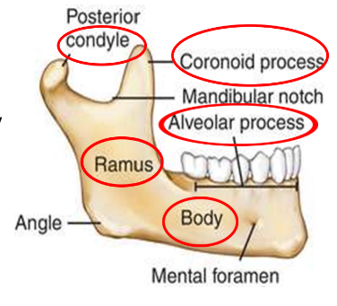
Body: Horizontal, curved like a horseshoe with two surfaces (external and internal) and two borders (alveolar and inferior).
Features include mental protuberance, mental foramen, and incisive fossa.
Rami: Vertical portions with lateral and medial surfaces.
Contains processes:
Condylar process (posterior): Forms the temporomandibular joint (TMJ).
Coronoid process (anterior): Attachment site for the temporalis muscle.
The mandibular notch separates these processes.
Development of the Mandible
Types of Ossification:
Intramembranous ossification: Direct bone formation in mesenchyme (most of the mandible).
The whole body of mandible except the anterior part – Ramus of mandible as far as mandibular foramen
Endochondral ossification: Uses cartilage as a precursor (e.g., symphysis and condylar process).
Anterior portion of the mandible (symphysis) – Part of ramus above the mandibular foramen – Coronoid process
Role of Meckel’s Cartilage (MC):
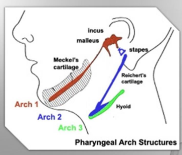
MC acts as a guide but does not contribute to the final mandible structure in humans.
Extends as a hyaline cartilage rod, with its proximal part connected to the ear region (otic capsule).
Lies medial to the developing mandible.
Formation Timeline:
6th Week: Mesenchymal condensation near the inferior alveolar nerve branches marks the initial development.
7th Week: Intramembranous ossification begins, forming the mandible around Meckel’s cartilage.
10th Week: Rapid ossification extends posteriorly, forming the ramus.
Secondary Growth Cartilages:
Appear between the 10th-14th weeks in utero and contribute to further growth:
Condylar cartilage (most significant).
Coronoid cartilage.
Symphyseal cartilage.
Mandibular Nerve and Ossification Center
Trigeminal Nerve:
Supplies sensory and motor functions to the mandible.
The mandibular branch divides near Meckel’s cartilage into:
Lingual nerve (medial).
Inferior alveolar nerve (IAN) (lateral), further dividing into mental and incisive branches.
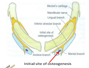
Ossification Center:
Located near the bifurcation of the IAN into mental and incisive branches.
Ossification spreads anteriorly, posteriorly, and upwards to form the body, ramus, and mandibular canal.
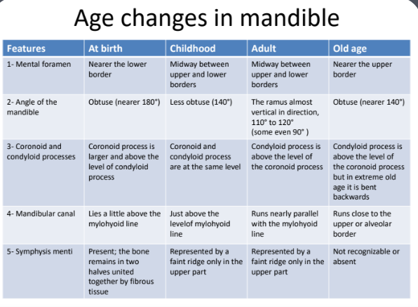
Age-Related Changes in the Mandible
At Birth:
Mandible is underdeveloped with large mandibular canal.
The angle is obtuse (~175°), and the coronoid process is more prominent than the condyle.
Childhood:
Mandibular halves fuse at the symphysis in the first year.
The angle reduces to ~140° by the fourth year as the teeth separate the jaws.
Adulthood:
The body of the mandible thickens and elongates.
The angle ranges between 110°-120°, nearly vertical.
Old Age:
Bone resorbs with tooth loss, and the alveolar process diminishes.
The angle increases to ~140° again, and the mandibular canal lies closer to the alveolar border.