proteins lecture 5
Another class of proteins that do not adhere to the paradigm is metamorphic proteins . These proteins appear to exist in an ensemble of structures of approximately equal energy that are in equilibrium. Small molecules or other proteins may bind to different members of the ensemble, resulting in various complexes, each having a different biochemical function.
the relasionship between protein structure and function
Tertiary Structure: The protein folds into a compact three-dimensional structure, with a heme group at its core. The heme group contains an iron atom that binds to oxygen.
Oxygen Binding: The primary function of myoglobin is to bind and store oxygen in muscle cells, providing a local oxygen reserve for periods of increased demand during muscle activity.
Heme Group: The heme group in myoglobin contains an iron ion in the ferrous (Fe^2+) state. This iron ion can reversibly bind to molecular oxygen (O2) forming an oxygenated myoglobin complex.
Structure-Function Relationship:
Heme Pocket: The specific structure of the heme pocket in myoglobin is crucial for its oxygen-binding function.
Iron Coordination: The iron ion in the heme group is coordinated by a histidine residue (proximal histidine) in the protein structure. This histidine helps stabilize the binding of oxygen to the iron.
Proximal and Distal Histidines: Myoglobin has a proximal histidine (coordinated to the iron) and a distal histidine that plays a role in regulating the binding and release of oxygen. The distal histidine helps prevent the oxidation of the iron.
Hydrophobic Core: The hydrophobic core of myoglobin stabilizes its structure, preventing unwanted interactions with water molecules and maintaining the integrity of the heme pocket.
Reversibility: The structure of myoglobin allows for the reversible binding and release of oxygen. The iron ion can bind to oxygen in environments with higher oxygen concentrations and release it in environments with lower concentrations.
Specificity: The specific amino acid sequence and three-dimensional structure of myoglobin determine its high specificity for oxygen binding and release.
Ligand Binding to Myoglobin:
Oxygen Binding: The binding of oxygen to myoglobin involves the reversible formation of a coordinated complex between the iron in the heme group and oxygen.
Dissociation: Under conditions of lower oxygen concentration, myoglobin releases oxygen due to changes in the protein conformation.
Porphyrin Ring + Fe²⁺ = Heme:
A porphyrin ring is a large, cyclic structure composed of four pyrrole subunits. It is a planar, conjugated system.
When a central iron ion (Fe²⁺) coordinates with the nitrogen atoms in the porphyrin ring, it forms a complex known as heme. This coordination involves coordination bonds between the iron and the nitrogen atoms of the porphyrin ring.
Heme + Protein = Myoglobin:
The heme group in myoglobin is embedded within the protein structure, and the iron ion in the heme is coordinated to specific amino acid residues, including a proximal histidine.
Porphyrin Ring+Fe2+→Heme→Protein→Myoglobin
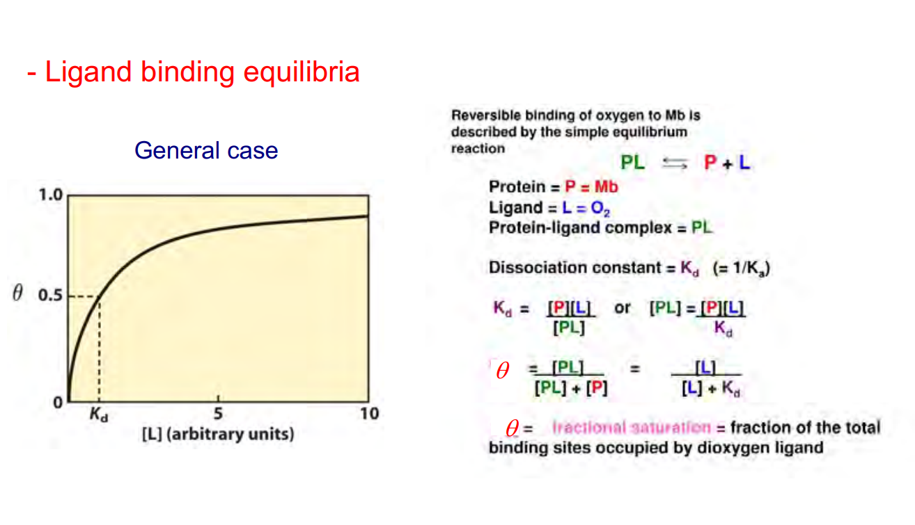 this is the gerenral case for ligand binding equilbria
for o2 and yoglobin there is a steeper curve that reaches saturation or near saturation more rapidly
hemoglobin and myoglobin are very similar in three D structure
hemoglobin consists of four Mb like subunits - 2 alpha chains and 2 beta chains
within the hemoglobin there are strong interactions between alpha one and beta one and alpha two and beta two
hemoglbon can exsist in two distinct states - the t state and the r state - both with different conformtations and different oxygen binding
they change from one state to another depending on the oxgyen concentration
T State (Tense State or Deoxyhemoglobin):
Low Oxygen Affinity: In the absence of oxygen binding, hemoglobin adopts the T state, also known as the tense state or deoxyhemoglobin state.
Quaternary Structure: Hemoglobin in the T state has a more compact quaternary structure, with subunits held in a relatively stabilized, tense conformation.
Oxygen Release: In tissues with lower oxygen concentration (e.g., active muscles), hemoglobin is more likely to release its bound oxygen due to the preference for the T state under these conditions.
R State (Relaxed State or Oxyhemoglobin):
High Oxygen Affinity: When oxygen binds to one of the four heme groups in hemoglobin, it induces a conformational change, shifting the protein to the R state, also known as the relaxed state or oxyhemoglobin state.
Quaternary Structure: Hemoglobin in the R state has a more relaxed quaternary structure, where subunits are in a more favorable, open conformation.
Cooperative Binding: The binding of one oxygen molecule increases the affinity of the remaining subunits for oxygen. This phenomenon is known as cooperative binding, enhancing the overall oxygen-binding capacity of hemoglobin.
Oxygen Transport: In the lungs, where oxygen concentration is high, hemoglobin primarily exists in the R state, facilitating the uptake of oxygen for transport through the bloodstream.
2,3 BPG stabelises the t state
h+ and co2 also promote the release of oxygen
a molecular disease of Hb is sickle cell anemia where the hemoglobin cannot bind to oxygen correctly - it is more prodominant in black men and women due to the evolution of the disease in countries where malaria was present
there are sticky patches on the surface of the molecule due to an alpha substrate
add these in
mechanism of action for tripsin
1. Binding and Activation:
Trypsin is initially produced in an inactive form called trypsinogen, primarily in the pancreas. Activation occurs through the cleavage of a peptide bond by another protease, typically enteropeptidase or trypsin itself in an autocatalytic process.
2. Catalytic Site:
Trypsin has a catalytic triad composed of three amino acids: Histidine (His), Aspartate (Asp), and Serine (Ser). These residues play a crucial role in its enzymatic activity.
3. Substrate Binding:
Trypsin specifically cleaves peptide bonds adjacent to positively charged amino acids like lysine or arginine in a polypeptide chain.
4. Mechanism:
Initiation: A substrate protein binds to the active site of trypsin.
Activation: The Ser residue in the catalytic triad undergoes nucleophilic attack on the peptide bond carbonyl carbon of the substrate.
Transition State Formation: This creates a tetrahedral intermediate.
Cleavage: The tetrahedral intermediate collapses, leading to the breaking of the peptide bond.
Release: The products of the cleavage are released from the active site, and trypsin remains available to cleave other peptide bonds.
5. Specificity:
Trypsin exhibits strict specificity, cleaving peptide bonds specifically on the carboxyl side of lysine or arginine residues unless they are followed by proline.
6. Regulation:
Its activity is regulated by endogenous inhibitors like serpins (serine protease inhibitors) and by the tight control of trypsinogen activation to prevent premature activation and damage to the pancreas.
7. Biological Significance:
Trypsin plays a crucial role in the digestion of dietary proteins, breaking them down into smaller peptides and amino acids that can be absorbed by the intestine for nutrition.
It is also utilized in laboratory settings for protein digestion, peptide mapping, and protein sequencing due to its specificity and efficiency in cleaving peptide bonds.
Trypsin's mechanism of action involves a precise molecular process governed by its active site residues, allowing it to cleave specific peptide bonds in proteins, facilitating digestion and other biochemical processes.
the catalystic tried - talk about its crutial
1.What is the initial inactive form of trypsin produced in the pancreas?
A. Trypsinase
B. Trypsinogen
C. Protease-I
D. Enteropeptidase
E. Serpin
2.What are the three crucial amino acids forming the catalytic triad in trypsin?
A. Histidine, Aspartate, Serine
B. Lysine, Arginine, Serine
C. Glycine, Cysteine, Histidine
D. Asparagine, Glutamine, Threonine
E. Valine, Leucine, Isoleucine
3.Trypsin specifically cleaves peptide bonds adjacent to which types of amino acids?
A. Negatively charged amino acids
B. Hydrophobic amino acids
C. Positively charged amino acids
D. Aromatic amino acids
E. Sulfur-containing amino acids
4.What is the role of the Ser residue in the catalytic triad during trypsin's mechanism of action?
A. Formation of a tetrahedral intermediate
B. Binding to the active site
C. Nucleophilic attack on the substrate
D. Catalyzing peptide bond formation
E. Initiating the transition state
5.How does trypsin exhibit specificity in cleaving peptide bonds?
A. Specifically cleaving on the amino side of lysine or arginine residues
B. Specifically cleaving on the carboxyl side of lysine or arginine residues
C. Cleaving randomly along the peptide chain
D. Avoiding cleavage near proline residues
E. Cleaving equally on both sides of lysine or arginine residues
6.What primarily regulates trypsin's activity?
A. Serpins
B. Enteropeptidase
C. Activation of trypsinogen
D. Tight control of enzymatic inhibitors
E. Formation of tetrahedral intermediates
7.What is the biological significance of trypsin in the body?
A. Maintenance of pancreatic health
B. Digestion of carbohydrates
C. Breakdown of nucleic acids
D. Digestion of dietary proteins
E. Facilitation of lipid absorption
8.Which molecular process governs trypsin's cleavage of specific peptide bonds in proteins?
A. Formation of tetrahedral intermediates
B. Activation of trypsinogen
C. Interaction with serpins
D. Specificity of active site residues
E. Nucleophilic attack on substrate carbonyl carbon
9.Trypsin plays a significant role in laboratory settings for:
A. Nucleic acid sequencing
B. Protein digestion
C. Lipid synthesis
D. Carbohydrate metabolism
E. Hormone production
10.How does trypsin's enzymatic activity benefit intestinal absorption?
A. Breaks down nucleic acids into smaller units
B. Facilitates absorption of lipids
C. Converts carbohydrates into simpler sugars
D. Breaks down dietary proteins into smaller peptides and amino acids
E. Supports the digestion of fiber for nutrient absorption
11.What primarily characterizes metamorphic proteins?
A. They possess a singular stable structure
B. They exist in an equilibrium of structures of similar energy
C. They have a fixed biochemical function
D. They lack the ability to bind with small molecules
E. They lack any tertiary structure
12.Which element within the heme group of myoglobin participates in reversible binding with oxygen?
A. Nitrogen
B. Iron
C. Carbon
D. Hydrogen
E. Sulfur
13.What is the primary function of myoglobin within muscle cells?
A. ATP synthesis
B. Storage of glucose
C. Oxygen binding and storage
D. Regulation of calcium levels
E. Facilitation of cell division
14.What is the specific role of the distal histidine in myoglobin?
A. Stabilizing the heme pocket
B. Coordinating the iron ion
C. Preventing oxidation of iron
D. Facilitating the binding of oxygen
E. Structurally stabilizing the hydrophobic core
15.What stabilizes the structure of myoglobin, preventing unwanted interactions with water molecules?
A. Heme group
B. Hydrogen bonds
C. Hydrophobic core
D. Oxygen molecules
E. Distal histidine
16.What feature of myoglobin enables the reversible binding and release of oxygen?
A. Hydrophobic core
B. Proximal histidine
C. Specific amino acid sequence
D. Tertiary structure
E. Distal histidine
17.What specifically determines the high specificity of myoglobin for oxygen binding and release?
A. Proximal histidine coordination
B. Hydrophobic core stability
C. Distal histidine positioning
D. Tertiary structure flexibility
E. Amino acid sequence and three-dimensional structure
18.What occurs when myoglobin binds with oxygen?
A. A complex involving iron and nitrogen forms
B. The iron ion loses coordination with the porphyrin ring
C. Protein conformation remains unchanged
D. The iron ion becomes oxidized
E. Distal histidine dissociates from the iron ion
19.What complex is formed when a central iron ion coordinates with nitrogen atoms in a porphyrin ring?
A. Oxygenated myoglobin
B. Hydrophobic core
C. Distal histidine
D. Heme
E. Reversible protein structure
20.What components form myoglobin in association with the heme group?
A. Porphyrin ring and protein
B. Hydrophobic core and distal histidine
C. Oxygen and iron
D. Nitrogen and hydrogen
E. Carbon and sulfur
21.Hemoglobin and myoglobin share a similarity in their:
A. Quaternary structure
B. Oxygen-binding capacity
C. Alpha and beta chains
D. Tense state conformation
E. Cooperative binding properties
22.In the absence of oxygen binding, hemoglobin adopts the T state, characterized by:
A. Relaxed quaternary structure
B. Strong interactions between alpha and beta chains
C. High oxygen affinity
D. A compact and tense conformation
E. Promotion of oxygen release in active muscles
23.What induces the transition of hemoglobin to the R state?
A. Low oxygen concentration
B. Cooperative binding of carbon dioxide
C. 2,3-BPG stabilization
D. Binding of oxygen to a heme group
E. Formation of sticky patches on the surface
24.The R state of hemoglobin is associated with:
A. A more stabilized, tense conformation
B. A decrease in oxygen affinity
C. A compact quaternary structure
D. Enhanced oxygen release in active muscles
E. An open and relaxed quaternary structure
25.Which molecule primarily promotes the release of oxygen from hemoglobin?
A. Oxygen
B. 2,3-BPG
C. Carbon monoxide
D. Hydrogen peroxide
E. Nitric oxide
26.Sickle cell anemia results in an inability of hemoglobin to:
A. Bind to oxygen correctly
B. Form the R state conformation
C. Release carbon dioxide
D. Maintain a compact quaternary structure
E. Exhibit cooperative binding with oxygen
27.What is the significance of 2,3-BPG in hemoglobin function?
A. It enhances oxygen binding in the R state
B. It stabilizes the T state conformation
C. It increases affinity for carbon dioxide
D. It promotes cooperative binding of hydrogen ions
E. It mediates the transition between T and R states
28.The molecular disease sickle cell anemia is more predominant in populations where:
A. Malaria was absent
B. There's a higher concentration of oxygen
C. There's a low presence of carbon monoxide
D. There's a lack of sticky patches on hemoglobin
E. Malaria was prevalent and led to evolutionary adaptations
29.What promotes the transition of hemoglobin to the T state?
A. Oxygen binding to heme groups
B. Stabilization by carbon dioxide
C. The presence of 2,3-BPG
D. Formation of sticky patches
E. Low concentrations of hydrogen ions
30.The sticky patches present on the surface of hemoglobin are attributed to:
A. Beta chains
B. Carbon monoxide binding
C. Stabilization by 2,3-BPG
D. Evolutionary adaptations to oxygen
E. Alpha substrate interactions
answers - B. Trypsinogen
A. Histidine, Aspartate, Serine
C. Positively charged amino acids
C. Nucleophilic attack on the substrate
A. Specifically cleaving on the amino side of lysine or arginine residues
B. Enteropeptidase
D. Digestion of dietary proteins
D. Specificity of active site residues
B. Protein digestion
D. Breaks down dietary proteins into smaller peptides and amino acids
B. They exist in an equilibrium of structures of similar energy
B. Iron
C. Oxygen binding and storage
D. Facilitating the binding of oxygen
C. Hydrophobic core
B. Proximal histidine
E. Amino acid sequence and three-dimensional structure
A. A complex involving iron and nitrogen forms
D. Heme
A. Porphyrin ring and protein
A. Quaternary structure
D. A compact and tense conformation
D. Binding of oxygen to a heme group
E. An open and relaxed quaternary structure
B. 2,3-BPG
A. Bind to oxygen correctly
B. It stabilizes the T state conformation
E. Malaria was prevalent and led to evolutionary adaptations
C. The presence of 2,3-BPG
E. Alpha substrate interactions