Macromolecules 2
GENERAL INFORMATION
Functional Groups
Carbohydrates
Hydroxyl: OH, found in alcohol (Like glycerol)
Carboxyl: COOH, found in amino acids
Carbonyl: Found in Aldehydes and Ketones. (Ketone has double bonded O in the middle, Aldehydes has it towards the end)
Amino: NH2, Found in amino acids
Phosphate: Found in P04, found in phospholipids and ATP
Caron can make 4 types of covalent bonds →4 electrons on the outer shell
Carbon can make single, double, and triple bonds →More bonds = Stronger hold
Organic compounds have carbon →Organic compounds: Carbohydrates, fats and protein
Dehydration Synthesis: When a hydrogen molecule is taken away to get two or more molecules together (ex: formation of monosaccharides) SUBTRACT H2O
Hydrolysis: Opposite of above, a hydrogen molecule is added, breaking the molecules apart. ADD H2O
CARBOHYDRATES
Monosaccharide: 1 carbohydrate monomer. SIMPLE SUGAR
Disaccharide: 2 carbohydrate monomers. SIMPLE SUGAR
Polysaccharide: Several monomers put together. COMPLEX SUGAR
Isomer: Isomers have the same molecular formula, and the structural arrangement of atoms is different
ALPHA VS BETA GLUCOSE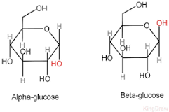
Alpha: OH is facing down C-1
Beta: OH is up on C-1
GALACTOSE VS GLUCOSE
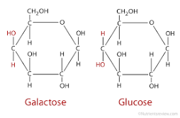 Galactose: OH is up on the C-4, Similar to BETA
Glucose: OH is down on the C-1, Similar to ALPHA
Starch: Energy storage for plants
Glycogen: Energy storage in animal muscle and liver
Cellulose: Plant cell wall
Chitin: Fungi cell wall
Peptidoglycan: Bacteria cell wall
Formulas for Sugars
Glucose + Glucose = Maltose Glucose pentagon (C6, H12, O6) - in blood
Glucose + Fucrose = Sucrose Fructose hexagon (C6, H12, O6) - in fruits
Glucose + Galactose = Lactose Galactose hexagon (C6, H12, O6) - in milk
Ribose (C5, H10, O5)
Deoxybose (C5, H10, O4)
LIPIDS
Purpose of Fats: Energy Storage, cushion organs and insulate body
Nonpolar: Electrons are shared equally
Overall NONPOLAR (because they have a greater number of carbon-hydrogen bonds than carbohydrates)
Triglycerides = Glycerol + 3 Fatty Acids (fatty acids are nonpolar)
Saturated VS Unsaturated:
Unsaturated are healthier because they have a bend in them, they DO have double bonds. Not solid because since they are curved, they are not packed as tight. Lower LDL, raise HDL
Saturated and unhealthier because they DO NOT have double bonds. Solid at room temperature. Raise LDL
Increase space between carbons, decrease density (less=healthier)
Cis and Fatty Acids
A Cis fat has hydrogen atoms located on the same side. Trans-fat acids, it has 2 hydrogen atoms on opposite sides. Bent shape
Trans fats are unhealthier, they raise bad cholesterol (LDL) which can cause heart attacks. In contrast, cis fats raise good cholesterol (HDL). Striaght shape
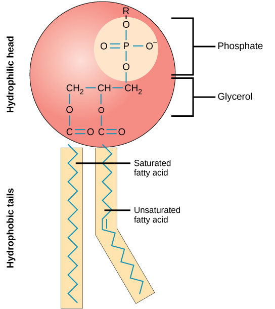
Phospholipids: 1 Glycerol + 2 Fatty Acids + Phosphate
Head →Phosphate, Glycerol, Carbonyl, Hydrophilic
Tails →Hydrophobic/Nonpolar, fatty acid chains. Hydrophobic because nonpolar, they balance out so they cant make bonds with water
Arranged in a bilayer in the cell membrane
 Stereoids
3 hexagons + 1 pentagon
Ex: Cholesterol: important in regulating cell membrane fluidity and is the basic steroid from which steroid hormones such as testosterone, estrogen, and progesterone are made
 Waxes
WaxesLong chain alcohol (an alcohol with many carbons) + a fatty acid
Coat the surface of leaves to prevent water loss
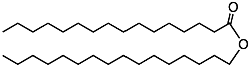 Trygleride Structure
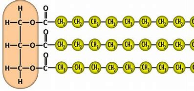 Ester Linkage: Covalent Bonds found in lipids (The double-bonded O and the C)
Glycerol: The orange part of the tryglericde, an alcohol
PROTEINS
Greatest variety in function → Depends on R-group
Function of proteins
Messenger Proteins
Enzyme Proteins
Structural Proteins
Defensive Proteins
Transportive Proteins
Structure of Amino Acid: Carboxyl, Amino, R group, central Carbon, and Hydrogen make up each amino acid
Amino acids are categorized as nonpolar, polar, acidic, or basic based on R groups
Levels of proteins
Peptide Bond (carboxyl of one amino acid attaches to the amino group of another amino acid)
Dipeptide (2 amino acids joined together by a peptide bond)
A polypeptide (linear arrangement of many amino acids)
Levels of organization
primary - Peptide bonds
secondary - Hydrogen bonds
tertiary (determines the overall shape and unique function of the final protein) - R group bonds
quaternary (determines the overall shape and unique function of the final protein for proteins
made up of more than one chain) - R group bonds
ENZYME’S
Catalyst: A substance that increases the rate of a chemical reaction without itself undergoing any permanent chemical change
Enzyme: Substrates are attracted to their active site and are broken down to speed up their activation rate
High Energy Intermediate: a short-lived, high-energy, highly reactive molecule
Induced Fit: The enzyme changes its shape to fit the substrate
Lock and key: Opposite of the above
enzymes usually end in a ase
Can be reused
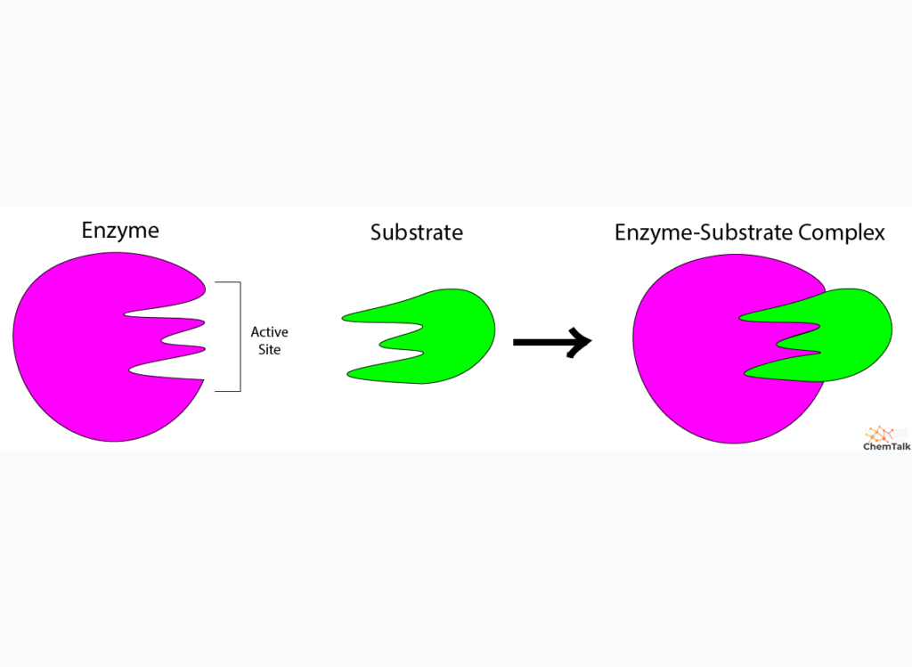
Special cases
Pepsin: More acidic, In stomach acids (1.5-2.5)
Trypsin: More basic. Small intestine, ph 7.5-8.5
Denaturation: When the conditions are not met and the 3rd and 4th level groups begin to unfold and might result in permanent damage
Factors that affect enymes
Temperature: Ideal temp 32 C. If too high or low denaturation can occur
Ph: Ideal Ph, neutral (7). If to high → denaturation. Special cases are pepsin and trypsin
Enzyme concentration: Too many substrates, not enough enzymes (Limiting Factor)
Substrate concentration: Too many enzymes, not enough substrates (Limiting Factor)
Competitive Inhibitor: When a molecule blocks the active site of an enzyme
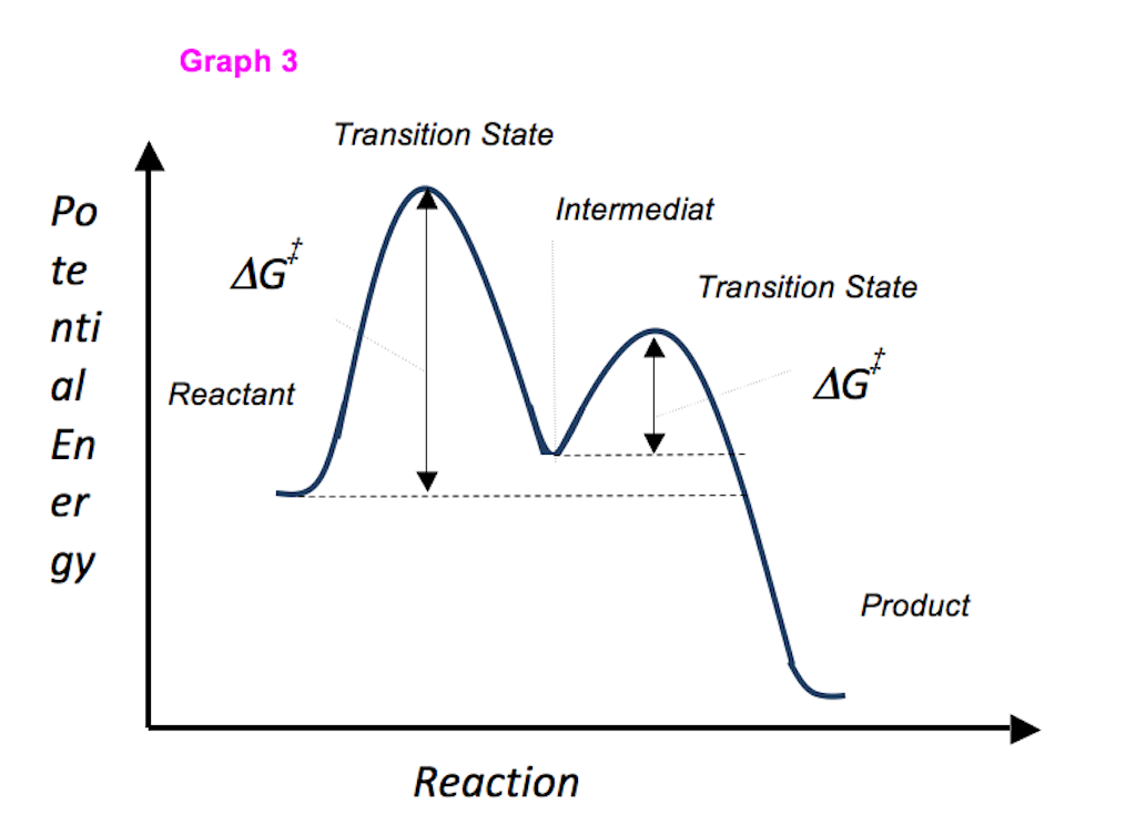 High energy intermediate/ transition state on a graph
MACROMOLECULE LABS
Iodine- tests for starch; negative: brown; positive: dark purple
Biuret- Tests for proteins, Negative: royal blue, Positive: violet/purple
Benedict: Needs to be heated. Tests for reducing sugars (all monosaccharides but only some disaccharides; sucrose is negative) Negative: aqua blue. Positive green, orange, or red, more red more sugar
Sudan IV- Tests for Fats. Negative- no red fat globules. Positive- red boundary around fat globules