The Six Short External Rotators of the Hip
The six short external (lateral) rotators of the hip, arranged from superior to inferior, include the following muscles:
Piriformis: This muscle originates from the sacrum and insert into the greater trochanter of the femur. It plays a crucial role in hip external rotation and abduction, especially when the hip is flexed.
Superior Gemellus: Located above the obturator internus, this muscle aids in the external rotation of the hip and stabilizes the hip joint during movement.
Obturator Internus: This muscle is unique as it crosses the greater sciatic notch. It assists in externally rotating the hip and also helps abduct the thigh when the hip is flexed.
Inferior Gemellus: Positioned below the obturator internus, it contributes to the external rotation of the hip, working alongside the superior gemellus and obturator internus for hip stabilization.
Obturator Externus: This muscle is situated on the outside of the pelvis and works to externally rotate the femur, especially during activities involving hip motion.
Quadratus Femoris: As the most inferior muscle of this group, it is essential for stabilizing the femoral head within the acetabulum and aids in external rotation.
These muscles are integral for hip stability and mobility, working collectively to control the movement of the femur. Proper function of the lateral rotators is crucial in activities such as walking, running, and other dynamic movements.
Understanding the order and anatomy of these muscles is vital, particularly for orthopedic surgeons during posterior hip surgeries. Their location is closely related to significant neurovascular structures including the sciatic nerve, which runs close to the piriformis muscle, the superior and inferior gluteal vessels, and the medial femoral circumflex artery. This artery is the principal blood supply to the femoral head, making it critical to avoid any damage to these structures during surgical procedures. An in-depth knowledge of these muscles enhances surgical precision and minimizes the risk of complications.
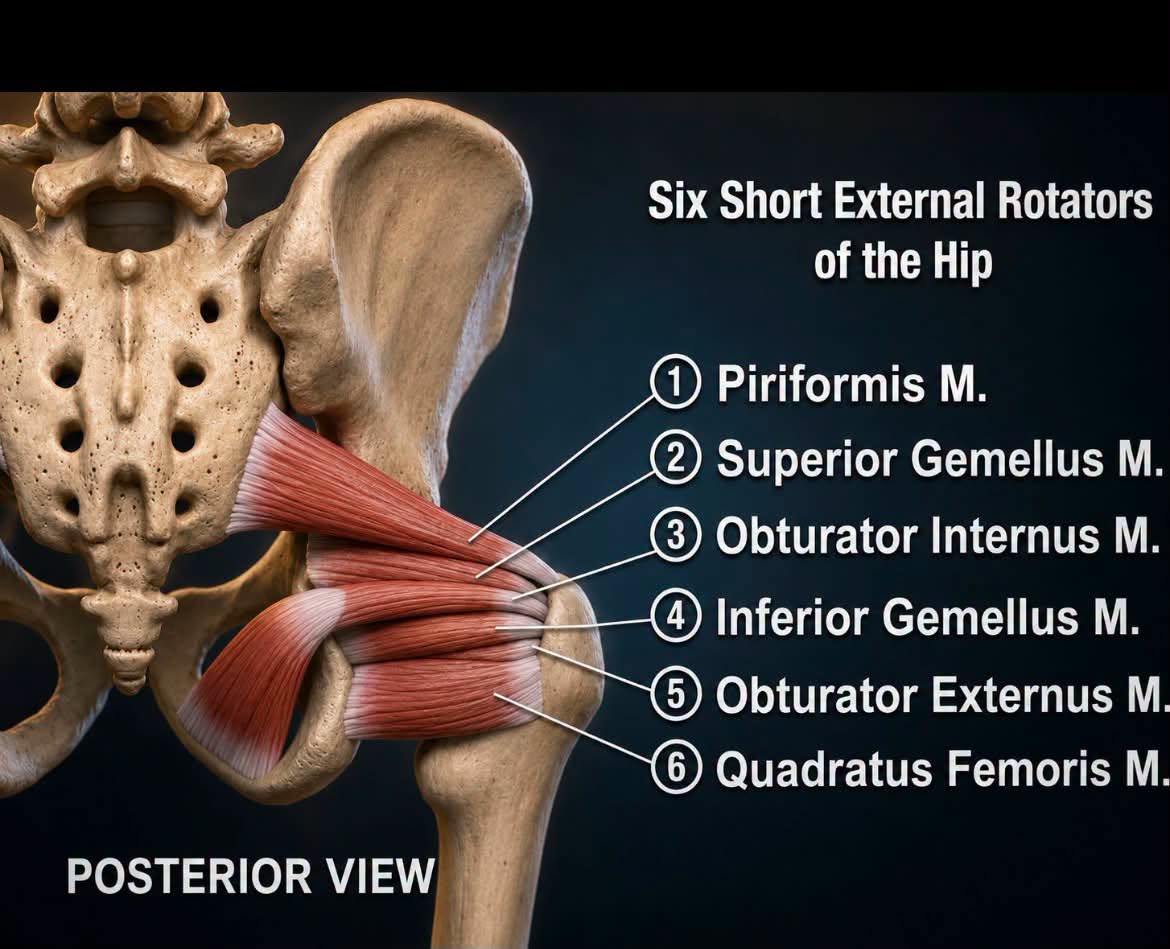
The six short external (lateral) rotators of the hip are arranged from superior to inferior as: piriformis, superior gemellus, obturator internus, inferior gemellus, obturator externus, and quadratus femoris. These muscles externally rotate the hip and help stabilize the femoral head within the acetabulum.
Knowing their order is essential during posterior hip surgery. They are important anatomical landmarks because of their close relationship to the sciatic nerve, the superior and inferior gluteal vessels, and the medial femoral circumflex artery, the principal blood supply to the femoral head. A thorough understanding of these muscles helps the surgeon perform a safe posterior approach to the hip and avoid injury to these critical structures
/