Gradients Explained
Gradients in MRI:
Main Magnetic Field (B₀):
MRI scanners have a strong main magnetic field (B₀) along the z-axis. This field aligns hydrogen protons in the body and determines their precessional frequency.
Purpose of Gradients:
Gradients are additional magnetic fields superimposed on the main field.
They vary in strength along specific directions (x, y, z axes) and are used for spatial encoding. This means they allow us to determine where in the body the MRI signal comes from.
How Gradients Work:
Important: protons precess frequency is determined by the strength of the B field. By applying gradients to the B field, protons at different points recess at different frequencies.
Gradient coils are electromagnets that create a magnetic field that either adds to or subtracts from the main magnetic field.
For example:
If a gradient increases the field on one side, protons in that area precess faster.
If the field decreases on the other side, protons there precess slower.
This creates a gradient in precessional frequencies along the axis where the gradient is applied.
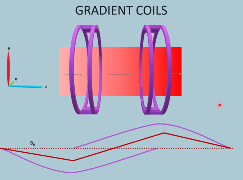
There are two different coils that apply their own fields and gradients. The first coil applies a field that oppose B_0 and the second coil applies a field that strengthens B_0. Both of these act in the z - axis. Protons will precess at different frequencies due to the changing strengths of the B field. Gradients can also be applied along perpendicular axis.
Why is this useful? When the RF pulse is applied to the protons at a specific frequency, only protons precessing at that frequency will be tilted, and consequently produce a signal. This means the signal only comes from a specific set or slice of protons that are experiencing a certain magnitude of B field, that causes it to precess at the same frequency of the pulse. These gradients can be shifted so each pulse would return a different slice.
Why Gradients Are Important:
Each gradient (x, y, or z) changes the magnetic field in a specific plane. This means the MRI system can isolate signals from:
A single slice of the body (z-gradient, for slice selection).
A specific point within the slice (x- and y-gradients, for localization within the slice). Basically, by applying multiple gradients in different axis, we can target a specific area with a specific frequency, experiencing a specific field strength.
Turning Gradients On and Off:
Gradients are rapidly switched on and off (e.g., in milliseconds) to manipulate the field in controlled ways.
This allows the scanner to encode spatial information into the MRI signal based on changes in precessional frequency and phase.
Superposition of Fields:
The gradient fields do not replace B₀ but are added to it. The direction of B₀ remains unchanged; only its strength varies depending on the gradient.
This process of applying gradients is essential for constructing detailed MRI images by determining where in the body the detected signal originates.