Test
1. Differentiate between each part of the nervous system:
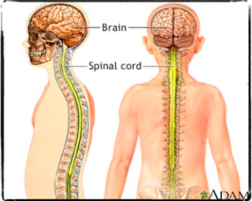
a) Central and Peripheral
Central | Peripheral |
|---|---|
|
|
b) Somatic and Autonomic
Somatic | Automatic |
|---|---|
(voluntary control)
| (involuntary control)
|
2. Describe the structure of a neuron. What is the resting membrane potential of a neuron?
Structure of Neuron
Dendrites
Highly branched projections of cytosol that carry signals towards nerve cell body
Cell Body (Soma)
Contains nucleus and is site of cell’s metabolic reactions and will process inputs from dendrites
Axon
Long, thin extension of cytosol that conducts signals away from nerve cell body - can be bundled together to form nerve fibers
Axon Terminal
Small swelling at end of axon - communicates with next neuron, muscle, or gland by releasing chemical signal into synapse
Myelin Sheath
Schwann cells form tightly wrapped insulated covering over axon of nerve cells - act as electrical insulators due to high lipid content
Node of Ranvier
Regularly occurring gap between sections of myelin sheath along axon - allows electrical impulse to travel faster along axon
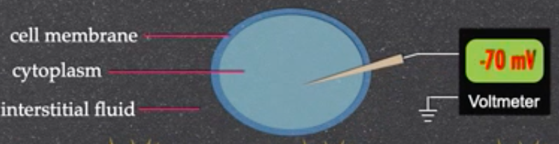
Resting Membrane Potential of Neuron
3. Describe how an action potential is generated. Be able to label a graph showing this process.
Description
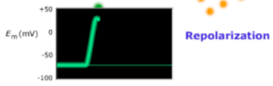
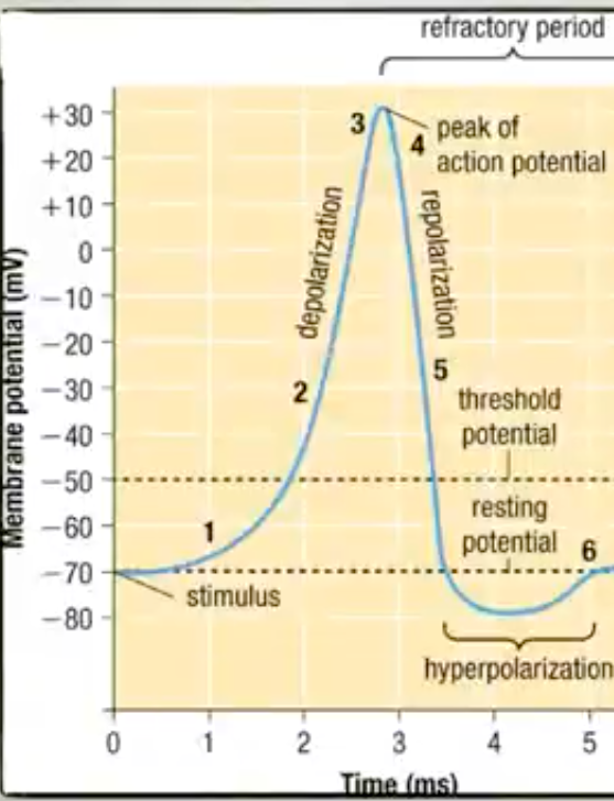
Resting State → Depolarization
Sodium channels open allowing Na+ ions to rush in
Causes depolarization (inside neuron positively charged)
Depolarization → Repolarization → Undershoot
Sodium channels close and potassium channels open
K+ ions rush out of neuron so inside of neuron becomes negative once again - repolarization
More negative than -70mV (undershoot - hyperpolarized)
Undershoot → Resting State
Ionic distribution is largely reversed via the sodium and potassium pumps so Na+/K+ pump restores resting membrane potential of neuron to -70mV
Graph
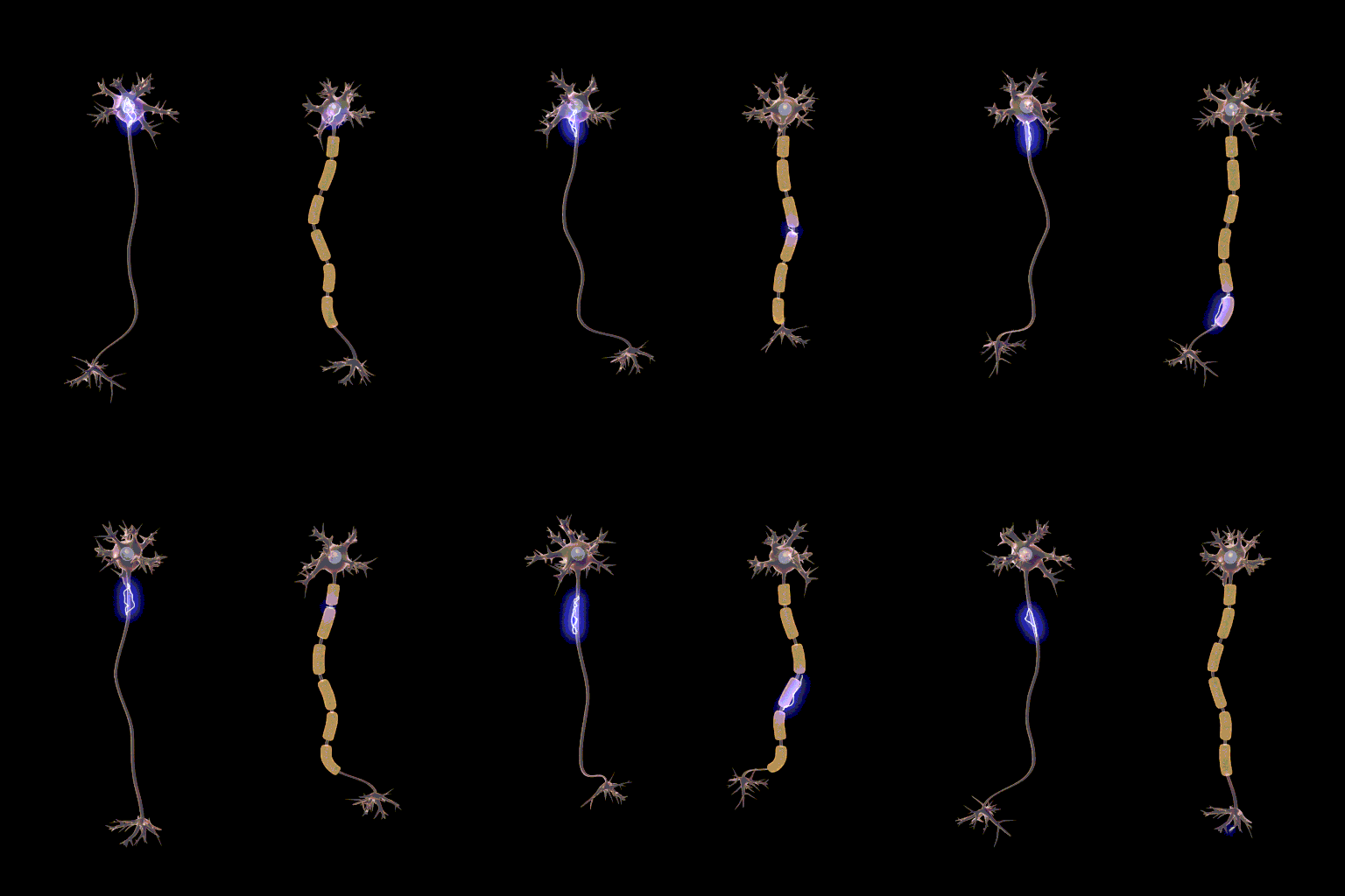
4. Describe how an action potential (nerve impulse) travels down a myelinated and unmyelinated neuron.
Action potential propagates (moves) down axon as wave of depolarization
Unmyelinated | Myelinated |
|---|---|
|
|
think of an exposed vs. covered wire, rubber around the metal controls the electrons on the inside allowing for smoother and quicker movement
5. Discuss the importance of the sodium-potassium pump to maintaining resting membrane potential.
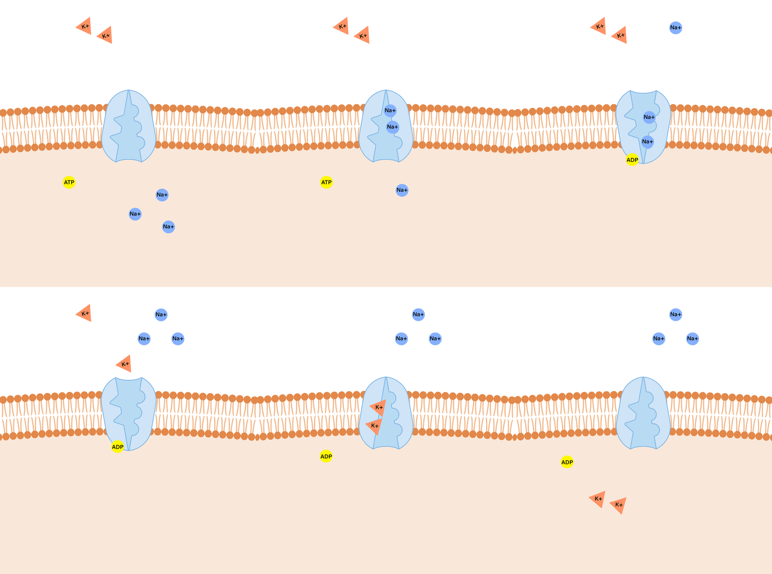 For every 3 Na+ ions pumped out, only 2 K+ ions are pumped in which leads to a net loss of positive charge from inside cell
makes inside of cell negative in relation to outside
Pump then maintains the resting membrane potential (RMP) at -70mV
6. Describe in detail, the mechanism by which a nerve impulse is sent between neurons.
7. Nerve impulses are all-or-none events, what does this mean? What does threshold potential mean?
This relates to Threshold Potential. Essentially, at -55 mV (Threshold), a neuron has enough energy to open voltage-gated ion channels or fire. It’s all or nothing as if not reached, neuron will not fire (no AP). Threshold reached when combined stimulus from nearby dendrites exceeds minimum level of depolarization
8. Describe in detail the way in which the endocrine system regulates blood sugar levels.
9. Describe the different types of Diabetes and treatment.
Type I | Type II | Gestational | |
|---|---|---|---|
Effects |
|
|
|
Aids |
|
|
|
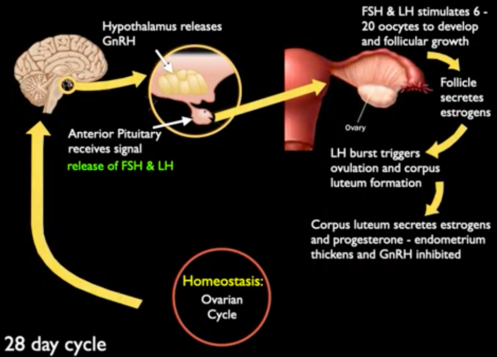
10. Describe the function of the male and female reproductive hormones, sex organs, process of gamete formation, as well as the ovarian cycle.
Female Reproductive Hormones
Hormone | Gland | Function |
|---|---|---|
Gonadotropin-releasing hormone (GnRH) | Hypothalamus | Controls release of FSH & LH by anterior pituitary - synthesis and release of sex hormones |
Follicle Stimulating Hormone (FSH) | Anterior Pituitary | Synthesis and secretion of progesterone, stimulates development of oocytes |
Luteinizing Hormone (LH) | Anterior Pituitary | Release of egg cell from ovaries |
Estrogen | Ovaries | Maturation of sex organs, sexual characteristics, and sex drive |
Progesterone | Ovaries | Prepares uterus for implantation of fertilized egg and growth and development of an embryo |
Male Reproductive Hormones
Hormone | Gland | Function |
|---|---|---|
Gonadotropin-releasing hormone (GnRH) | Hypothalamus | Controls release of FSH & LH by anterior pituitary - synthesis and release of sex hormones |
Follicle Stimulating Hormone (FSH) | Anterior Pituitary | Release of testosterone in testes |
Luteinizing Hormone (LH) | Anterior Pituitary | Secrete proteins and other molecules for spermatogenesis |
Testosterone | Testes | Maturation of sex organs, sexual characteristics, sex drive, and spermatogenesis |
Female Sex Organs
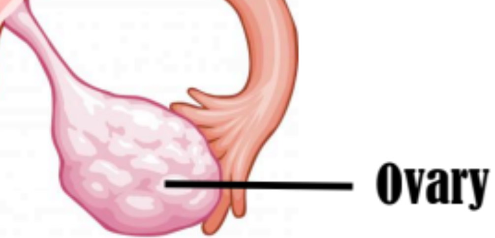
Ovary
Females have a pair of ovaries
Eggs (ova) produced and released here
Estrogens and progestins produced
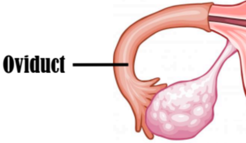
Oviduct (Fallopian Tube)
Hollow tube with muscular walls lined with cilia
propel egg
Connects ovary and uterus
fertilization occurs here
Egg travels through oviduct (fallopian tube) to get to uterus
Uterus
Hollow structure with walls made of smooth muscle
Fertilized egg (zygote) implants into lining (endometrium)
Cervix
Lower portion of uterus - cervix
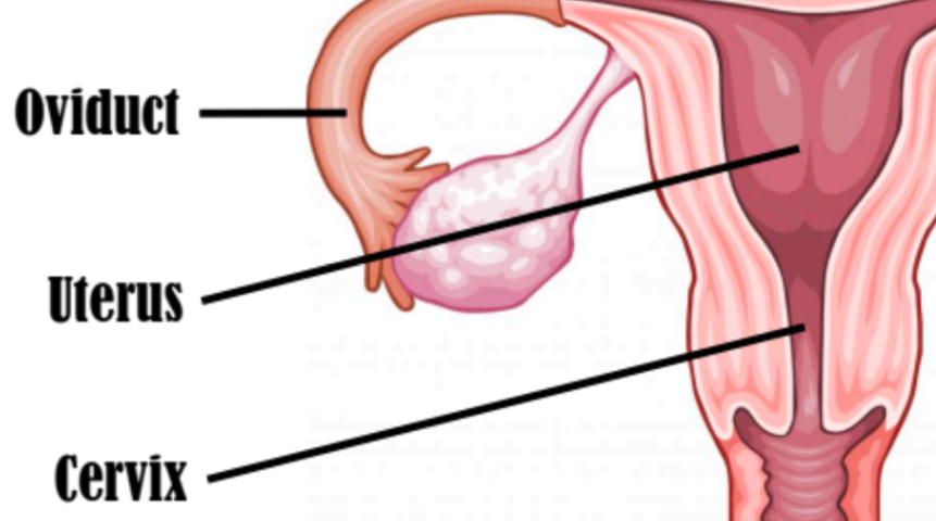
Vagina
Muscular canal connected to cervix
At birth, baby passes from the uterus through the vagina to the outside

Male Sex Organs
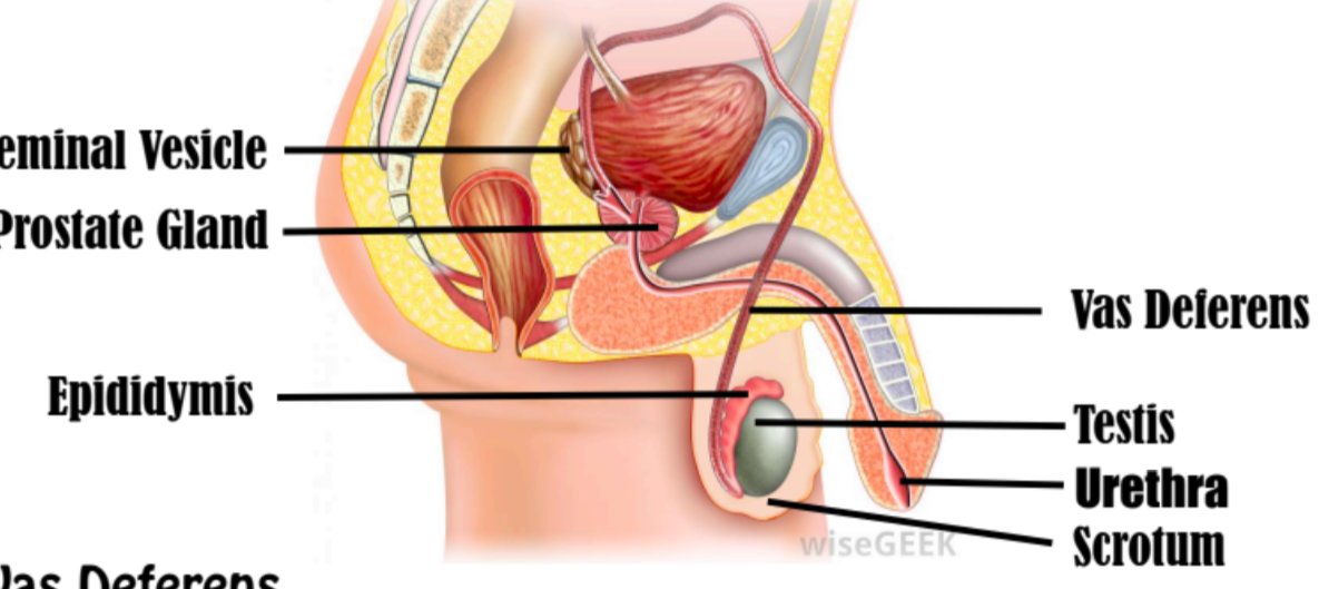
Testes
Males have two testes, suspended in a bag-like scrotum
125m of seminiferous tubules - spermatogenesis occurs
Scrotum helps regulate body temperature
Epididymis
Sperm flows from seminiferous tubules into epididymis (coiled storage tubule attached to surface of each testis)
Rhythmic muscular contractions of epididymis move sperm into vas deferens

Vas Deferens
Thick-walled muscular tube that extends through abdominal cavity - sperm travels through until passes out urethra
Fluid from seminal vesicle & prostate mix with sperm - semen
Female Gamete Formation
Oogenesis - process of producing and releasing eggs
Oocyte - immature egg that only undergone 1st meiotic division
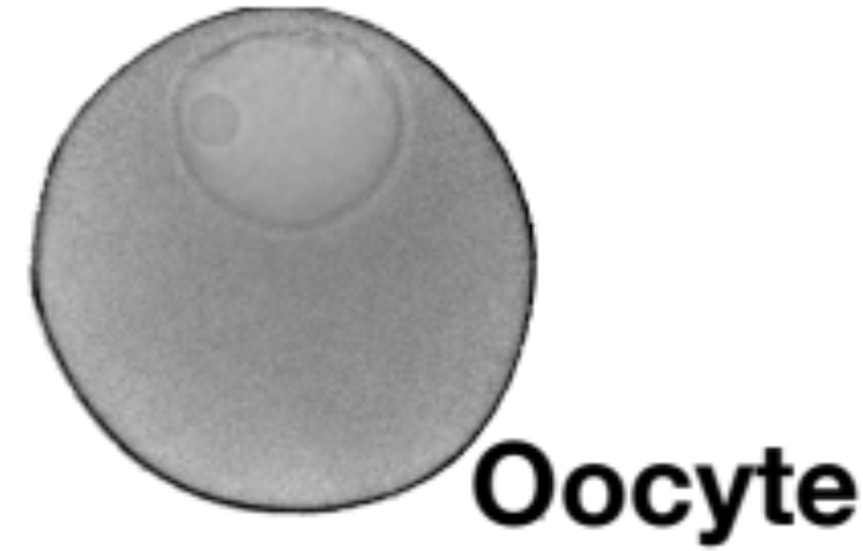
Born with ~1 million oocytes arrested at Prophase I
~380 ovulated before menopause
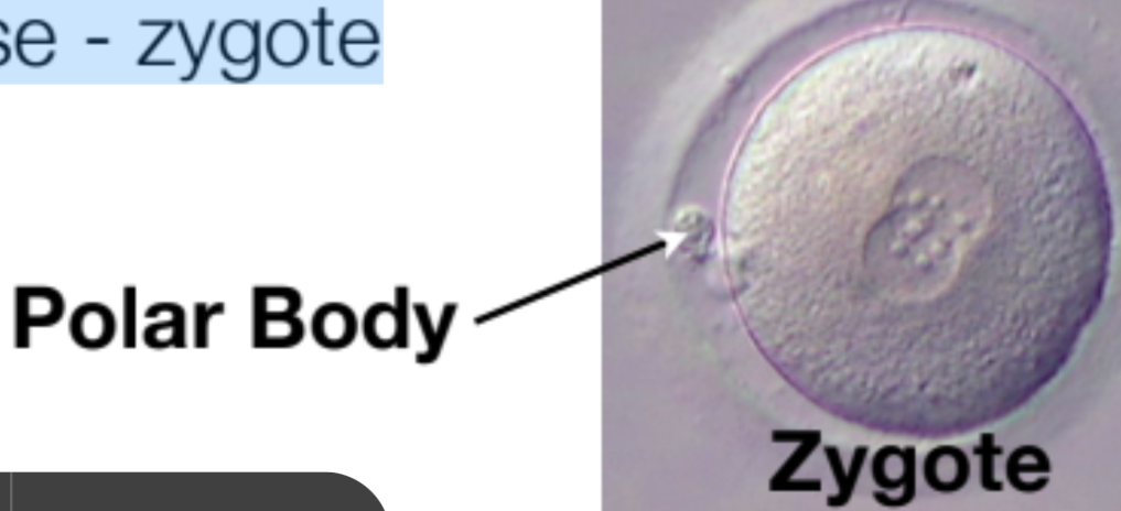
2nd meiotic division occurs only if oocyte is penetrated by sperm
mature ovum and polar body formed
Nuclei of ovum and sperm fuse - zygote
Male Gamete Formation
125m of seminiferous tubules in testes
Spermatogenesis occurs
Sperm flows from seminiferous tubules into epididymis
Rhythmic muscular contractions of epididymis move sperm into Vas Deferens
Sperm travels through until passes out urethra
Fluid from seminal vesicle & prostate mix with sperm - semen
Ovarian Cycle
11. Explain in detail how the immune system works to protect your body from illness and infection
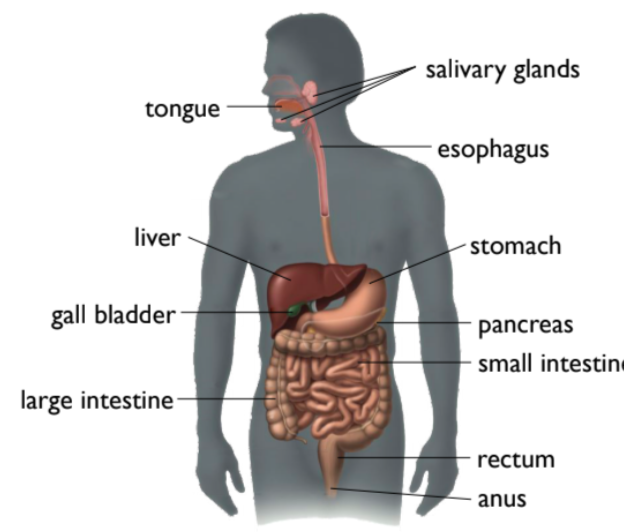
12. What is the function of the digestive system? What organs does it include? What is structure and function of each organ discussed in class? Label.
Function
specialized organs help to break down the food that we eat into nutrients that can then be delivered to the body’s cells
Organs Involved
teeth
small, hard bone structures
breaks down food for physical digestion
salivary glands
starts chemical breakdown
alimentary canal
pushes food towards stomach via peristalsis
epiglottis
stops food from entering lungs
stomach
organ that contracts to move food
uses muscles as valves
chemical breakdown occurs
liver
responsible for detoxification
metabolism of nutrients
production of bile for digestion
storage of glycogen
pancreas
releases various enzymes to further help breakdown food
gallbladder
small, pear-shaped organ located beneath liver
stores and concentrates bile produced by the liver
releasing it into the small intestine to aid in digestion of fats.
small intestine
is where bile is released into break down fats
most absorption of nutrients occurs here
has villi to increase surface area
capillaries to absorb water-based nutrients
lacteals to absorb fat-based nutrients
large intestine
absorption of water, vitamins, and minerals occurs here
has billions of E. coli to help with absorption
rectum
removes food waste
Diagram
13. Explain in detail physical digestion, chemical digestion, and absorption.
Physical Digestion
intended to break down large forms of macromolecules into smaller forms to increase surface area for chemical reactions
organs involved include: teeth, stomach, gallbladder, liver, small intestine
Chemical Digestion
intended to break down small macromolecules (carbs, fats, proteins) even further than physical digestion, into its monomers to allow for absorption in the body
enzymes help speed up the process of these chemical reactions
organs involved include: salivary glands, stomach, small intestine, liver, pancreas, bile
Absorption
how the body gets something useful out of food and transports it into the body
occurs in the small and large intestine
small intestine absorbs larger molecules like macromolecules via villi
villi are hair-like projections that increase surface area for absorption
it has capillaries for water-based nutrients and lacteals for fat-based ones
large intestine absorbs water, vitamins, and minerals via E. coli, the bacteria
14. Describe how food passes through this system.
Imagine an all-in-one food called Seangoop. When ingested:
It enters the mouth and begins to physically break down
Enzymes in the saliva begin breaking down the fats and carbs into smaller forms
It is a ball (bolus) and is swallowed
It goes down the esophagus by peristalsis
it enters (and it cut off) by the stomach by its valves
the acids and enzymes in the stomach begin to chemically breakdown the fats and proteins
when it enter the small intestine, the fats are liquified (called “chyme”) and bile is secreted by the duodenum (first section of small intestine)
the carbs break down from maltose to glucose, the proteins from short aa chains → amino acids, and fats → glycerol and fatty acid tails
the villi in your small intestine begin to absorb the glucose, glycerol+fatty acids, and amino acids
the remaining water, vitamins, and minerals of Seangoop are absorbed by the E. coli of the large intestine
the food enters your rectum and exits your body by the anus
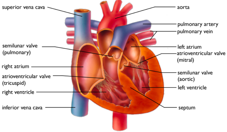
15. What is the function of the circulatory system? What organs/tissues does it include? What is the structure and function of each organ discussed? Label.
Function
transports nutrients
transports urea and other wastes
transports respiratory gases
transports hormones
transports special proteins to fight infection and clot wounds
Organs Involved+Function and Structure
Blood vessels
moves blood form and to heart, lungs and rest of body
Heart
made of cardiac muscle
branching and has ability to contract without being stimulated by a nerve (myogenic)
4 chamber muscle responsible for the pumping of blood throughout the body and the lungs
made of artery, veins, atriums and ventricles
its right side is smaller as it bring blood from the body to the lung to get oxygenated (the lung is close by
its left side is larger as it brings blood from the lungs to the body to deliver nutrients
Blood
carries compounds throughout the body needed for it to survive
Lungs
oxygenate deoxygenated blood
Diagram
16. What are components of our blood? What does each do?
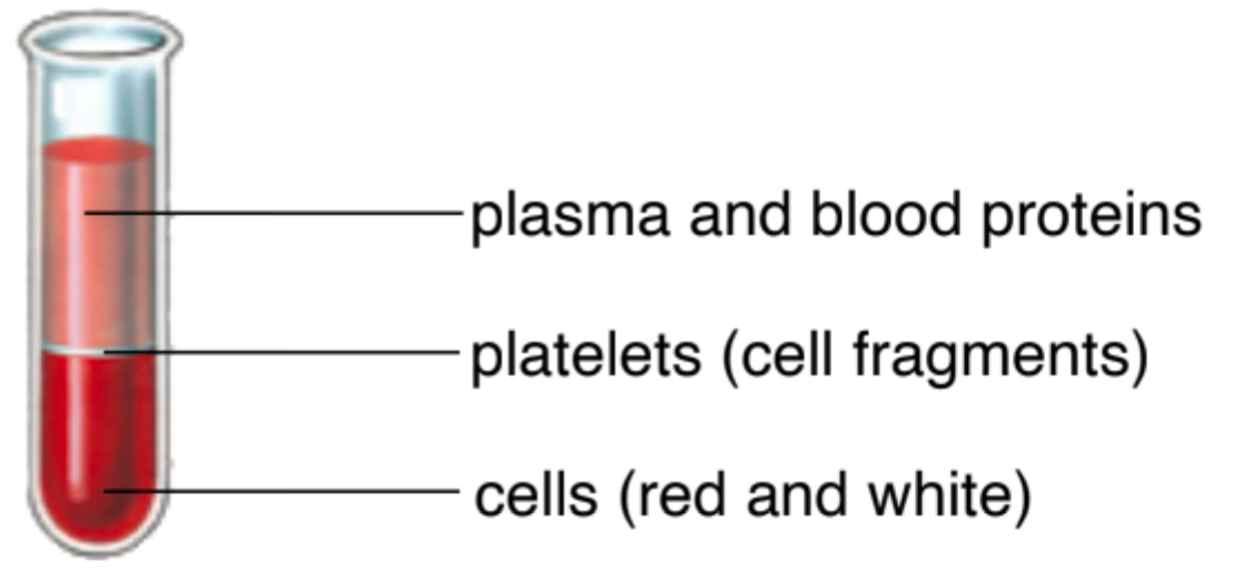
Plasma is about 90% water and 10% dissolved substances (nutrients, vitamins, minerals, antibodies, hormones, clotting proteins, CO2, urea)
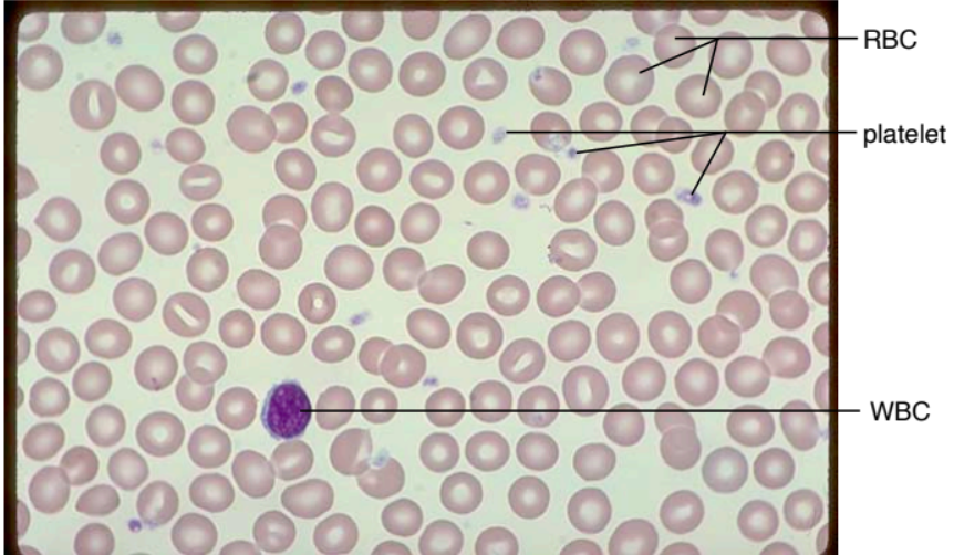
Red blood cells (RBC), also called erythrocytes, and are
enucleated and biconcave in shape
They are filled with hemoglobin, which transports oxygen
White blood cells (WBC), also called leucocytes and lymphocytes, are slightly larger than RBC and have a nucleus and fight infections
Platelets, also called thrombocytes, are cell fragments that are involved in forming blood clots
17. Compare the three types of blood vessels.
Artery | Capillary | Vein |
|---|---|---|
|
|
|
18. Explain how blood flows through the heart in detail.
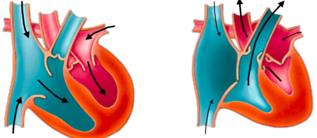
atria contract
blood flows from atria to ventricles
ventricles contract
blood flows from right ventricle to lungs blood flows from left ventricle to aorta (body)
19. Describe heart rate, heartbeat, and blood pressure in detail.
Heart Rate
Sinoatrial (SA) node is a neuromuscular mass in right atrium that sets heart’s tempo
Stimulation by sympathetic nerves increases heart rate, while parasympathetic stimulation slows heart rate
Secretion of adrenaline speeds up heart rate