Lecture 3 - Tissue supporting the nervous system
-note: VENTRICLES, CIRCLE OF WILLIS
🧠 DETAILED MULTI-PARAGRAPH SUMMARY
The brain is a highly sensitive organ that requires strict protection and regulation due to its complex functions and vulnerability to toxins and physical damage. This protection is provided by several systems, including the meninges, cerebrospinal fluid (CSF), and the blood–brain barrier. Together, these structures ensure mechanical protection, nutrient delivery, and waste removal while tightly controlling what enters and exits the brain.
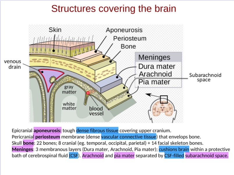
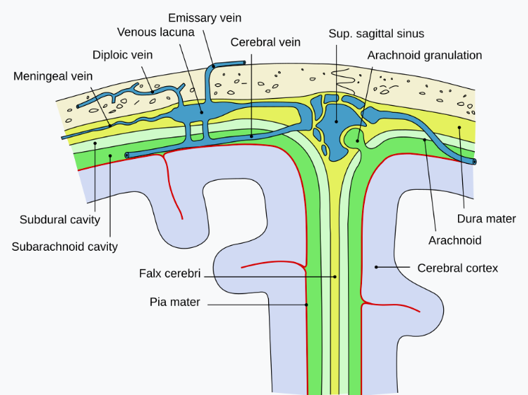
The meninges are three protective layers surrounding the brain: the dura mater, arachnoid mater, and pia mater. The dura mater is the tough outer layer composed of two sublayers (periosteal and meningeal), which also contain venous sinuses that drain blood and CSF. Beneath this lies the arachnoid mater, a web-like structure containing the subarachnoid space filled with CSF. This fluid provides cushioning and protection against mechanical injury. The innermost layer, the pia mater, is a thin, delicate, and impermeable membrane that tightly adheres to the brain surface, preventing uncontrolled fluid entry while allowing regulated passage of blood vessels.
Cerebrospinal fluid (CSF) plays several essential roles. It acts as a shock absorber, maintains buoyancy of the brain, supplies nutrients, and removes metabolic waste. CSF is produced mainly by the choroid plexus within the brain ventricles. Specialised cuboidal epithelial cells filter blood plasma in a controlled manner to form CSF. Approximately 600 mL of CSF is produced daily, with a total volume of about 150 mL, meaning it is renewed multiple times per day.
The ventricular system consists of four interconnected cavities: two lateral ventricles, the third ventricle, and the fourth ventricle. CSF flows through these ventricles and exits into the subarachnoid space, where it circulates around the brain and spinal cord. It is eventually reabsorbed into the venous sinuses via arachnoid granulations. The flow of CSF is facilitated by cilia lining the ventricles and by arterial pulsations.
A recently discovered system called the glymphatic system highlights the role of CSF in waste clearance. CSF enters the brain along perivascular spaces (Virchow–Robin spaces), mixes with interstitial fluid (ISF), and helps remove toxic substances such as metabolic waste and proteins. This system is particularly active during sleep and is crucial for maintaining brain health.
The brain’s blood supply is provided by two major arterial systems: the internal carotid arteries and the vertebral arteries, which form the basilar artery. (2Va=Ba)These vessels branch extensively to supply oxygen and nutrients. The blood–brain barrier (BBB), formed by endothelial cells with tight junctions, astrocytes, and pericytes, strictly regulates substance entry. Small molecules like water and gases diffuse passively, while larger molecules require active transport mechanisms such as carrier proteins or receptor-mediated transcytosis.
Transport across the BBB is highly selective. Glucose enters via transporters (e.g., GLUT1), while larger molecules like proteins require receptor-mediated mechanisms. This selectivity poses challenges for drug delivery, leading to strategies such as “Trojan horse” methods, where therapeutic agents are linked to molecules that can cross the barrier (L-dopa).
📌 BULLET-POINT SUMMARY
Meninges
Dura mater: tough outer layer, venous sinuses
Arachnoid mater: web-like, contains CSF
Pia mater: thin, impermeable, adheres to brain
Cerebrospinal Fluid (CSF)
Cushions brain
Provides nutrients
Removes waste
Produced by choroid plexus
~600 mL/day, 150 mL total volume
Plexus epithelium filters liquid from pia mater blood vessels to form CSF
Ventricular System
2 lateral ventricles
3rd ventricle
4th ventricle
CSF flows → subarachnoid space → venous sinuses
Glymphatic System
CSF enters via perivascular spaces
Mixes with interstitial fluid
Removes toxins
Blood Supply
Internal carotid arteries
Vertebral → basilar artery
Blood–Brain Barrier
Tight junctions
Astrocyte support
Selective transport:
Passive diffusion (small molecules)
Active transport (glucose, ions)
Receptor-mediated transport (proteins)
Clinical Notes
Meningitis = infection of meninges
BBB limits drug delivery
Sleep enhances waste clearance
✏ DETAILED FILL-IN-THE-BLANK SUMMARY
The three meningeal layers are ___dura_______, _archonoid_________, and ___pia_______.
The outermost meningeal layer is the __dura________ mater.
The middle layer is called the ___archonoid_______ mater.
The innermost layer is the __pia________ mater.
CSF is mainly found in the __subarchnoid________ space.
CSF is produced by the __choriod_____ plexus.
The total volume of CSF is about ___150_______ mL.
CSF is produced at approximately ___600_______ mL/day.
CSF flows through structures called __vemtricles________.
The first pair of ventricles are the ___lateral_______ ventricles.
The third ventricle is located in the ____decencelphan_____.
CSF exits via the ___4_______ ventricle.
CSF drains into the __venous________ sinuses.
The BBB is formed by ___endothelial______ cells.
Tight junctions prevent __paracelluar________ movement of substances.
Glucose crosses the BBB via ___GLUT-1_______ transporters.
Large molecules cross via ___receptor_______-mediated transcytosis.
The glymphatic system removes ___waste_______ from the brain.
CSF enters brain via ___perivascular______ spaces.
The main arteries supplying the brain are __internal caronoid________ and ___vertebral_______ arteries.
Here are the correct filled answers:
1. The three meningeal layers are dura mater, arachnoid mater, and pia mater.
2. The outermost meningeal layer is the dura mater.
3. The middle layer is called the arachnoid mater.
4. The innermost layer is the pia mater.
5. CSF is mainly found in the subarachnoid space.
6. CSF is produced by the choroid plexus.
7. The total volume of CSF is about 150 mL.
8. CSF is produced at approximately 600 mL/day.
9. CSF flows through structures called ventricles.
10. The first pair of ventricles are the lateral ventricles.
11. The third ventricle is located in the diencephalon.
12. CSF exits via the fourth ventricle.
13. CSF drains into the venous sinuses.
14. The BBB is formed by endothelial cells.
15. Tight junctions prevent paracellular (between-cell) movement of substances.
16. Glucose crosses the BBB via GLUT1 (facilitated diffusion) transporters.
17. Large molecules cross via receptor-mediated transcytosis.
18. The glymphatic system removes metabolic waste from the brain.
19. CSF enters brain via perivascular (Virchow–Robin) spaces.
20. The main arteries supplying the brain are internal carotid and vertebral arteries.
📝 40 EXAM-STYLE MCQs
1. The meninges consist of how many layers?
a) 1
b) 2
c) 3
d) 4
e) 5
2. The outermost meningeal layer is:
a) Pia mater
b) Arachnoid mater
c) Dura mater
d) Endothelium
e) Pericyte layer
3. The arachnoid mater is named due to its:
a) Thickness
b) Spider-like appearance
c) Vascularity
d) Elasticity
e) Rigidity
4. CSF is primarily located in the:
a) Epidural space
b) Subdural space
c) Subarachnoid space
d) Intracellular space
e) Synaptic cleft
5. The pia mater is:
a) Thick and fibrous
b) Highly vascular
c) Impermeable and delicate
d) Detached from brain
e) Filled with CSF
6. CSF is produced by:
a) Astrocytes
b) Neurons
c) Choroid plexus
d) Microglia
e) Oligodendrocytes
7. The total CSF volume is approximately:
a) 50 mL
b) 100 mL
c) 150 mL
d) 300 mL
e) 600 mL
8. CSF is renewed approximately:
a) Once daily
b) Twice daily
c) 4 times daily
d) Weekly
e) Monthly
9. The lateral ventricles connect to the third ventricle via:
a) Aqueduct
b) Foramen
c) Canal
d) Sinus
e) Plexus
10. The fourth ventricle drains into the:
a) Bloodstream
b) Subarachnoid space
c) Spinal cord
d) Cortex
e) Thalamus
11. The glymphatic system primarily removes:
a) Oxygen
b) Glucose
c) Waste products
d) Hormones
e) Lipids
12. Perivascular spaces are also called:
a) Synaptic clefts
b) Virchow–Robin spaces
c) Ventricular canals
d) Neural gaps
e) Axonal spaces
13. The BBB is formed mainly by:
a) Neurons
b) Astrocytes only
c) Endothelial cells
d) Microglia
e) CSF
14. Tight junctions in BBB prevent:
a) Active transport
b) Passive diffusion
c) Paracellular movement
d) Ion exchange
e) Oxygen entry
15. Which molecule crosses BBB via transporter?
a) Oxygen
b) Glucose
c) CO₂
d) Water
e) Lipids
16. Receptor-mediated transcytosis is used for:
a) Gases
b) Small ions
c) Large proteins
d) Water
e) Sodium
17. The internal carotid arteries supply:
a) Spinal cord
b) Brain
c) Liver
d) Heart
e) Kidneys
18. Vertebral arteries form the:
a) Carotid artery
b) Basilar artery
c) Jugular vein
d) Sinus
e) Cortex
19. Astrocytes contribute to BBB via:
a) Axons
b) Synapses
c) End-feet
d) Dendrites
e) Nodes
20. CSF flow is aided by:
a) Neurons
b) Cilia
c) Muscles
d) Hormones
e) Enzymes
21–40 (same format, continuing difficulty):
CSF provides → cushioning
CSF density is similar to → brain tissue
Venous sinuses drain → CSF + blood
Meningitis affects → meninges
BBB restricts → toxins
Lipid-soluble molecules cross via → diffusion
Interstitial fluid originates from → blood
CSF mixes with → interstitial fluid
Choroid plexus cells are → cuboidal
BBB transport is → selective
Tight junctions are between → endothelial cells
Glymphatic system is active during → sleep
CSF exits via → arachnoid granulations
Brain floats in → CSF
CSF protects against → mechanical shock
BBB uses → transport proteins
Receptor example → transferrin receptor
Trojan horse method → drug delivery
BBB limits → large molecules
Brain protection systems → meninges + CSF + BBB
✅ ANSWER KEY
1-c,
2-c,
3-b,
4-c,
5-c,
6-c,
7-c,
8-c,
9-b,
10-b
11-c,
12-b,
13-c,
14-c,
15-b,
16-c,
17-b,
18-b,
19-c,
20-b