The Cardiovascular System
The cardiovascular system is composed of the heart, blood vessels, and blood, and plays a crucial role in maintaining homeostasis by transporting oxygen, nutrients, hormones, and waste products throughout the body. It is vital for regulating body temperature and pH levels, as well as ensuring the efficient functioning of all organs.
Anatomy and Physiology of the heart
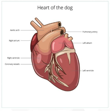
The heart is often considered the most important organ of the body. It is the pump of the cardiovascular system; the cardiovascular system is the body system that has the life sustaining task of feeding all the body tissue.
The heart is a hollow muscular organ located inside the central part of the chest cavity in between the lungs.
The Pericardium
is a membrane that surrounds the heart. This membrane has two layers:
Fibrous pericardium: this is the first layer and tough external later of the peridcardium
Serous pericardium: this is the second and inner layer of the pericardium. This later is further divided into two more layers called the parietal layer. Which lines the heart’s double-walled pericardium and the visceral layer, which covers the heart itself, providing protection and reducing friction as the heart beats.
Between the parietal layer and the visceral later of the pericardium is a space filled with liquid. This space is called the pericardial space and the liquid is called the pericardial fluid. When the heart beats, the pericardial fluid allows the heart and the pericardium to interact smoothly by preventing friction.
The Heart Wall The heart wall consists of three distinct layers: the epicardium, which is the outermost layer that also forms part of the pericardium; the myocardium, which is the thick middle layer composed of cardiac muscle that enables the heart to pump blood effectively; and the endocardium, the innermost layer that lines the heart chambers and valves, ensuring smooth blood flow.
lets look at each layer:
epicardium : The epicardium is a thin, transparent layer that provides a protective covering for the heart and contains blood vessels, nerves, and lymphatics that supply the heart muscle.
myocardium: The myocardium is the muscular layer responsible for the contractile function of the heart, facilitating the pumping of blood throughout the body with each heartbeat, and it is particularly thick in the left ventricle to generate sufficient pressure for systemic circulation.
Endocardium: The endocardium is the innermost layer of the heart, lining the heart chambers and covering the heart valves; it plays a critical role in minimizing friction during blood flow and preventing blood clots.
Blood Supply

Heart Valves
Four heart valves control the blood flow in the heart. The two valves that control blood flow between the atria and the ventricles are known as the atrioventricular valves. The other two valves are known as semilunar valves, and they control blood flowing from the ventricles into the pulmonary artery and arterial system.
Here is how each valves controls blood flow in the heart:
The right atrioventicular valve or right AV controls blood flow from the right atrium to the right ventricle, preventing backflow into the atrium during ventricular contraction.
The left atrioventicular valve or left AV controls blood flow from the left atrium to the left ventricle, ensuring that blood does not flow back into the atrium as the ventricle contracts.


Heart Rhythm : The heart rhythm is regulated by electrical impulses originating from the sinoatrial (SA) node, which triggers the contraction of the atria and subsequently the ventricles, maintaining a coordinated heartbeat.
Here are important terms to know relating to heart rhythms:
Palpitation: A sensation of rapid or irregular heartbeat, often felt as a pounding or fluttering in the chest, which can occur due to various factors including stress, anxiety, or certain medical conditions.
Fibrillation: A serious cardiac condition characterized by rapid and unsynchronized contractions of the heart muscle, leading to ineffective pumping of blood, which can result in life-threatening situations if not addressed promptly.
Flutter: A type of arrhythmia that involves rapid contractions of the heart's atria, which may lead to an irregular heartbeat and can affect blood flow, potentially increasing the risk of stroke and other complications.
Bradycardia: A slower than normal heart rate, typically defined as fewer than 60 beats per minute, which can be caused by various factors such as aging, heart disease, or the use of certain medications. While often asymptomatic, bradycardia may lead to dizziness, fatigue, or fainting in some individuals.
Tachycardia: A condition characterized by an abnormally fast heart rate, typically defined as exceeding 100 beats per minute. Tachycardia can result from stress, exercise, fever, or underlying heart conditions, and may lead to symptoms such as palpitations, shortness of breath, and chest pain.
Paroxysmal: A term used to describe episodes of rapid heart rate that occur suddenly and unexpectedly, often at rest. These episodes can be recurrent and may resolve on their own, but they can also lead to serious complications if not properly managed.
Normal Sinus Arrythmia: A variation in heart rhythm that occurs naturally, particularly during breathing. In normal sinus arrhythmia, the heart rate increases with inhalation and decreases with exhalation, reflecting the influence of the autonomic nervous system, and is generally considered a benign condition without significant health implications.
Asystole: A state of flatline on the electrocardiogram (ECG), representing a complete absence of electrical activity in the heart. It is a critical condition that requires immediate medical intervention, as it indicates that the heart is not pumping blood, leading to a lack of circulation to vital organs.
Syncope: A temporary loss of consciousness due to a decrease in blood flow to the brain. Syncope can result from various causes, including vasovagal response, dehydration, or cardiac issues, and often requires evaluation to determine underlying factors.
Gallop: An abnormal heart sound characterized by a three-part rhythm, often described as "large animal galloping." It can indicate heart failure or volume overload, and is typically assessed during a physical examination using a stethoscope.
Blood Flow in the Heart
Throughout the cardiac cycle, blood flows through the heart and is then pumped to all parts of the animal’s body. Blood inside the heart flows through each heart chamber and heart valve.
Lets follow blood as it flows through the heart:
Blood first enters the heart through two large veins called the cranial and caudal venae. These veins open directly into the right atrium of the heart. Blood then flows from the right atrium through the right AV or tricuspid valve into the right ventricle.
From the right ventricle, blood flows through the pulmonary semilunar valve into the pulmonary artery. It then flows through the pulmonary artery to the lungs where it is oxygenated.
Once oxygenated, blood flows out of the lungs and through the pulmonary veins back to the heart. This freshly oxygenated blood enters the left atrium, completing the pulmonary circuit and preparing for the systemic circulation. From the left atrium, blood is then pushed through the left AV or mitral valve into the left ventricle, where it is finally pumped out to the rest of the body through the aorta. This entire process ensures that oxygen-rich blood is delivered to tissues, while carbon dioxide is removed, maintaining proper metabolic function. Once the blood flows through the body it repeats this cycle through the heart.
Electrical Activity in the Heart
The heart's electrical system controls the rhythm and timing of the heartbeat, ensuring efficient blood flow.
Sinoatrial (SA) Node: Often referred to as the natural pacemaker, it initiates electrical impulses that spread through the atria, causing them to contract.
Atrioventricular (AV) Node: This node receives impulses from the SA node and delays them slightly to allow the ventricles to fill with blood before contracting.
Bundle of His and Purkinje Fibers: These structures carry electrical impulses into the ventricles, causing them to contract and pump blood to the lungs and the rest of the body.
This coordinated electrical activity is crucial for maintaining an effective cardiac cycle.
Factors influencing heart rhythm include autonomic nervous system activity and various hormones, which can increase or decrease heart rate based on the body's demands.


Electrocardiograms
are essential tools for assessing heart rhythm and electrical activity, allowing medical professionals to detect arrhythmias, myocardial infarctions, and other cardiac abnormalities. Proper interpretation of these readings is vital for accurate diagnosis and treatment, guiding interventions that may include medication, lifestyle modifications, or surgical procedures.

Heart sounds include: lub-dub, murmurs, splits, crackles, clicks, and rumbles.. here is what they each mean:
Lub-dub: The normal heart sounds produced by the closing of the heart valves during the cardiac cycle. The "lub" (S1) is the sound of the mitral and tricuspid valves closing, while the "dub" (S2) is the sound of the aortic and pulmonic valves closing.
Murmurs: Abnormal sounds caused by turbulent blood flow across damaged or malformed heart valves, which can indicate underlying heart conditions.
Splits: Occur when the closure of the heart's valves is not perfectly synchronized, leading to a delay in the second heart sound (S2) that is often heard during inspiration.
Crackles: Sounds typically associated with fluid in the lungs, which may indicate heart failure or other pulmonary issues affecting the heart's function.
Clicks: Abnormal sounds that may arise from structural heart issues, such as prosthetic valves or mitral valve prolapse.
Rumbles: Low-frequency sounds that may indicate increased blood flow across heart valves or structural abnormalities such as stenosis.


Anatomy & Physiology of Blood Vessels
Arteries: Blood vessels that carry oxygenated blood away from the heart, with thick, elastic walls to withstand high pressure.
Veins: Blood vessels that return deoxygenated blood to the heart, equipped with valves to prevent backflow, resulting in a lower pressure system.
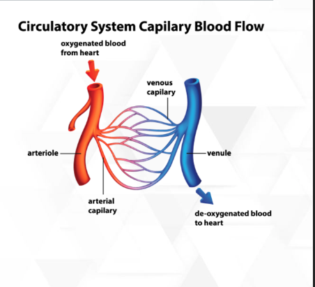
Capillaries: Microscopic blood vessels where the exchange of oxygen, carbon dioxide, nutrients, and waste products occurs between blood and tissues.
Functions of Blood Vessels: Each type of blood vessel plays a crucial role in maintaining overall circulatory efficiency and homeostasis, ensuring that nutrient and gas exchange is optimized throughout the body.
Blood Vessel Structure: Blood vessels are composed of three primary layers: the tunica intima (inner layer), the tunica media (middle layer), and the tunica externa (outer layer), each contributing to the overall function and integrity of the vessel.

The Arteries are the blood vessels that carry oxygenated blood away from the heart.
Lets take a look at the many types of arteries in the body:
aorta: the largest artery in the body, it distributes oxygenated blood from the heart to the rest of the body.
celiac artery: supplies blood to the stomach, liver, spleen, and other organs in the abdominal cavity.
Renal Artery: supplies blood to the kidneys, playing a crucial role in filtering blood and regulating blood pressure.
Ovarian Arteries: supply blood to the ovaries, which are essential for female reproductive health and hormone production.
Testicular Arteries: supply blood to the testes, playing a key role in male reproductive health and the production of sperm and testosterone.
Subclavian artery: supplies blood to the arms and shoulders, as well as parts of the neck and head, facilitating proper circulation to these vital areas.
arterioles: smaller branches of arteries that lead to capillaries, regulating blood flow and pressure within tissues, thus playing a critical role in nutrient and oxygen exchange.
The capillaries
: the smallest blood vessels, where the exchange of oxygen, carbon dioxide, nutrients, and waste occurs between blood and surrounding tissues, ensuring cellular metabolism and maintaining homeostasis.
CRT or capillary refill time is a clinical assessment used to evaluate peripheral perfusion by measuring the time it takes for color to return to a nail bed after it has been compressed, providing insight into cardiovascular health and circulation efficiency.
Alveolar capillaries: specialized capillaries found in the lungs that play a critical role in gas exchange, allowing oxygen to enter the bloodstream and carbon dioxide to be expelled, thereby facilitating respiration.
The Veins
carry deoxygenated blood back to the heart, except for pulmonary veins which transport oxygenated blood from the lungs. They have thinner walls compared to arteries and often contain valves that prevent backflow, assisting in the return of blood against the effects of gravity. The venous system is crucial for maintaining efficient circulation, ensuring that blood is directed back to the heart for reoxygenation and redistribution throughout the body.
Here are a few of the larger veins in an animal’s body:
jugular vein: a major vein that drains blood from the head and neck region back to the heart.
femoral veins: veins that transport deoxygenated blood from the lower limbs back to the heart, playing a vital role in the circulation of blood in the legs.
renal veins: veins that carry deoxygenated blood away from the kidneys to the inferior vena cava, helping to regulate blood pressure and maintain fluid balance in the body.
Azygous vein: a single vein that drains blood from the thoracic wall and various organs into the superior vena cava, assisting in the collateral circulation of blood between the upper and lower body.
pulmonary veins: vessels that transport oxygenated blood from the lungs to the left atrium of the heart, essential for replenishing the oxygen supply in the bloodstream.

Blood Pressure : the force exerted by circulating blood upon the walls of blood vessels, which is crucial for proper organ function and overall health. It is measured using two values: systolic pressure (the pressure during heartbeats) and diastolic pressure (the pressure in between heartbeats), and is regulated by factors such as cardiac output, blood volume, and vascular resistance.
it is measured by a sphygmanometer, which provides readings in millimeters of mercury (mmHg), allowing healthcare professionals to monitor and assess cardiovascular health.
There are a few important terms to know regarding blood pressure:
Hypertension: a condition characterized by persistently elevated blood pressure, typically defined as having a systolic reading of 130 mmHg or higher, and/or a diastolic reading of 80 mmHg or higher. This condition can lead to serious health issues, including heart disease and stroke, if not managed properly.
Hypotension: a condition characterized by abnormally low blood pressure, generally defined as having a systolic reading lower than 90 mmHg and/or a diastolic reading lower than 60 mmHg. While it may not always cause symptoms, hypotension can lead to dizziness, fainting, and in severe cases, shock.
Antihypertensives: medications used to treat high blood pressure by various mechanisms, including reducing blood volume, relaxing blood vessels, or decreasing heart rate, ultimately aiming to lower the risk of cardiovascular complications.
Pulse: the rhythmic expansion and contraction of arteries as blood is pumped by the heart, typically measured at the wrist or neck, indicating heart rate and overall cardiovascular health.


Terms for Testing, Pathology and Procedures of the Cardiovascular System
Electrocardiogram (ECG): a test that records the electrical activity of the heart, helping to identify irregularities in heart rhythm and structure.
Echocardiogram: an ultrasound examination that visualizes heart structures and assesses function, often used to detect heart valve issues or heart failure.
Angiography: a procedure that uses X-rays to visualize blood vessels after injecting a contrast dye, allowing for the assessment of blockages or abnormalities in the vascular system.
Cardiac Catheterization: a minimally invasive procedure that involves threading a thin tube through a blood vessel to the heart, enabling direct measurement of pressures within the heart chambers and the assessment of coronary arteries, often helping to diagnose and treat various heart conditions.
Radiography: the use of X-rays to create images of the heart and blood vessels, providing valuable information for diagnosing conditions such as heart enlargement, pulmonary congestion, or congenital heart defects.
Pathological Conditions of the Blood and Blood Vessels
Aneurysm: a bulging or dilation in the wall of a blood vessel, commonly occurring in arteries, which can lead to rupture and severe internal bleeding if not monitored or treated.
Angiopathy: a term used to describe diseases of the blood vessels, which can result from various factors including diabetes, hypertension, or infections, leading to impaired circulation and potential complications like ischemia.
Atherosclerosis: a condition characterized by the buildup of fatty deposits, cholesterol, and other substances in the arterial walls, leading to narrowing and hardening of the arteries, which increases the risk of cardiovascular events such as heart attacks and strokes.
Embolus: a blood clot or other debris that travels through the bloodstream and can obstruct a blood vessel, leading to serious complications such as stroke or pulmonary embolism.
Hypercania: an abnormal increase of carbon dioxide in the blood, which can result from respiratory issues and lead to various complications including respiratory acidosis and decreased oxygen delivery to tissues.
Hypocapnia: a condition characterized by lower than normal levels of carbon dioxide in the blood, often caused by hyperventilation, and can lead to respiratory alkalosis and associated symptoms such as dizziness, tingling, and muscle spasms.
Hypoxia: a condition in which there is insufficient oxygen available to tissues, which can result from various causes such as respiratory disease, anemia, or high altitudes, and may lead to symptoms like confusion, shortness of breath, and cyanosis.
Occlusion: a blockage in a blood vessel that can restrict blood flow to tissues, potentially resulting in ischemia and increased risk of tissue damage.
Hemangioma: a benign tumor made up of blood vessels, which can occur in various parts of the body and may cause complications if they interfere with blood flow or compress surrounding tissues.
Thrombus: a blood clot that forms within a blood vessel, obstructing blood flow and potentially leading to conditions such as stroke or myocardial infarction if it dislodges and travels to vital organs.
Thromboembolism: a condition where a thrombus (blood clot) breaks loose from its site of formation and travels through the bloodstream, potentially causing blockages in other blood vessels, which may result in serious complications such as pulmonary embolism or stroke.
Vasculitis: inflammation of the blood vessels, which can lead to narrowing or blockage of the vessels, resulting in reduced blood flow and potential damage to the affected organs.
Pathological Conditions of the Heart and Valves
Aortic Insufficiency: a condition in which the aortic valve does not close properly, allowing blood to flow back into the left ventricle from the aorta during diastole, which can lead to heart failure and other complications if left untreated.
Atria Septic Defect: a congenital heart defect characterized by an opening in the atrial septum, which separates the heart's two upper chambers (the atria). This can result in oxygen-rich blood mixing with oxygen-poor blood and may lead to increased pulmonary blood flow, potentially causing heart failure and other complications.
Cardiac Tamponade: a serious condition resulting from the accumulation of fluid in the pericardial space, which exerts pressure on the heart and impairs its ability to pump effectively, leading to decreased cardiac output and potentially life-threatening arrhythmias.
Cardiomegaly: a condition characterized by an enlarged heart, which can result from various factors including hypertension, heart valve disease, or cardiomyopathy. This enlargement can lead to impaired heart function and may increase the risk of heart failure and arrhythmias.
Cardiomyopathy: a group of diseases that affect the heart muscle, leading to decreased heart function and potentially resulting in heart failure. It can be caused by genetic factors, long-term high blood pressure, or previous heart attacks. Symptoms may include shortness of breath, fatigue, and swelling in the legs and feet.
Carditis: inflammation of the heart, which can affect the heart's inner lining (endocarditis), the heart muscle (myocarditis), or the outer lining (pericarditis). It is often caused by infections, autoimmune diseases, or exposure to certain medications. Symptoms can vary widely, ranging from fever and fatigue to chest pain and difficulty breathing.
Congestive Heart Failure: a condition in which the heart is unable to pump effectively, leading to a buildup of fluid in the lungs and surrounding tissues. This can result from various underlying conditions, including coronary artery disease, high blood pressure, or previous myocardial infarctions. Symptoms typically include persistent cough, rapid or irregular heartbeat, and sudden weight gain due to fluid retention.
Cor Pulmonale: a type of heart failure that occurs due to chronic lung disease, where the right side of the heart cannot pump blood effectively to the lungs. This condition often arises from chronic obstructive pulmonary disease (COPD), pulmonary hypertension, or other lung ailments. Symptoms may include shortness of breath, fatigue, and swelling in the legs and abdomen.
Heart Block: a condition where the electrical signals in the heart are impaired, leading to a slow or irregular heartbeat. This can be caused by aging, heart tissue damage, or certain medications. Symptoms may include dizziness, fainting, and chest pain.
Mitral Stenosis: a narrowing of the mitral valve opening, which can obstruct blood flow from the left atrium to the left ventricle. This condition is often caused by rheumatic fever, congenital defects, or calcium buildup on the valve. Symptoms may include shortness of breath, fatigue, and palpitations.
Mitral Valve Insufficiency: a condition where the mitral valve does not close properly, allowing blood to flow backward into the left atrium during contraction of the left ventricle. This can occur due to degeneration of valve tissue, ischemic heart disease, or injury to the heart. Symptoms may include shortness of breath, especially during exertion, fatigue, and swelling in the legs or feet.
Mitral Valve Prolapse: a condition in which the mitral valve leaflets bulge or prolapse into the left atrium during the heart's contraction, often leading to mitral regurgitation. This may be caused by inherited connective tissue disorders, such as Marfan syndrome, or it can occur in otherwise healthy individuals. Symptoms can include palpitations, chest pain, and, in some cases, dizziness.
Myocarditis: an inflammation of the heart muscle, which can result from viral infections, autoimmune diseases, or exposure to toxins. Symptoms may vary widely but often include chest pain, fatigue, and arrhythmias. In severe cases, myocarditis can lead to heart failure or sudden cardiac death.
Pericarditis: an inflammation of the pericardium, the fibrous sac surrounding the heart. It can be caused by infections, autoimmune diseases, or after heart surgery. Common symptoms include sharp chest pain that worsens with deep breathing, fever, and a feeling of pressure in the chest.
Regurgitation: a condition where the heart's valves do not close properly, allowing blood to flow backward within the heart chambers. This can occur in conditions such as mitral valve prolapse or aortic regurgitation, leading to symptoms like fatigue, shortness of breath, and palpitations. Over time, severe regurgitation can result in heart enlargement and heart failure.
Shock: a life-threatening condition that occurs when the body does not get enough blood flow, leading to a lack of oxygen in the tissues. It can result from severe blood loss, infections (septic shock), or heart problems, and symptoms may include rapid heartbeat, confusion, and cold, clammy skin. Immediate medical attention is crucial to prevent organ damage and other serious complications.
Ventricular Septal Defect: a congenital heart defect characterized by an abnormal opening in the interventricular septum, which can lead to increased blood flow to the lungs and decreased oxygen delivery to the rest of the body. Symptoms might include difficulty breathing, fatigue, and a failure to thrive in infants. Treatment options vary and may include monitoring, medication, or surgical intervention to repair the defect.
Pulmonic Stenosis: a congenital heart defect that involves narrowing at the entrance to the pulmonary artery, which can obstruct blood flow from the right ventricle to the lungs. Symptoms can include a heart murmur, shortness of breath during physical activity, and fatigue. Treatment may involve balloon valvuloplasty or surgical repair, depending on the severity of the stenosis.
Cardiovascular System Procedures
Angioplasty: a minimally invasive procedure used to open narrowed or blocked blood vessels in the heart. This is achieved by inserting a thin tube called a catheter into the affected vessel, which is then inflated to widen the artery and improve blood flow. In some cases, a stent may be placed to keep the artery open after the procedure.
Angiorrhaphy: a surgical procedure that involves the suturing of an artery to repair damage or control bleeding, often used in cases of traumatic injury or after vascular surgery.
Arteriectomy: a surgical procedure that involves the excision of a portion of an artery, typically performed to remove obstructions or lesions that obstruct blood flow, thereby restoring proper circulation.
Arteriotomy: a surgical procedure that involves making an incision into an artery to gain access for various treatments or examinations, often performed to facilitate procedures such as bypass surgery or to sample blood directly from the artery.
Central Venous Pressure: a clinical measurement that reflects the pressure in the thoracic vena cava, near the right atrium of the heart; it is an important indicator of the patient's volume status and cardiac function.
Defibrillation: a medical procedure that involves delivering a controlled electrical shock to the heart in order to restore a normal rhythm, commonly used in cases of life-threatening arrhythmias such as ventricular fibrillation or pulseless ventricular tachycardia.
Hemostasis: the process that prevents and stops bleeding, involving a complex interaction between blood vessels, platelets, and various proteins; it is vital for wound healing and maintaining blood volume.
Stent: a medical device implanted in a narrowed or obstructed blood vessel to keep it open and ensure proper blood flow, commonly used in the treatment of coronary artery disease.
Transfusion: the process of transferring blood or blood components from one person into another, typically used to treat anemia, blood loss from surgery, or trauma, as well as to improve the oxygen-carrying capacity of the blood.
Valvototomy: a surgical procedure that involves cutting open a valve in the heart to relieve obstruction and improve blood flow, often performed in cases of valvular heart disease.
Route of Administration: the method by which a medication or other therapeutic substance is delivered to the body, which can include oral, intravenous, subcutaneous, or topical routes.
Abbreviations of the Cardiovascular System:
CAD: Coronary Artery Disease, a condition characterized by the narrowing or blockage of coronary arteries, typically due to atherosclerosis.
CHF: Congestive Heart Failure, a chronic condition where the heart struggles to pump blood effectively, leading to a buildup of fluid in the lungs and surrounding tissues.
MI: Myocardial Infarction, commonly known as a heart attack, occurs when blood flow to a part of the heart is blocked, causing tissue damage.
HTN: Hypertension, a condition where the force of the blood against the artery walls is consistently too high, increasing the risk of heart disease and stroke.
AF: Atrial Fibrillation, an irregular and often rapid heart rate that can lead to poor blood flow, resulting in an increased risk of stroke and other heart-related complications.
ASD : Atrial Septal Defect, a congenital heart defect characterized by an opening in the septum that divides the heart's two upper chambers, leading to abnormal blood flow between them.
AV: Atrioventricular block, a condition where the electrical signals between the atria and ventricles are impeded, resulting in a slower heart rate and potential symptoms of heart failure.
BP : Blood Pressure, the force exerted by circulating blood on the walls of blood vessels, which is crucial for maintaining adequate blood flow to organs and tissues.
LV: Left Ventricle, the chamber of the heart responsible for pumping oxygenated blood to the body through the aorta, playing a vital role in the overall circulatory efficiency.
RV: Right Ventricle, the chamber of the heart that receives deoxygenated blood from the right atrium and pumps it to the lungs via the pulmonary arteries, essential for oxygenating blood before it circulates to the rest of the body.
MM: Mucus Membrane , a thin layer of tissue that lines various cavities in the body and covers the surfaces of organs, which plays a vital role in maintaining moisture, protecting underlying tissues, and facilitating the exchange of gases and nutrients within the cardiovascular system.
MS : Mitral Stenosis, a heart valve disorder characterized by the narrowing of the mitral valve opening, which impedes blood flow from the left atrium to the left ventricle, potentially leading to increased pressure in the lungs and associated symptoms such as shortness of breath and fatigue.
PDA : Patent Ductus Arteriosus, a congenital heart defect in which the ductus arteriosus fails to close after birth, resulting in an abnormal blood flow between the aorta and the pulmonary artery, which can lead to heart enlargement and increased pulmonary blood flow.
PMI : Point of Maximum Intensity, the location on the chest where the heart's sound is the loudest, typically found at the fifth intercostal space in adults, and is used in physical examinations to assess cardiac function and detect potential abnormalities.
PVC: Premature Ventricular complex: a common cardiac arrhythmia characterized by early depolarization of the ventricles, which can lead to palpitations and may indicate underlying heart disease, especially if occurring frequently.
SA: Sinoatrial Node, the natural pacemaker of the heart, located in the right atrium, which generates electrical impulses that initiate each heartbeat and help regulate the heart rate.
VSD: Ventricular Septal Defect, a congenital heart defect characterized by an abnormal opening in the ventricular septum that separates the left and right ventricles, potentially leading to increased blood flow to the lungs and heart failure if not managed properly.
VT: Ventricular Tachycardia, a type of arrhythmia where the ventricles beat rapidly, which can lead to insufficient blood flow to the body and may be life-threatening, often associated with underlying heart conditions.
CPR: Cardiopulmonary Resuscitation, an emergency procedure performed to manually preserve intact brain function until further measures are taken to restore spontaneous blood circulation and breathing in a person experiencing cardiac arrest.
CRT: Cardiac Resynchronization Therapy, a treatment for heart failure that uses a specialized pacemaker to improve the timing of heart contractions, thereby enhancing the efficiency of the heart's pumping ability.
CVP : Central Venous Pressure, a measurement of the pressure in the thoracic vena cava near the right atrium of the heart, used as an indicator of the heart's function and fluid volume status.
ECG/EKG Electrocardiogram (ECG or EKG), a test that records the electrical activity of the heart over a period of time, providing valuable information about heart rhythm, size, and overall electrical activity.
HR: Heart Rate, the number of times the heart beats per minute, which is a vital sign that reflects the body's overall cardiovascular health and helps assess the effectiveness of the heart's function.
Disease Terminology:
Congenital means: a condition that is present from birth, which may be due to genetic factors or environmental influences during fetal development.
Hereditary: relating to traits or conditions that are passed down from parents to their offspring through genes, often playing a crucial role in the predisposition to various cardiovascular diseases.
Anomaly: a deviation from the normal structure or function of the heart or blood vessels, which can contribute to various cardiovascular complications.
Idiopathic: a term used to describe a condition or disease for which there is no known cause, often seen in various cardiovascular disorders, where the origin remains unclear despite extensive research.
Iatrogenic: resulting from the medical treatment or intervention itself, which can sometimes lead to unintended cardiovascular issues or complications during or after procedures.


