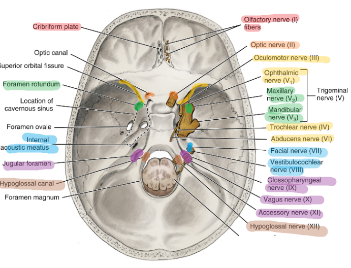
Cranial Nerve
Overview of the Exits of the Cranial nerve
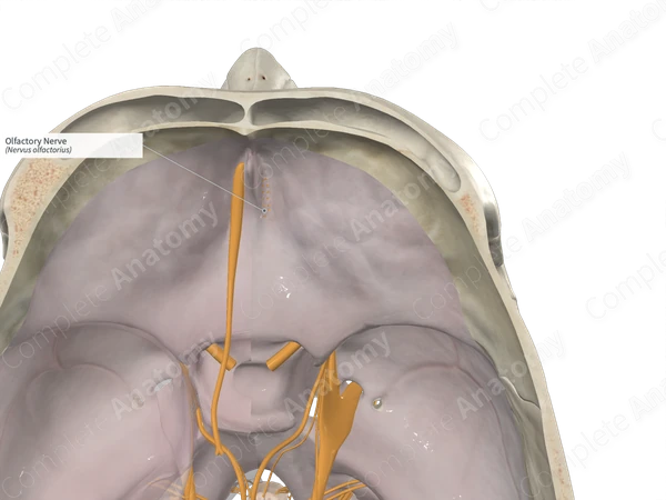
CN I → cribriform plate
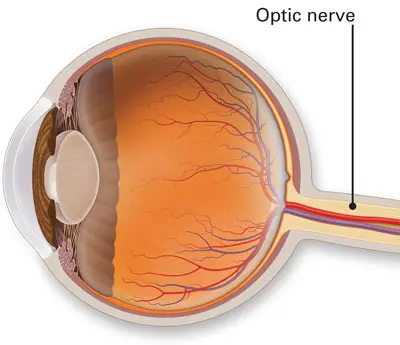
CN II → optic canal
CN III → superior orbital fissure
CN IV → superior orbital fissure
CN V →
CN V1 → superior orbital fissure
CN V2 → foramen rotundum
CN V3 → foramen ovale
CN VI = superior orbital fissure
CN VII → internal acoustic meatus
CN VIII → internal acoustic meatus
CN IX → jugular foramen
CN X → jugular foramen
CN XI → jugular foramen
CN XII → hypoglossal canal
Acronym:
Some
Say
Marry
Money
But
My
Brother
Says
Big
Brains
Matter
More
Olfactory Nerve (I)
Function: smell from the nasal mucosa to the brain
Classification: Afferent
Location: cribriform plate of the ethmoid bone
Optic Nerve (II)
Function: sight from the retina to the brain
Classification: Afferent
Location: optic canal in the sphenoid bone
Oculomotor Nerve (III)
Function: movement of the eye muscle
Classification: Efferent
Location: superior orbital fissure of the sphenoid bone
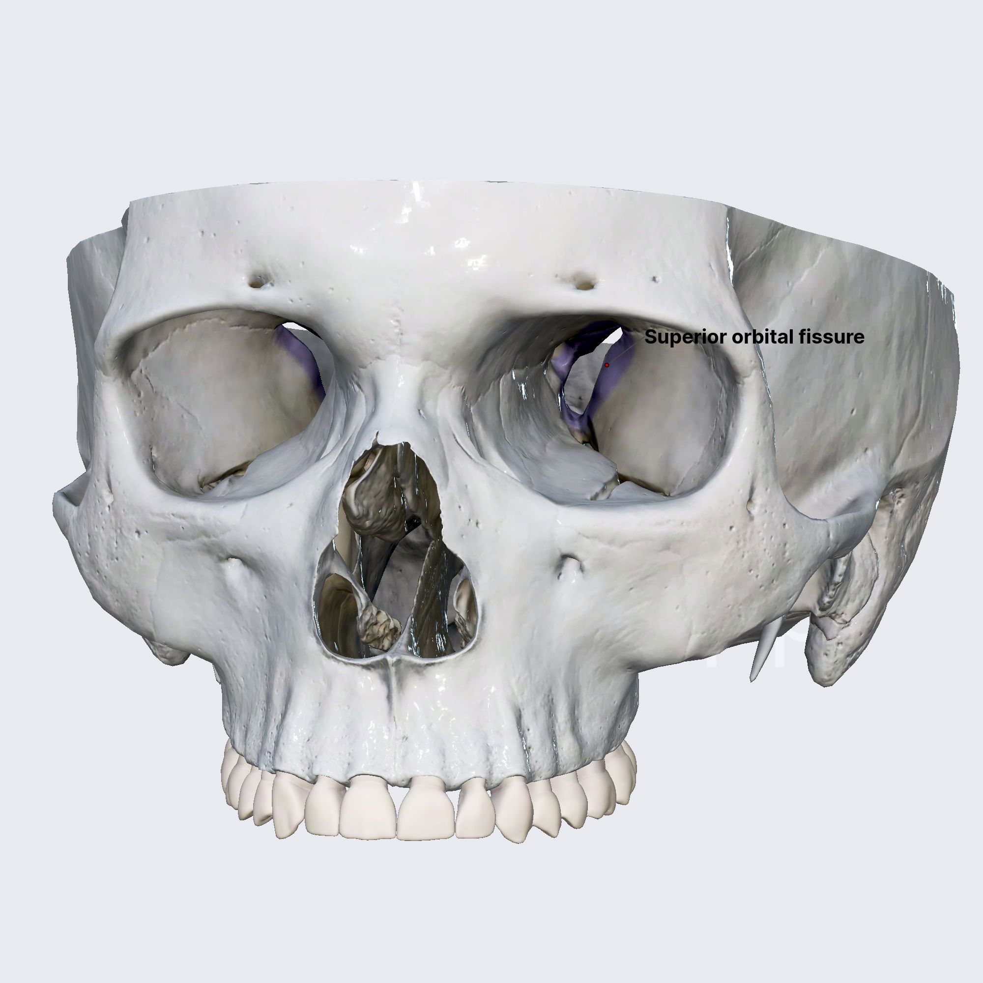
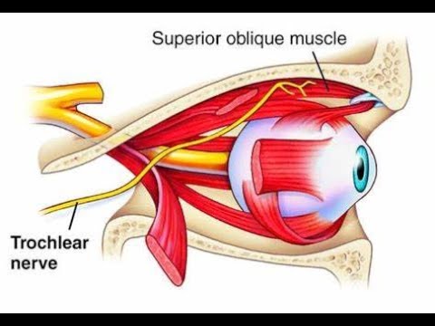
Trochlear nerve (IV)
Function: the superior oblique muscle of the eye
Classification: Efferent
Location: superior orbital fissure of the sphenoid bone
Trigeminal Nerve (V)
Note: Largest cranial nerve and MOST significant due to innervation of tissue, structures, and organs of the head and neck
Three division: Ophthalmic V1, Maxillary V2, and Mandibular V3
both afferent and efferent
Division | Type | Function | Area Supplied | Illustration |
|---|---|---|---|---|
Ophthalmic (V1) | Afferent | Eyes, skin of the forehead, eyelids, nose  | superior orbital fissure 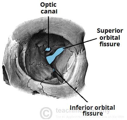 | |
Maxillary (V2) | Afferent | Cheek, lower eyelid, nose, upper lip; all maxillary teeth  | foramen rotundum 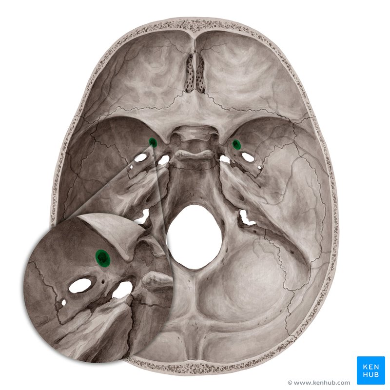 | |
Mandibular (V3) | Afferent & Efferent | Sensory: pain, temperature, touch, and pressure (lower face, including mandibular teeth) Motor: mastication 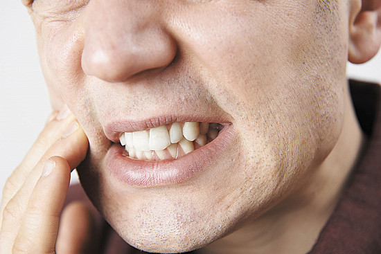 | foramen ovale 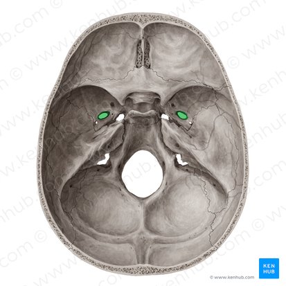 |
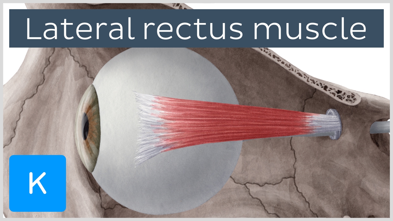
Abducens Nerves (VI)
Function: movement of the lateral rectus eye muscle
Classification: Efferent
Location: superior orbital fissure
Facial Nerve (VII)
Classification: afferent and efferent
Note: significant because of its innervation through the parotid salivary gland
Clinical Application: Bell’s Palsy
Component | Structures |
|---|---|
Motor | muscles of facial expression, lacrimal glands, and submandibular and sublingual glands 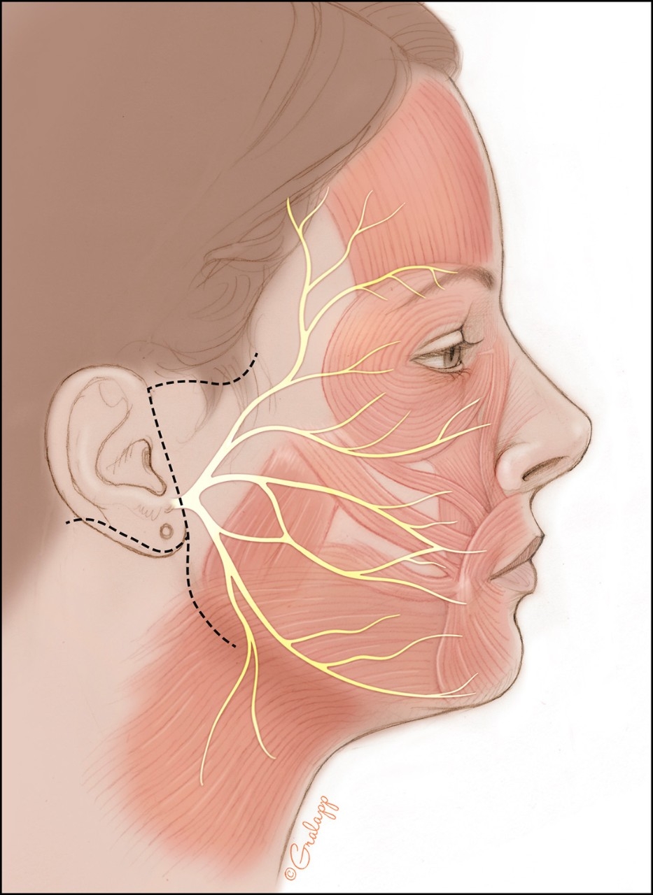 |
Pathway (2): | Lacrimal Gland: facial nerve → pterygopalatine ganglion → lacrimal gland 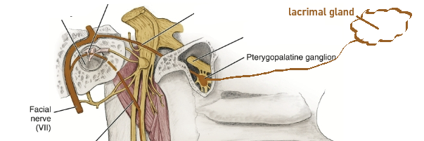 Salivary Glands: facial nerves → chorda tympani (branch) → submandibular ganglion, submandibular and sublingual salivary glands 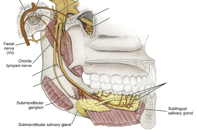 |
Sensory | anterior 2/3 of tongue 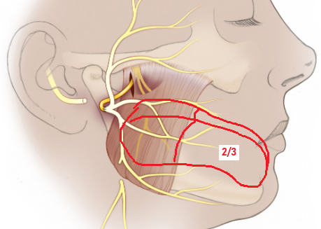 |
Overall Pathway: | enters internal acoustic meatus → facial canal (temporal bone) → stylomastoid foramen (exits) |
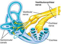
Vestibulocochlear Nerve (VII)
Function: receptors in the inner ear (within the temporal bone) for equilibrium and hearing
Classification: afferent
Location: internal acoustic meatus
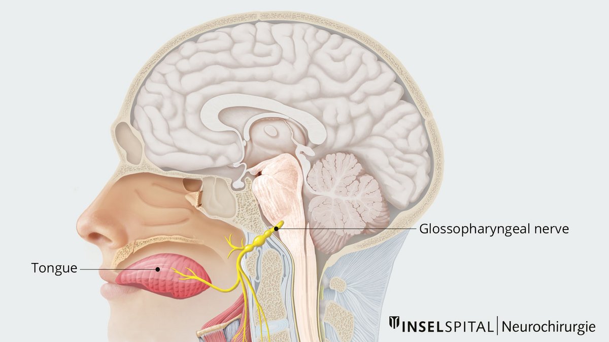
Glossopharyngeal Nerve (IX)
Function: signal the optic ganglion to make the parotid gland secrete
Classification: afferent and efferent
afferent → posterior 1/3 of the tongue, pharyngeal mucosa, and tonsils
efferent → stylopharyngeal muscle
Location: jugular foramen
NOTE: innervates the tongue and pharynx
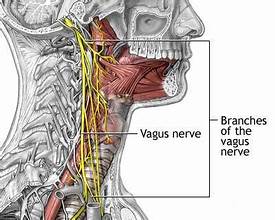
Vagus Nerve (X)
Classification: afferent and efferent
afferent → epiglottis
efferent → pharynx, larynx, soft palate, smooth muscle, cardiac muscle
Location: jugular foramen
NOTE: affects body >
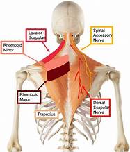
Accessory nerve (XI)
Two Parts (motor)
cranial → larynx, soft palate, and pharynx muscle (swallowing)
spinal → sternocleidomastoid and trapezius muscle
Location: hypoglossal canal
CN IX, X, XI (pharyngeal plexus)
Hypoglossal Nerve (XII)
Classification: Efferent
Function: innervates tongue
hypoglossus
styloglossus
genioglossus
Location: hypoglossal canal