Cognitive Neuroscience
Definition (#f7aeae)
Important (#edcae9)
Extra (#fffe9d)
How does cognitive neuroscience some into play:
Links brain and other aspects of nervous system to the cognitive process and behaviors. Ex: thoughts, emotions, motivations.
The brain can be reactive and also directive. (Brain is the boss but also listens to the body)
Psychology helps us to understand the mind and behaviors, cognitive neuroscience allows us to explain how the brain/neural activities are correlated with those mental processes.
Aim of cognitive neuroscience:
To study localization of functions to the specific areas of the brain that controls specific skills/behaviors.
Starts from the basic functional units, neuron.
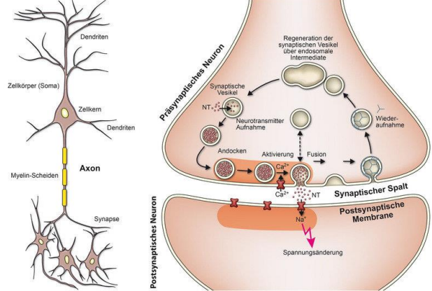
Neuron: Building blocks of the nervous system.
Neurons:

Soma: Connects the life of a neuron to the axon.
Dendrites: receive information from other neurons and process those in the soma
Axon: Helps to travel electrochemical signals to the neuron end and transmit to the other neuron.
Myeline sheaths and node of Ranvier in some neurons helps to speed up the transmission of information.
Learning is the formation of the new neural connections, in the branching structures of the dendrites.
Synapses:
The gap between the terminal buttons of one or more neurons and the dendrites of another or more neurons.
Estimated billions of neurons are connected to each other to process information simultaneously yet functions separately depending on the localization of the brain.
Learning increases the size and number of synapses.
Cognitive decline reduces it.
What do neurons transmit:
Acetylcholine: Excitatory in the brain believed to be involved in memory formation. Found in the hippocampus.
Dopamine: Involves in attention, learning, motivation, reward and reinforcement. Too little leads to tremors and limb rigidity in Parkinson, and too much found in schizophrenia.
Serotonin: Arousal, sleep, dreaming, defects are linked to depression. Drugs that blocks serotonin will increase aggression.
GABA (gamma aminobutyric acid): Involves in learning and memory and focus.
When such chemicals go haywire – results in psychopathological conditions (due to chemical imbalance).
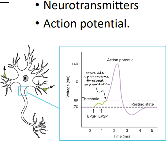
Structure of the brain:
Postmortem Dissection:
Common practice for centuries, how we studied about the brain.
Researchers carefully study those behaviors who have brain damage.
During postmortem look for lesions, areas of tissue damage.
Famous case study: Phineas Gage.
Animal studies:
Single cell recording activity by inserting electrodes to the brain had helped us to understand neurons firing patterns.
Advanced in Brain-computer interface (BCI) research.
Debates about ethical considerations.
But the benefit is greater than the risk.
Possibilities:
We can study parts individually and understand how they function independently or as a whole.
We need to have better techniques to study brains and cognitive processes.
Anatomy:
Cerebral cortex: Sensory information processing, cognitive processing, motor information processing.
Basal ganglia: Motor system.
Limbic system: Emotions, learning, motivation; amygdala involves in aggression
Thalamus: Transmit sensory information to the related areas in the cortex.
Superior and inferiour caliculi: For visual reflexes and hearing;
Cerebellum: Balance and coordination.
Pons: Involves in consciousness.
Medulla oblongata: Crossing of the nerves from one side of the body to the opposite side of the body.

Cortical Structures and Functions:

Functional Areas of the Cerbellum Cortex:

Frontal lobe:
Cognitive functions such as attention (sustained attention, divided attention).
Right hemisphere lesions are associated with deficits in sustained attention.
Executive functions such as working memory, response inhibition, goal-directed behaviors.
ADHD: Associated with poor working memory and response inhibition → frontal lobe dysfunctions.
Parietal lobe:
Receiving and processing sensory information (touch, pressure, heat, cold), body perception, movements, spatial coordination.
Individuals with damage to the right parietal lobe show abnormalities in body image and spatial relations, such as failing to attend to part of the body.
Patients with damage to the left parietal lobe experience difficulty with writing (agraphia), an inability to recognize familiar objects (agnosia), and language disorders (aphasia).
Bilateral damage can lead to problems with visual attention and motor abilities.
Temporal lobe:
If damaged, problems:
Speech perception: Posterior superior temporal gyrus.
Speech production: Posterior superior temporal gyrus.
Hearing: Posterior superior temporal gyrus.
Episodic memory: Medial structures.
Phonological: Dorsal and posterior structures.
Semantic: Ventral temporal lobe.
Social: 2 areas in the temporal lobe.
Visual: Temporal-occipital junction.
Occipital lobe:
Vision, visual memories, and visual perceptions.
Found excitation-inhibition abnormalities in the visual cortex in MDD.

Non-Invasive Techniques to Measure Brain Activities:
Electroencephalograph (EEG):
Records continuous electrical activities in the brain as waves.
Could identify response specific activities such as event-related potentials (ERP) or spectral brands (alpha beta gamma, etc).

ERP abnormalities are associated with psychopathological conditions such as depression, ADHD, Language impairments, attention.

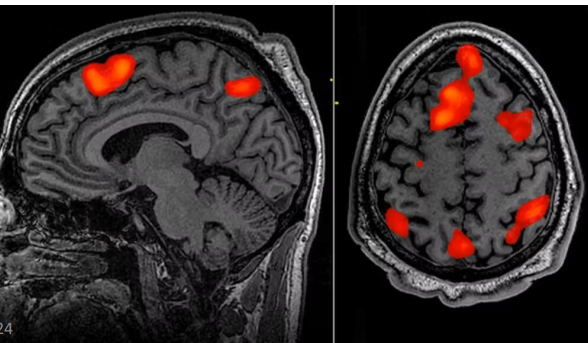
fMRI:
Functional magnetic resonance imaging.
Uses a large magnetic machine.
To measure the brain activity while performing a task.
Differences in brain activation patterns are observable between healthy and psychopathological states.
PET & CT scans:
Brain imaging techniques.
CT scans used x-ray image methods.
CT scans are used to find which part of the brain is stimulated for memory, encoding, recall or decoding.
PET use a radio-active isotope injected to the blood stream.
PET can be used to diagnose cancer and cannot be used often.
Measure the area of activation in the brain during the isotope.

DTI:
Diffusion tensor imaging (DTI): An MRI technique that uses anisotropic diffusion to estimate the axonal (white matter) organization of the brain.
Fiber tractography (FT): A 3D reconstruction technique to assess neural tracts using data collected by diffusion tensor imaging.

DTI can facilitate early detection of pathology:
Tumors
Alzheimer disease - detection of early disease.
Schizophrenia.
Focal cortical dysplasia.
Multiple sclerosis - plaque assessment.
Early identification of musculoskeletal and peripheral nerve pathology.
TMS:
Transcranial Magnetic Stimulation.
Brain stimulation methods using magnetic pulses.
Temporarily help to adjust neural activities.
Can improve symptoms of MDD, hemi-spatial neglect.

Brain Disorder:
Brain disorders can impair cognitive functions.
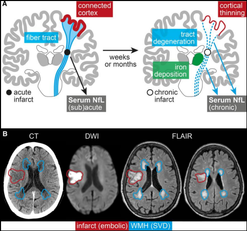
Stroke:
Flow of blood disrupts going to the brain.
Due to the damage of specific areas, there can be damage to cognitive functions such as speech comprehension, thought processing, emotional regulations, etc.
Brain tumor:
Affects cognitive functions.
Can go undiagnosed until there is an issue.
Can lead to personality changes, aggression issues.
Traumatic Brain injuries (TBI):
Can be due to many reasons.
Can lead to movement disorders, speech disorders, impaired cognition.
Ex: Phinease Gage.