Bio Unit 4 - Kognity Notes
B2.2 - Organelles & Compartmentalization
Compartmentalization allows cells to work more effectively than they would be able to without compartments
The metabolic reactions of catabolism must be separated from the metabolic reactions of anabolism
Organelles are subunits of cells that perform specific functions
Compartmentalization: the organization of different functions and processes within specific areas or structures within the cell that are separated by plasma membranes
allows for the development of specialized cell structures such as the chloroplasts and mitochondria
Organelles can be found in both prokaryotic and eukaryotic cells
membrane-bound organelles are only found in eukaryotic cells
allows separation of chemical reactions and other cellular processes ——> allows the cell to increase the rate of chemical reactions
Lysosomes: compartments that break down and recycle waste materials within the cell
The cytoplasm and nucleus are separated in eukaryotic cells to prevent protein synthesis before post-transcriptional modification of mRNA
Isolating transcription in the nucleus and translation in the cytoplasm allows the nucleus to carry out post-transcriptional modification of mRNA before it joins with a ribosome.
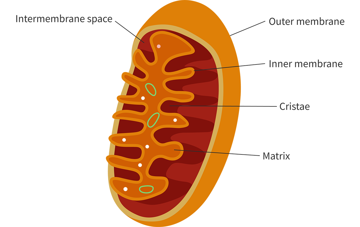
Mitochondria’s main function is to produce ATP through the breakdown of molecules (especially Glucose)
The outer membrane is permeable to many small molecules and ions ——> it contains transport proteins that assist in moving larger molecules into the mitochondria
The inner membrane is highly folded and forms structures called cristae
Cristae: increases the surface area for cellular respiration & increases the efficiency and speed of cell respiration by increasing the number of enzymes available for various reactions
create an enclosed space between the inner membrane called the matrix ——> the matrix space contains a lot of enzymes and other molecules in high concentration
Intermembrane Space: small space between the inner membrane and outer membrane of mitochondria.
Chloroplasts contains three distinct membranes: the outer membrane, the inner membrane and thylakoid membranes
This creates three distinct compartmentalised areas: the intermembrane space, the stroma and the thylakoid space
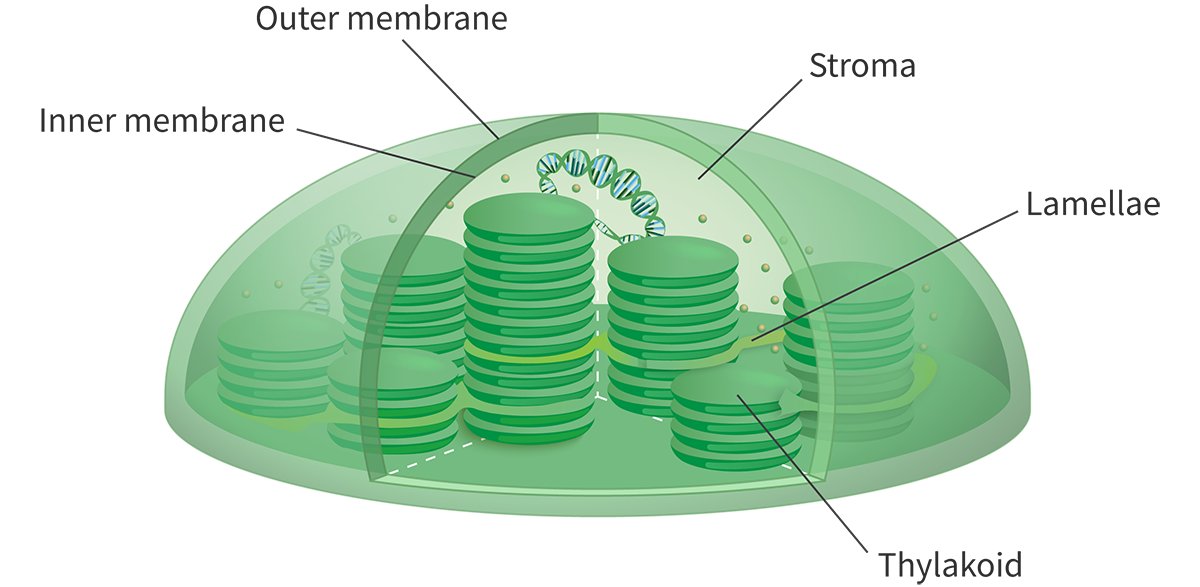
The thylakoid membranes form thylakoids (look like stacked pancakes) ——> where the light-dependent reactions of photosynthesis take place
Many thylakoids together form a stack and this stack-like structure is referred to as a granum (plural, grana)
Lamellae: thylakoid membranes that connect grana
The nucleus contains two membranes (the inner and outer membranes) ——> have several essential functions that regulate the cell’s activities
outer membrane has ribosomes attached to it and is continuous, joined with the rough endoplasmic reticulum
double membrane acts as a barrier between the genetic material inside the nucleus and the rest of the cell
Molecules enter and exit through the nuclear pores of the inner membrane
Nuclear pores: integral proteins that serve as channel proteins that also regulate mRNA leaving the nucleus for the rough endoplasmic reticulum or free ribosomes
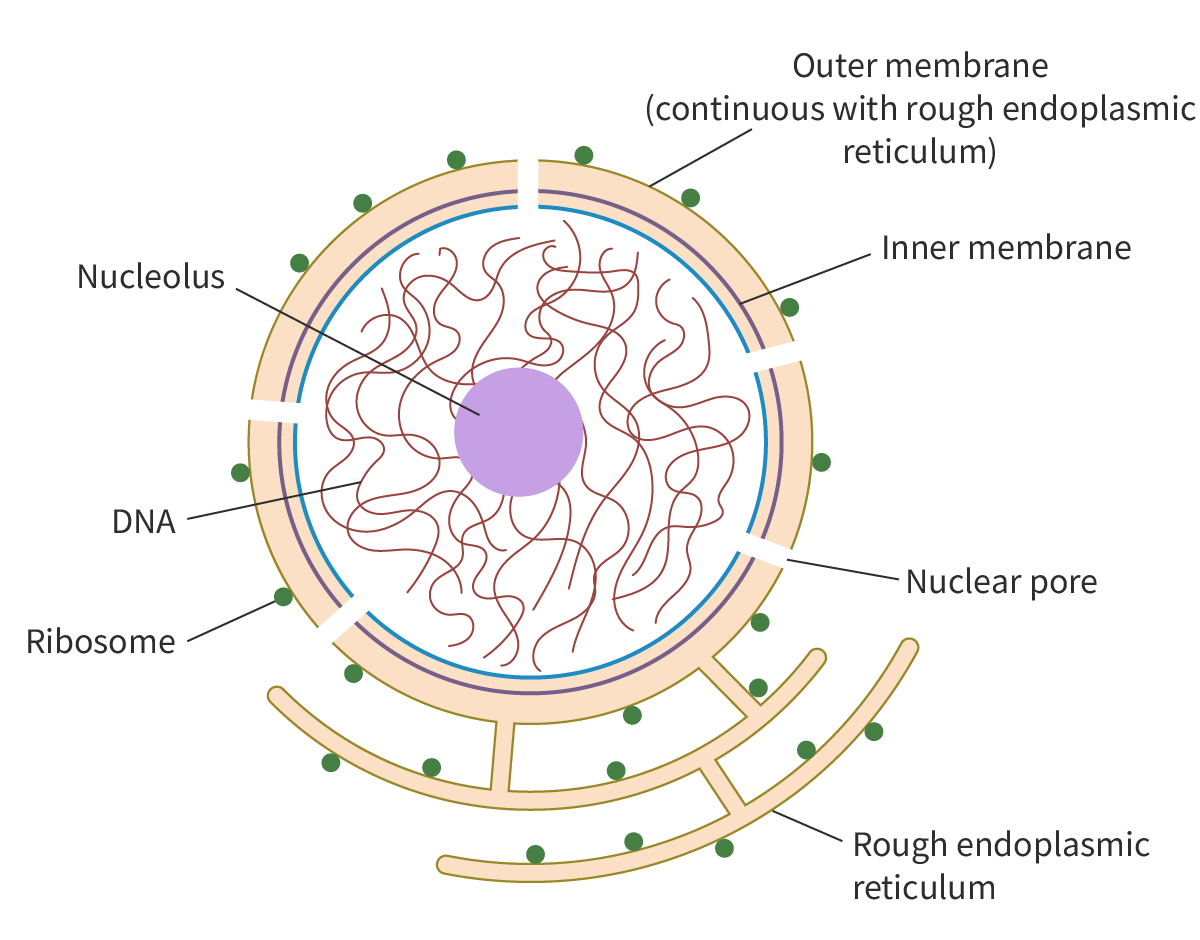
In cell division, the nuclear membrane reforms with the help of many smaller vesicles
Ribosomes, the endoplasmic reticulum, Golgi apparatus and vesicles play a key role in moving proteins around the cell
Ribosomes: translate the mRNA from the nucleus into proteins (can be bound or free.)
If the ribosome is joined to the ER, it is referred to as a bound ribosome, and the endoplasmic reticulum is referred to as the rough ER (RER)
If there are no ribosomes on the surface of the ER, it is referred to as smooth ER (SER)
If the ribosome is located in the cytoplasm, it is referred to as a free ribosome
In bound ribosomes, the ribosome is bound to the cytosolic side of the RER and the proteins that it produces end up inside the RER ——> exported for use outside of the cell
there are more bound ribosomes in the cell than free ribosomes
ER Signal Sequence: a short sequence on a protein that directs the protein towards the endoplasmic reticulum
Golgi Apparatus: a stack of flattened, membrane-bound sacs that are organised into cis, medial, and trans compartments (involved in sorting & modifying proteins)
cis: newly modified proteins
medial: proteins destined for use w/in the cell
trans: proteins destined for export outside of the cell ——> packaged into vesicles for secretion
Vesicles: small, membrane-bound cell structures that play a key role in cellular processes such as the transport and storage of materials
Clathrin: protein involved in forming vesicles and transporting cargo from the Golgi apparatus
D2.3 - Osmosis
Water is known as the “universal solvent” because of its ability to dissolve so many other molecules (solutes)
When an ionic compound is introduced into water, the slightly positively charged hydrogen atoms of water will be attracted to the negative ions and the slightly negatively charged oxygen atoms will be attracted to the positively charged ions
water molecules surround the ions, creating hydration shells —> presence of hydration shells leads to the separation of solute particles and their uniform distribution throughout the solution, a process called dissolution
Hydration Shells: the water molecules surrounding and forming hydrogen bonds with dissolved ions in a solution
Dissolution: The process of solute particles moving and becoming evenly spread out in a solvent
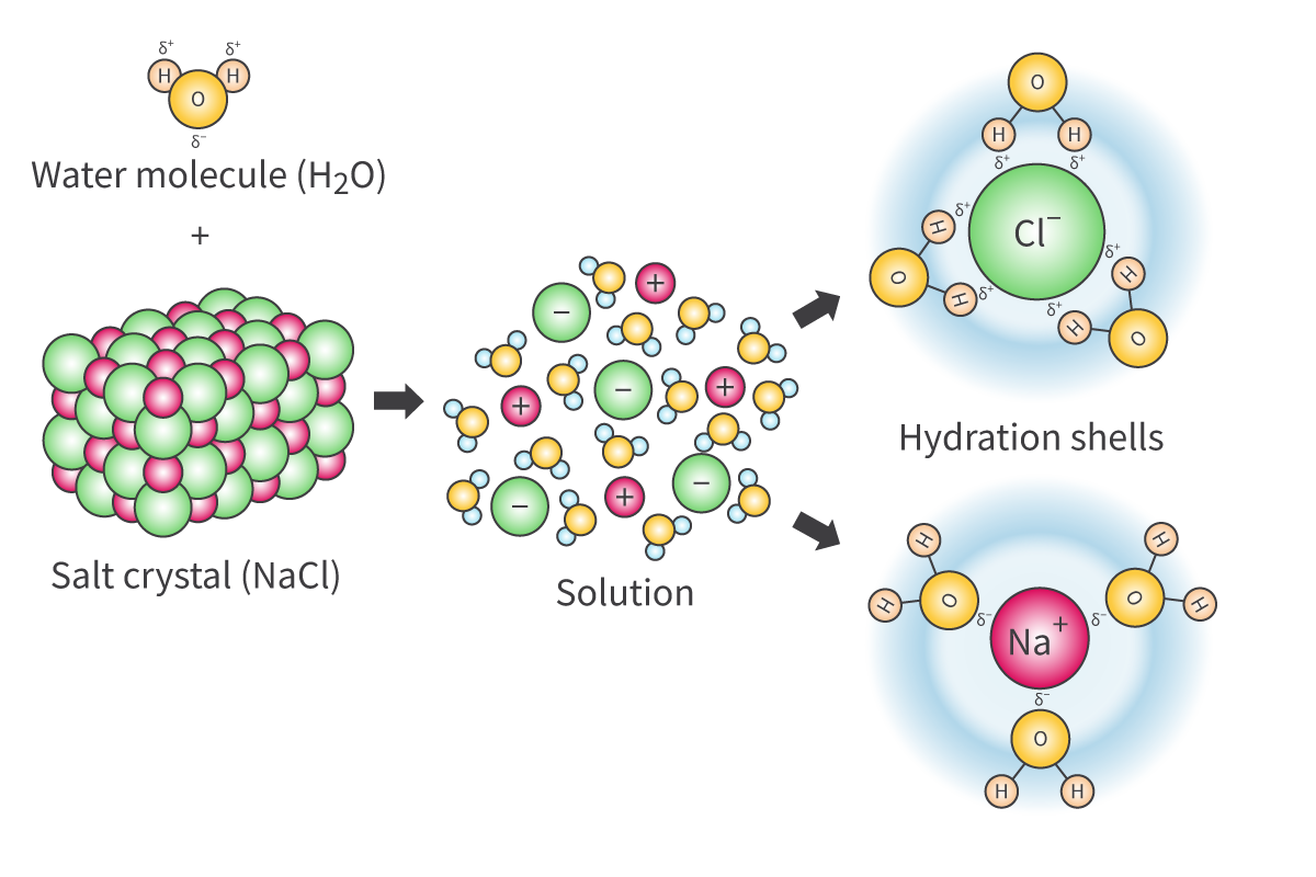
Solution: the homogenous (evenly distributed) mixture formed when a solute is dissolved in a solvent
The attraction between water and dissolved solutes causes water to move from lower solute concentration to regions of higher solute concentration
Water will move across the membrane by osmosis until both solutions have equal solute concentrations, resulting in a state of dynamic equilibrium
Hypertonic: higher solute concentration
Hypotonic: lower solute concentration
Water flows from the hypotonic solution to the hypertonic solution until they the solutions become isotonic (same solute concentration)
Osmosis: movement of water molecules across a selectively permeable membrane from an area of lower solute concentration to an area of higher solute concentration.
specific type of diffusion —> general process of particle movement from an area of higher concentration to an area of lower concentration
In hypertonic solution: net movement of water out of the cell —> cell shrinking
Plant cell: Plasmolyze
Animal Cell: Crenate
In hypotonic solution: net movement of water into the cell —> cell swells
Plant Cell: Turgid —> Turgid Pressure (normal)
Animal Cell: Lyse (burst)
In isotonic solution: no net movement —> equal movement into/out of the cell (dynamic equilibrium)
Plant cell: flaccid
Animal cell: normal
PPTF & ACLN
hyper, hypo, iso
Plant Tissue:
in hyper: loses water —> loss in length & mass
in hypo: gains water —> increase in length & mass
Standard Deviation: measure of the variability (spread) of a data set relative to the mean within a data set.
high standard deviation —> higher variability
low standard deviation —> lower variability in the data set
Standard Error: measure of the variability (spread) between multiple data sets —> used to determine how precise the data are
data set with a low standard error has a higher precision
data set with a high standard error has lower precision
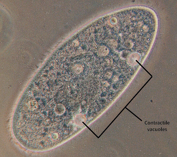
Contractile Vacuoles: Paramecium & Amoeba (freshwater unicellular organisms) use these specialized structures to survive their hypotonic environments
expel water from the cell to maintain a correct intracellular solute concentration & prevent cellular bursting
Homeostasis
multicellular organisms require isotonicity
Water Movement in Plant Cells:
Turgor Pressure: (in plant cells; in hypotonic solution) is exerted by the cytoplasm against the rigid cell wall
the cell wall prevents the cell from bursting and ensures the cell maintains its shape —> turgid
In hyper solution: water exits the cell & the membrane shrinks away from the cell wall (loss of turgor pressure) —> plasmolysis
Organs intended for transplants must be submerged in isotonic solutions to prevent damage to other organs & cells in the body
B2.1 - Membranes & Membrane Transport
Membranes are composed of:
Lipids —> like phospholipids, glycolipids and steroids
Proteins
small amounts of carbohydrates —> in the form of glycolipids and glycoproteins
The hydrocarbon tails of both layers extend inward to form a continuous hydrophobic interior —> important role in determining the permeability of the membrane
Simple diffusion: movement of molecules of a substance down a concentration gradient
one of the easiest ways for molecules to move across the membrane is by simple diffusion.
small polar molecules like water diffuse through the cell membrane, BUT simple diffusion alone cannot explain the rapid diffusion of large amounts of water
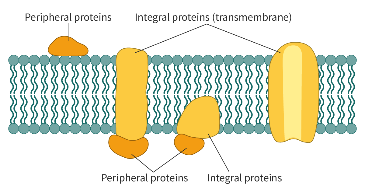
Two Types of Membrane Proteins: Integral proteins & Peripheral proteins
Integral Membrane Proteins: embedded in the phospholipid bilayer
transmembrane proteins —> amphipathic
Peripheral Proteins: found on the surface of the membrane and interact only with the hydrophilic regions of the integral proteins & the hydrophilic heads of the phospholipids
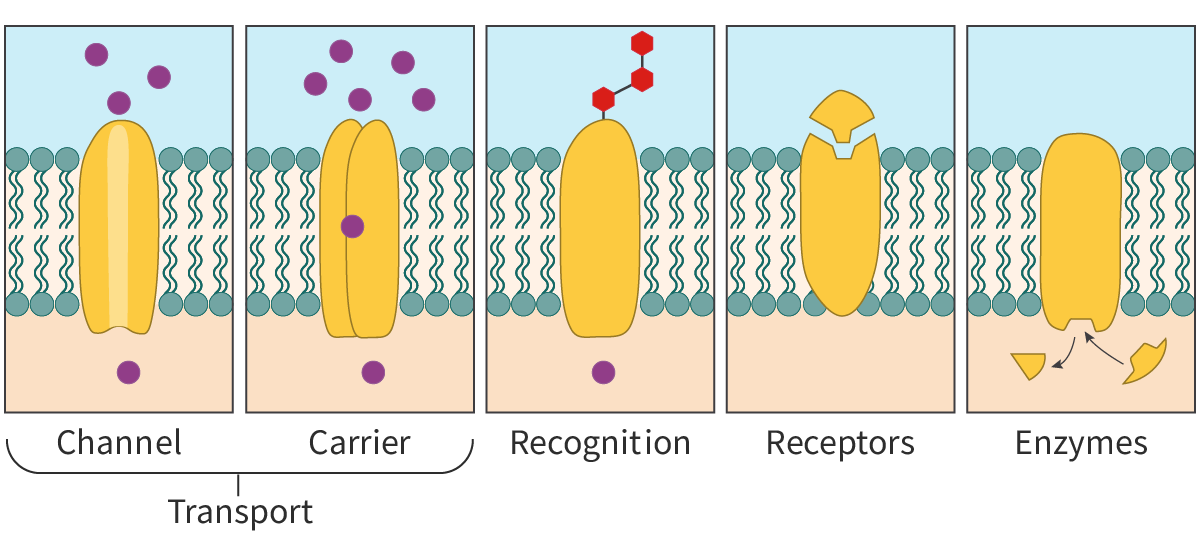
Functions of Membrane Proteins:
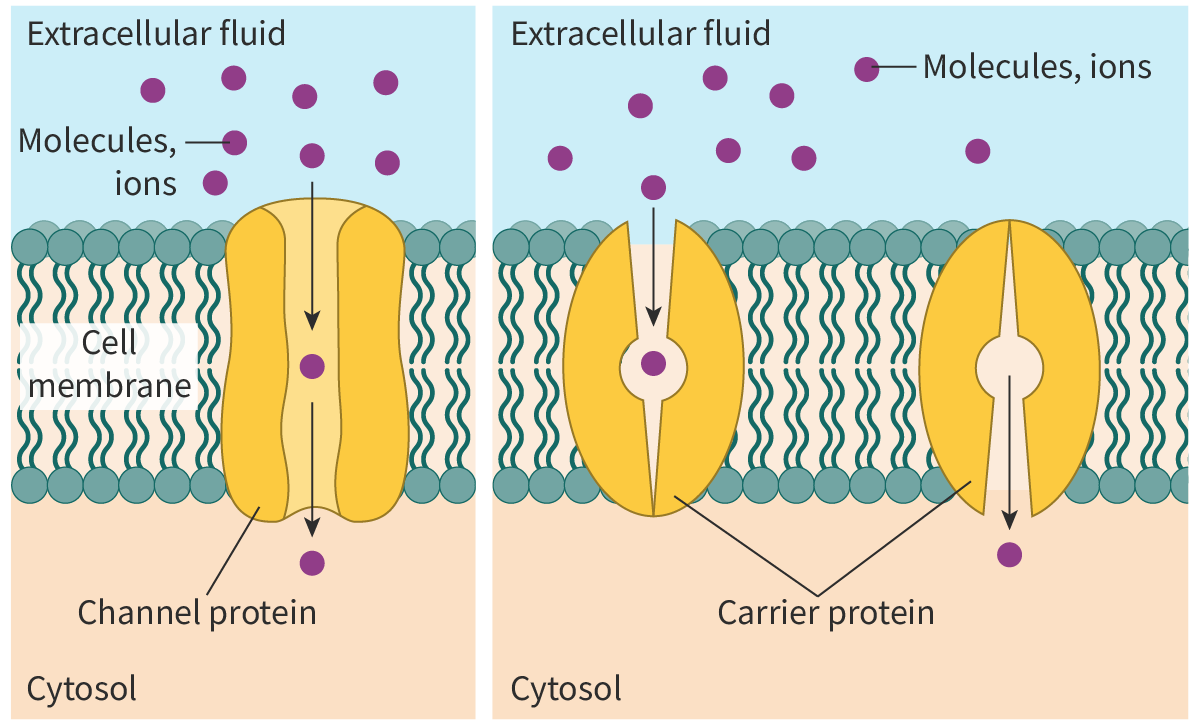
Transport proteins: membrane proteins facilitate the movement of molecules in and out of the cell
Channel proteins & Carrier proteins
Channel proteins: transmembrane proteins that form channels or pores for the passage of molecules
Carrier proteins: undergo a conformational change to transfer the molecules from one side of the membrane to the other
Recognition: membrane proteins help in cell–cell recognition acting as ‘name tags’ for the cells
essential in the functioning of the immune system —> helps to distinguish between self and non-self cells
Receptors: membrane proteins act as receptors for chemical signals & are binding sites for molecules like hormones and neurotransmitters
binding of these molecules triggers a chain of intracellular reactions
Enzymes: membrane proteins show enzymatic activity and catalyse reactions
EX: glucose-6-phosphatase is a membrane-bound enzyme found in the endoplasmic reticulum
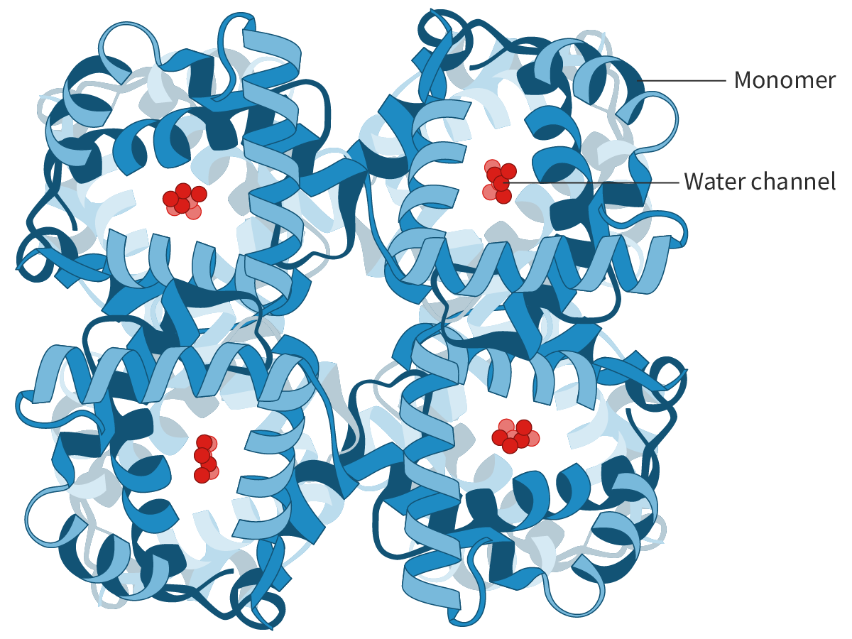
Aquaporins: channel proteins that facilitate the transport of water across cell membranes
Tetrameric Protein: quaternary protein w/ four subunits
each subunit has a water channel —> an aquaporin molecule has four identical water channels
the water channels are lined with specific hydrophilic side chains (of amino acid residues) which allows the passage of water molecules but not of ions
Facilitated Diffusion:
Facilitated Diffusion: the movement of a molecule down its concentration gradient with the help of specialised transport proteins (carrier and channel proteins) across the cell membrane
Most channels open or close in response to specific stimuli, such as:
changes in voltage (change in membrane potential) across the membrane (voltage-gated channels)
binding of small molecules to the channel proteins (ligand-gated channels)
mechanical forces like pressure
Carrier Proteins are transmembrane transport proteins that play an important role in facilitated diffusion.
the carrier protein binds to the solute molecules (molecules to be transported), undergoes a conformational change and transfers the molecules to the other side of the membrane
Carrier proteins have sites specific for the solute or class of solutes to be transported —> highly specific
In the inactivated state: an inactivation particle blocks the channel pore and prevents the movement of ions
Open conducting state
‘Resting’ State where the gate remains closed until it is activated by a change in membrane potential.
Active Transport:
when ions or molecules have to be moved against their concentration gradient they need to be pumped.
Active Transport: the net movement of particles through a cell membrane from a region of lower concentration to a region of higher concentration —> using energy from respiration (ATP)
exergonic reaction of breaking down ATP —> energy released
Two main types of active transport: Direct & Indirect
Direct active transport: the energy released by an exergonic reaction like the breakdown of ATP is used to directly transport molecules across the cell membrane
as energy is derived by the hydrolysis of ATP —> these transport proteins are called ATPases or ATPase pumps
Indirect active transport or cotransport: the movement of one solute down its concentration gradient drives the movement of the second solute against its concentration gradient
The selective permeability of the membrane is due to facilitated diffusion and active transport
simple diffusion is not selective —> facilitated diffusion/active transport involve transport proteins that exhibit selectivity
Active Transport uses Carrier Proteins
Channel Proteins allow movement down a concentration gradient
Glycolipids: the covalent bonding of carbohydrates to lipids
Amphipathic
The carbohydrate groups of these molecules are polar and extend into the extracellular environment
the non-polar lipid component lies embedded in the bilayer.
Glycoproteins: The covalent bonding of oligosaccharides (short carbohydrate chains) to the protein molecules
Functions of glycolipids and glycoproteins:
Cell recognition: they act as ‘markers’ on the cell surface and help cells of the body recognise each other —> help cells of the immune system to recognise foreign cells
Cell adhesion: Both glycolipids and glycoproteins help cells to attach and bind to other cells to form tissues
Cell-adhesion molecules or CAMs are cell-surface glycoproteins that play an important role in cell adhesion.
Cell signalling: they act as receptors for enzymes and other molecules helping in cell signalling (receiving and transmitting chemical signals)
Glycocalyx: sticky layer formed by the carbohydrate groups of the glycolipids and glycoproteins that protrude from the cell surface —> the glycocalyx in addition to its roles in cell signalling, cell adhesion and cell–cell recognition, helps in protecting the cell surface.
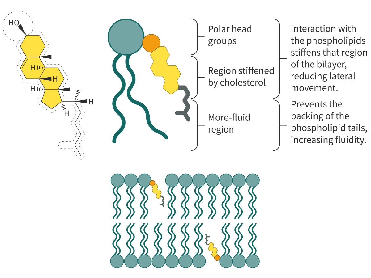
Cholesterol: amphipathic steroid —> the hydrophobic region comprises four steroid rings and a hydrocarbon side chain & the hydrophilic region is a polar hydroxyl group
cholesterol molecules interact with the corresponding hydrophilic and hydrophobic regions of adjacent phospholipid molecules —> hold the phospholipid molecules together
Formation of Vesicles:
The two bulk transport mechanisms utilised by cells are exocytosis and endocytosis —> active transport mechanisms (both processes require energy)
Endocytosis: particles are moved into the cell —> cell membrane progressively invaginates and engulfs the particles —> the membrane then pinches off to form a vesicle with the ingested particles
Phagocytosis: ingestion of large solid particles (“cellular eating”)
Pinocytosis: ingestion of liquids (“cellular drinking”)
Exocytosis: bulk transport of material to be secreted or excreted out of the cell
the material to be removed from the cell is enclosed in vesicles —> vesicles move to the plasma membrane and fuse with it, discharging its contents to the exterior
Gated ion channels in neurons:
Ion Channels: transmembrane proteins present on the cell membrane that form pores for the movement of ions across the membrane, down their concentration gradient
channels are usually gated —> open or close in response to stimuli:
voltage-gated channels
ligand-gated channels
mechanically gated channels (respond to mechanical cues such as sound waves and vibrations)
Neurons: cells of the nervous system that transmit information in the form of electrical impulses
Electrochemical gradient: the difference in chemical concentration and charge of ions across a membrane
Action potential and the role of the voltage-gated channels in neurons:
A stimulus causes the ‘activation gate’ of the voltage-gated sodium channels to open —> Sodium ions diffuse rapidly into the neuron
causes the interior of the neuron to be more positively charged than the exterior (depolarisation) —> generating an action potential
the action potential travels down the nerve fibre
same process repeats with voltage-gated potassium channels opening (potassium diffuses out —> repolarisation & neuron becomes negatively charged)
gated channels would open or close only when the voltage reaches a certain minimum value called the threshold value
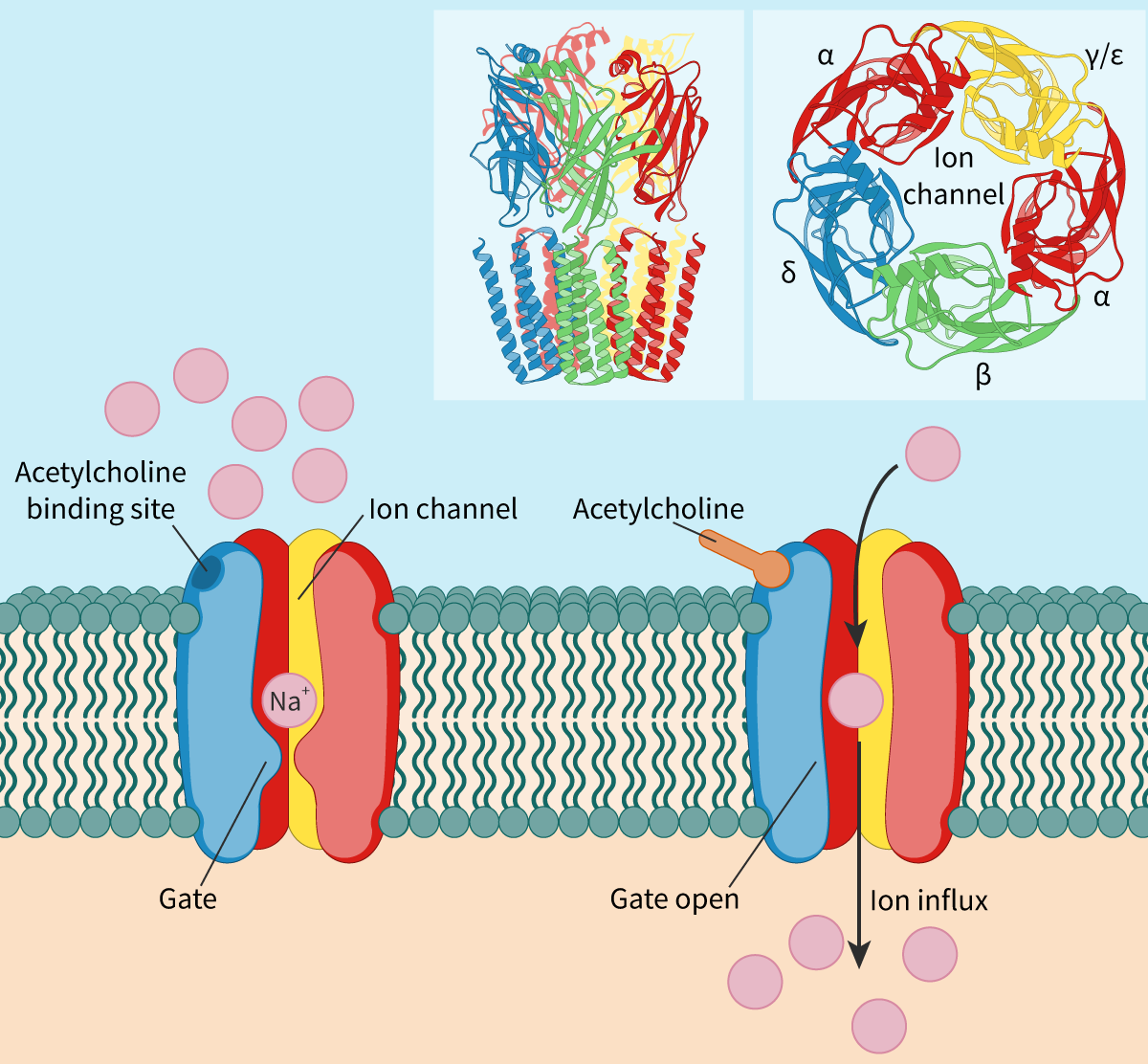
Nicotinic acetylcholine receptor: a ligand-gated channel:
When the ligand is a neurotransmitter, the ion channel is called a neurotransmitter-gated ion channel
neurotransmitter: A signaling molecule released by a neuron
Acetylcholine (neurotransmitter)
Nicotinic acetylcholine receptors (nAchR) (ligand-gated ion channels)
the binding of acetylcholine molecules results in a conformational change that opens the channel
depolarization/repolarization occurs w/ the entering & exiting of sodium ions & potassium ions (respectively)
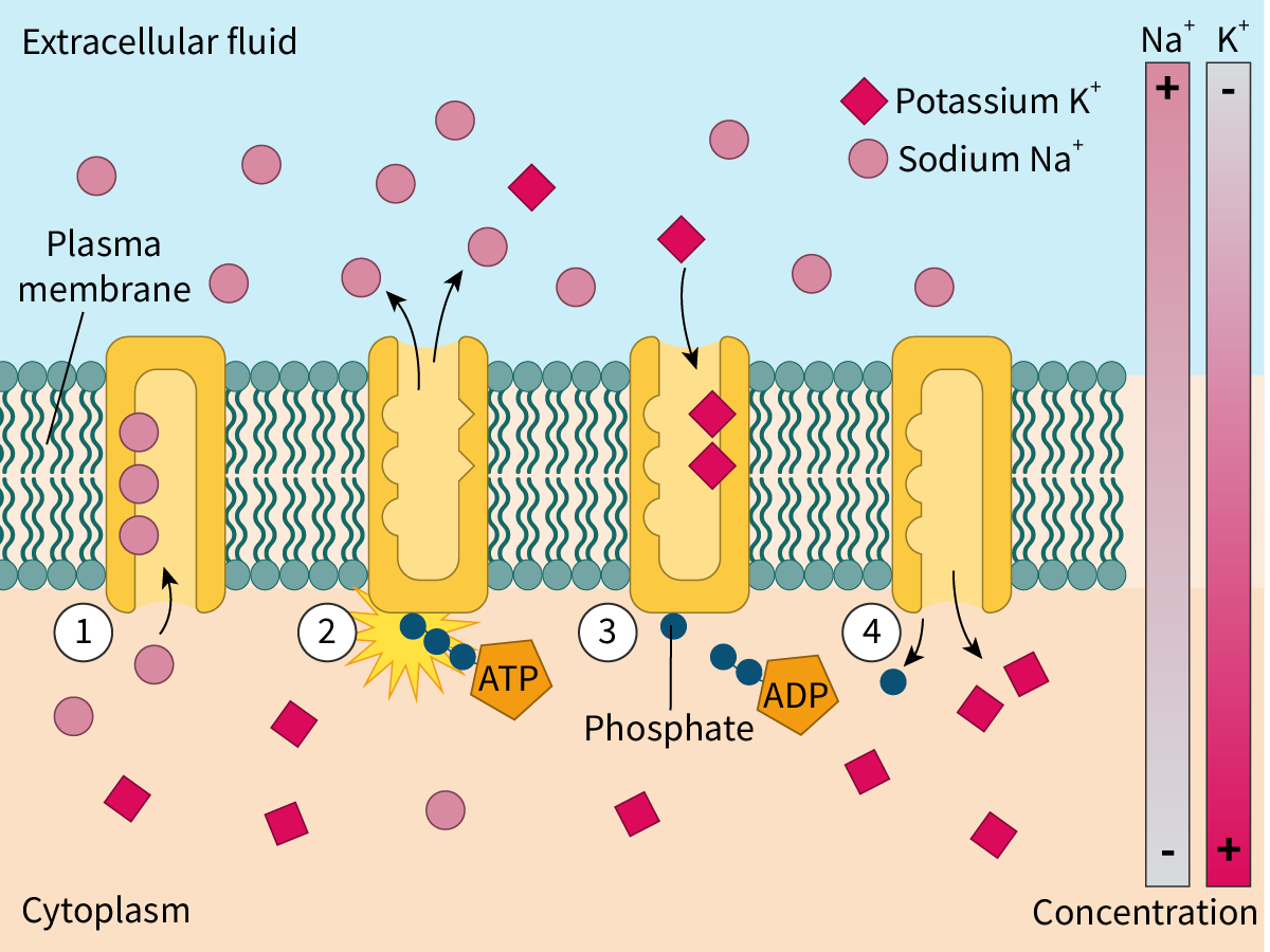
Direct active transport —> the sodium-potassium pump:
NaK Pump: found in the cell membranes of all animal cells and involves active transport
The Na+/K+ pump is an enzyme that generates energy by the breakdown of ATP
hydrolysis
also called ATPase
the energy released in the process is used to drive the transport of sodium and potassium ions against their concentration gradient
Most animal cells have a high intracellular concentration of K+
active transport of K+ ions into the cell
& Low intracellular concentration of Na+
active transport of Na+ ions out of the cell
The Na+/K+ pump uses ATP as an energy source —> direct active transport
Indirect Active Transport:
source of energy is NOT ATP
Indirect Active Transport: Mechanism involves the transport of two solutes (ions or molecules) —> one solute is transported down its concentration gradient & the other is transported against its concentration gradient
the favorable movement (down the concentration gradient) is coupled with an unfavorable movement (against the concentration gradient) and drives the latter
The Na+/K+ pumps are transmembrane pumps with three binding sites for sodium and two for potassium
1.) Initially, the Na+/K+ pump is open to the inside of the cell in a way that the sodium ions bind to all three of its binding sites
2.) The binding of sodium ions triggers the hydrolysis of ATP to ADP and a phosphate group —> Phosphate group attaches to the pump resulting in a conformational change —> The pump now opens to the exterior releasing the sodium ions
3.) At the same time, potassium ions attach to both binding sites, which causes the phosphate group to detach from the pump
4.) The pump undergoes a conformational change to regain its original form and once again opens to the interior of the cell
CAM’s and Cell Adhesion:
Adhesion: the binding of cells with each other to form tissues
Cell-adhesion molecules (CAMs): these glycoproteins mediate the binding of cells with other adjacent cells or with the extracellular matrix
play a crucial role in cell adhesion
different forms of CAM’s include:
cadherins
integrins
selectins
immunoglobulin super family
Cell junctions: connect cells to each other allowing intracellular transport and communication
play important roles in cell proliferation, cell migration and prevent unregulated movement of materials between cells
CAMs are essential for the formation of cell junctions.
Types of Cell Junctions in Animals:
1.) Adhesive Junctions: in epithelial cells and cardiac cells —> they facilitate cell-cell adhesion in tissues to ensure structural stability and allow the cells to withstand mechanical stress
AKA anchoring junctions
EX: Desmosomes
2.) Tight junctions: present in epithelial cells —> they form a tight seal between two neighboring cells and act as occluding junctions
this barrier prevents the unregulated movement of molecules across the barrier
3.) Gap junctions: found in several cell types throughout the body —> they are intracellular channels physically connecting neighboring cells for the movement of molecules
they help in the cell-cell transfer of small molecules —> AKA communicating junctions
The type of CAMs used depends on the types of cell junctions
B2.3 - What Determines Cell Size?
The human body’s cells range from the smallest being about 7.5 µm to significantly larger ones like the human egg cell or ovum (150 µm)
the egg cell is one of the largest in the human body, while the sperm cell is one of the smallest
The structure (size) of cells is linked directly to their function
The spherical human egg cell has evolved to be large and highly specialized so that it contains all the nutrient materials needed for the early development of the embryo.
The sperm does not need such nutritional content and so remains small
Many human cells are approximately spherical (but not all)
Some specialized nerve cells are incredibly long.
Neurons (in the sciatic nerve) are the longest in the human body —> their axons can exceed 1 m
these elongated cells have evolved as part of the mechanism for communication between the spinal cord and other more distant parts of the body
Red Blood Cells are some of the smallest cells in the human body
DON’T have a nucleus, which leaves space for packing in more hemoglobin for binding oxygen to transport around the body
have a highly flexible membrane that allows them to be repeatedly deformed and spring back in shape —> important as they move through the circulatory system.
AKA erythrocytes
White Blood Cells (less common) are larger than RBCs
they have nuclei of various shapes (unlike RBCs) which can aid in their identification
they can move in an amoeboid way (like an Amoeba) towards sites of infection and can squeeze out of blood vessels into surrounding tissues
Cells have control mechanisms involving cell surface receptors and growth factors in the surrounding environment to ensure that the maximum size of any given cell type is consistent within an organism
The larger the cell becomes, the more the SA:V ratio reduces
EX: think of it as a balloon being pumped up —> As the volume increases, the surface area does not increase at the same rate
The size that a cell achieves is a fine balance in being large enough to contain all the necessary organelles for the cell’s particular function without compromising the ability for efficient gas exchange and nutrients across its membrane