Renal scanning
The Kidneys: Anatomy and Physiology
Urinary system and kidney anatomy
Two kidneys do most of the work; ureters carry urine to the bladder, which stores it and empties into the urethra.
Kidneys: 10–12 cm long, 3 cm thick, 5–7 cm wide. Lie posteriorly between T12 and L3, partly protected by ribs 11 and 12, and are retroperitoneal. The right kidney sits slightly lower (liver above). Adrenal glands sit on top of each kidney.
They filter about 150 L of blood per day; each kidney has about 1 million nephrons. Imaging tip: the best view for nuclear medicine renal imaging is posterior.

Ureters
25–30 cm long, thick-walled, retroperitoneal; peristalsis (plus pressure and gravity) moves urine. Diameter varies from 1 to 10 mm. They enter the posterior bladder wall, not the top.
Filling bladder compresses the ureter openings (a physiological valve) to prevent backflow; if it fails, microbes can travel up to infect the kidneys.
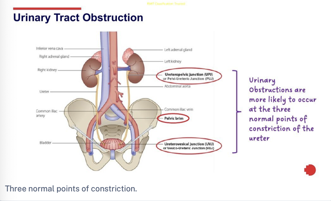
Three normal constrictions of the ureter (the commonest obstruction sites): pelvi-ureteric junction (PUJ), pelvic brim (crossing the iliac vessels), uretero-vesical junction (UVJ).

Layers and internal structure
Structure | Description |
|---|---|
Renal fascia | Outer dense connective tissue; anchors the kidney |
Adipose capsule | Middle fat layer; protects from trauma |
Renal capsule | Inner dense connective tissue; gives the kidney its shape |
Hilum | Entry/exit for renal artery, vein and ureter |
Renal cortex | Outer zone of the parenchyma (functional tissue); about 85% of nephrons sit here |
Renal medulla | Inner zone: renal pyramids and papillae. Cortex between pyramids = renal columns; pyramid + overlying cortex = renal lobe |
Collecting system | Minor calyces → major calyces → renal pelvis → ureter |

Functions of the kidneys
Function | Detail |
|---|---|
Hormones | Calcitriol (active vitamin D; calcium homeostasis; skin → liver → kidney), erythropoietin (red cell production), renin (starts RAAS) |
Blood glucose | Make new glucose (gluconeogenesis) in prolonged fasting, mainly in the proximal tubule |
Blood pH | Excrete variable H⁺ and conserve bicarbonate (HCO₃⁻) buffer |
Ionic composition | Adjust excretion of Na⁺, K⁺, Ca²⁺, Cl⁻, phosphate |
Blood volume and pressure | Conserve or eliminate water; renin–angiotensin–aldosterone system |
Excretion of waste | Metabolic waste, toxins and drugs via filtration and secretion |
Blood osmolarity | Keep close to 300 mOsm/L by regulating solute loss and water |
Nitrogenous wastes: urea and ammonia (amino acid deamination), creatinine (creatine phosphate in muscle), uric acid (nucleic acids), urobilin (haemoglobin). Blood creatinine is used to assess kidney function.
The nephron: the functional unit
Renal corpuscle = glomerulus (capillary ball) + glomerular (Bowman's) capsule. Filtrate enters the capsular space.
Renal tubule, in order: proximal convoluted tubule (PCT) → nephron loop (loop of Henle) → distal convoluted tubule (DCT). Several DCTs drain into one collecting duct, then papillary ducts, then the minor calyces.
The corpuscle and both convoluted tubules are in the cortex; the loop dips into the medulla. 85% are cortical nephrons with short loops, so most work happens in the cortex.
Three functions: (1) glomerular filtration, (2) tubular reabsorption(back to blood), (3) tubular secretion (blood to filtrate).

Blood supply
Kidneys receive 20–25% of resting cardiac output via the renal arteries (renal blood flow = volume per minute).
Renal artery → segmental arteries
→ interlobar arteries (through renal columns)
→ arcuate arteries (around pyramids)
→ cortical radiate arteries
→ afferent arteriole (one per nephron) → glomerulus
→ efferent arteriole → peritubular capillaries (cortex) and vasa recta (medulla)
→ cortical radiate veins → arcuate veins → interlobar veins → single renal vein → inferior vena cava (there are no segmental veins).

Glomerular filtration

Filtration membrane lets water and small solutes through but blocks most plasma proteins and blood cells.
Filtrate: about 180 L/day (men), 150 L/day (women). GFR (filtrate per minute, both kidneys) averages 125 mL/min (men), 105 mL/min (women). About 99% is reabsorbed, so only 1–2 L becomes urine.
Layer | Features |
|---|---|
1. Capillary endothelium | About 50× more porous than ordinary capillaries because of fenestrations: all plasma solutes escape, blood cells cannot |
2. Basement membrane | Gel-like mesh of collagen fibres and glycoproteins (sponge-like): passes most solutes, blocks larger plasma proteins |
3. Epithelium (podocytes) | Octopus-like cells with pedicels that interlock; gaps are filtration slits covered by a slit membrane that allows small molecules (water, glucose, amino acids, urea, ions, very small proteins) and blocks other proteins |
Pressures that drive filtration
Pressure | Effect | Normal value |
|---|---|---|
Glomerular blood hydrostatic pressure (GBHP) | Promotes filtration; high because the efferent arteriole is narrower than the afferent | 55 mmHg |
Capsular hydrostatic pressure (CHP) | Opposes filtration (fluid already in the capsule/tubule) | 15 mmHg |
Blood colloid osmotic pressure (BCOP) | Opposes filtration (plasma proteins such as albumin) | 30 mmHg |
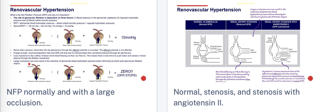
NFP = GBHP − (BCOP + CHP) = 55 − (30 + 15) = 10 mmHg. (The pathology lecture adds that if GBHP falls to about 45 mmHg, NFP reaches zero and filtration stops.)

GFR too high: needed substances move through too fast and are lost in urine. GFR too low: waste stays in the blood longer and nearly all filtrate is reabsorbed, so wastes are not excreted.
How GFR is regulated
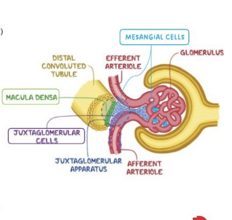
GFR is changed by adjusting blood flow in and out of the glomerulus or the capillary surface area (mesangial cells).
Juxtaglomerular (JG) apparatus (between DCT and afferent arteriole): macula densa cells (DCT wall), juxtaglomerular (granular) cells(afferent arteriole wall), extraglomerular mesangial cells.
Mechanism | How it works |
|---|---|
1. Renal autoregulation | Keeps renal blood flow and GFR constant at mean arterial pressure 80–180 mmHg. Two parts working together: |
2. Neural | Sympathetic fibres release norepinephrine (α1 receptors, plentiful on afferent arterioles) → vasoconstriction → lower GFR (e.g. intense exercise, hypovolaemic shock); conserves blood volume and diverts blood elsewhere |
3a. Angiotensin II | Low blood pressure/volume → renin → angiotensin II: constricts afferent and efferent arterioles and contracts mesangial cells (less filtration area) → lower GFR, less urine, conserves volume |
3b. ANP | Large rise in blood volume stretches the atria → ANP relaxes mesangial cells → more surface area → higher GFR, more urine, lower volume and pressure |
Tubular reabsorption and secretion
Reabsorption: selective return of about 99% of filtrate to blood. Occurs all along the tubule and collecting duct, but the PCT does most. Reabsorbed: glucose, amino acids, urea, Na⁺, K⁺, Ca²⁺, Cl⁻, HCO₃⁻, phosphate, most small proteins/peptides (by pinocytosis). Substances go into the interstitial space, then into peritubular capillaries or vasa recta.
Route | How |
|---|---|
Paracellular(passive) | Between tubule cells through tight junctions; PCT junctions are leakiest. Large ions/complexes and many wastes cannot pass |
Transcellular(active) | Through tubule cells using channels and transporters: Na⁺/K⁺ ATPase, sodium–glucose cotransporters, glucose transporters, Cl⁻, Ca²⁺ and phosphate transporters |

Solute reabsorption drives water reabsorption:
Obligatory (~90%) | Facultative (~10%) | |
|---|---|---|
How | Osmosis following solutes; keeps blood osmolarity constant | Controlled by ADH (vasopressin)according to body needs |
Where | Mainly PCT and loop of Henle (most water-permeable) | Last part of DCT and the collecting duct (principal cells) |
Mechanism | Water follows reabsorbed solutes | ADH (from posterior pituitary when osmolarity rises or volume falls) inserts aquaporin-2 into the apical membrane |

Tubular secretion
Active, one-way transport of wastes, drugs and excess ions from peritubular capillaries into the tubular fluid. Removes substances that did not filter (certain drugs, protein-bound waste, large molecules).
Mainly in the PCT, DCT and collecting ducts; organic anion and cation transporters (mostly PCT) secrete organic acids and bases, including drugs.
Hormonal control and the RAAS
Hormone | Trigger | Effect |
|---|---|---|
Angiotensin II | Low blood volume or pressure (via renin) | ↑ Na⁺/H⁺ antiporter activity in the PCT → more Na⁺ and water reabsorbed; thirst and sodium appetite; ↑ blood volume and pressure |
Aldosterone(adrenal) | ↑ angiotensin II; ↑ plasma K⁺ | ↑ Na⁺/K⁺ pumps and Na⁺ channels in principal cells of DCT/collecting duct → ↑ K⁺ secretion, ↑ Na⁺ and water reabsorption |
ADH | ↑ blood osmolarity or ↓ volume | Aquaporin-2 inserted in DCT/collecting duct → ↑ water reabsorption |
ANP | Large ↑ in blood volume (atrial stretch) | Suppresses Na⁺ and water reabsorption (PCT, collecting duct); inhibits aldosterone and ADH → ↑ urine, ↓ volume and pressure |
Renin release is triggered by
β1-adrenergic receptors on JG cells (renal sympathetic activity, "fight or flight").
Intrarenal baroreceptors: JG cells sense a fall in afferent arteriole pressure (reflects systemic pressure, except in renal artery stenosis).
Macula densa: very low Na⁺/Cl⁻ delivery stimulates renin; higher delivery reduces it.
RAAS steps
Reduced renal blood flow/low pressure (dehydration, haemorrhage) → JG cells release renin.
Renin converts angiotensinogen (from the liver) to angiotensin I.
ACE (angiotensin-converting enzyme, mainly in lung capillaries) converts it to angiotensin II.
Angiotensin II: systemic vasoconstriction (↑ pressure); PCT Na⁺/H⁺ antiporters (↑ Na⁺ and water reabsorption); lowers GFR (afferent/efferent constriction, mesangial contraction); stimulates aldosterone.
Aldosterone: ↑ Na⁺ reabsorption and K⁺ secretion in the distal nephron, so more water follows. Rising osmolarity also triggers ADH → more water reabsorbed.
Blood volume and pressure return toward normal.

Revision checklist
Kidney position T12–L3, retroperitoneal; layers: fascia, adipose capsule, renal capsule; cortex, medulla, collecting system.
Nephron order: corpuscle → PCT → loop of Henle → DCT → collecting duct; 85% cortical.
Blood path: renal artery → segmental → interlobar → arcuate → cortical radiate → afferent → glomerulus → efferent → peritubular/vasa recta → veins (no segmental veins).
Three filtration layers: endothelium (fenestrations), basement membrane, podocyte slit membrane.
NFP = GBHP − (BCOP + CHP) = 10 mmHg; GFR 125 (M) / 105 (F) mL/min; ~99% reabsorbed.
GFR control: autoregulation (myogenic + tubuloglomerular feedback, 80–180 mmHg), neural (norepinephrine), hormonal (angiotensin II lowers, ANP raises).
Reabsorption: PCT most; paracellular vs transcellular; water 90% obligatory, 10% facultative (ADH, aquaporin-2). Secretion: PCT organic anion/cation transporters.
Hormones: angiotensin II, aldosterone, ADH, ANP; RAAS: renin → angiotensin I → (ACE) → angiotensin II → aldosterone.
The Kidneys: Pathologies
Congenital renal anomalies
Faulty formation of the urinary system in utero. Several have little effect on function but raise the risk of infection, stones or obstruction.
Anomaly | What it is | Key points |
|---|---|---|
Renal agenesis | Inadequate formation of one or both kidneys | Single or no kidney at birth |
Renal hypoplasia | Abnormally small kidney(s) with fewer nephrons but normal form | |
Renal dysplasia | Abnormally formed kidney(s) | Function often impaired |
Horseshoe kidney | Kidneys joined into one arched kidney | Function often normal; risk of UTI, stones, obstruction |
Ureteral obstruction | Malformed ureter causing a blockage | Often at the PUJ or UVJ |
Vesicoureteral reflux | Ureters attach to the bladder incorrectly, so urine flows back up (valve fails) | Recurrent UTIs, renal cortical scarring, renal failure if severe |
Duplex collecting system | Two ureters draining one kidney | Function often normal; UTI risk |

Urinary tract obstruction
Causes: congenital abnormality, urethral stricture, kidney stone or enlarged prostate.
Urethral stricture: scar tissue (from chronic inflammation or injury) narrows the urethra.
Prostate enlargement: the male urethra passes through the prostate, so enlargement can constrict it and cause urinary retention.
Kidney stones (renal calculi): calcium, uric acid and other crystal-forming substances crystallise at high urine concentrations and solidify into insoluble stones.
Obstruction is most likely at the three normal ureteric constrictions: PUJ, pelvic brim, UVJ.
Hydronephrosis = swelling of the kidney from dilation of the renal pelvis and calyces due to obstructed urine flow. Severe cases can damage the kidneys and lead to renal failure.
Urinary tract infection (UTI)
Infection of any part of the urinary system. Most commonly E. coli(passed from anus to urethra); also mycoplasma and chlamydia (sexually transmitted). More common in females because the urethra is shorter.
Symptoms: painful or burning urination, urgent and frequent urination, feeling of a full bladder after voiding, smelly urine, low back pain, bedwetting.
Condition | Site inflamed |
|---|---|
Urethritis | Urethra |
Cystitis | Bladder |
Pyelonephritis | Kidney |
Chronic cystitis/urethritis can disturb bladder or urethral function and lead to vesicoureteral reflux (cortical scarring, renal failure if severe).
Chronic pyelonephritis forms scar tissue that severely impairs kidney function.
Renovascular hypertension
Caused by progressive narrowing of one or both renal arteries (renal artery stenosis), usually from atherosclerosis (plaque of cholesterol, fat, blood cells); also fibromuscular dysplasia, dissection or emboli.
One of the commonest causes of secondary hypertension; mostly older patients with systemic atherosclerosis and other risks (diabetes, hypertension, smoking, peripheral vascular and coronary disease).
Why it happens: the stenosis lowers pressure and flow in the afferent arteriole, but systemic pressure is normal. The juxtaglomerular cells assume the whole body is hypotensive, so they release renin → angiotensin II → systemic vasoconstriction and water retention → systemic hypertension.

Effect on GFR: why angiotensin II can "maintain" it
NFP = GBHP − (BCOP + CHP); normally 55 − (30 + 15) = 10 mmHg.
Stenosis blocks inflow through the afferent arteriole while the efferent arteriole is unaffected, so more blood leaves than enters. Glomerular pressure and GFR fall. If GBHP drops to about 45 mmHg or below, NFP is zero and filtration stops.
Angiotensin II constricts the afferent and efferent arterioles, restoring glomerular pressure and helping maintain GFR (although overall renal blood flow is still reduced). Elsewhere in the lecture it lowers GFR; here it protects it because the starting problem is low inflow.
The restriction in flow to the affected kidney remains. Chronic ischaemia leads to kidney atrophy (shrunken, abnormal kidney), loss of nephrons and fibrosis (ischaemic nephropathy).
Renal failure
Glomerular filtration is reduced or stops. Wastes stay in the blood and nearly all filtrate may be reabsorbed.
Problem | Cause |
|---|---|
Oedema | Salt and water retention (fluid restriction may be needed) |
Metabolic acidosis | Cannot excrete acids |
Raised urea and creatinine | Impaired excretion of metabolic waste |
High potassium | Cannot regulate ions; can cause cardiac arrest |
Anaemia | Less erythropoietin |
Low calcium absorption | Cannot convert vitamin D to calcitriol |
Acute renal failure (acute kidney injury, AKI) | Chronic renal failure | |
|---|---|---|
Course | Abrupt decrease or stop in kidney function; urine flow suppressed; most recover fully | Progressive, irreversibledecline in GFR |
Causes | Low blood volume (e.g. haemorrhage), reduced cardiac output, kidney stones, CT/fluoroscopy contrast, illness, trauma, renal artery stenosis, drugs (NSAIDs, antibiotics, chemotherapy) | Untreated acute failure, chronic pyelonephritis, diabetic nephropathy, polycystic kidney disease, traumatic tissue loss |
Chronic renal failure by cause
Type | Mechanism | Examples |
|---|---|---|
Pre-renal | Prolonged reduced blood flow to the kidneys | Chronic dehydration, heart failure, bilateral renal artery stenosis, some long-term medications |
Renal (intrinsic) | Direct damage to kidney tissue | Glomerulonephritis (glomerular inflammation; often an allergic reaction to streptococcal toxins), diabetic nephropathy (high blood sugar damages capillaries), hypertension |
Post-renal | Obstruction to urine outflow | Kidney stones, tumours, enlarged prostate |
Stages
Stage (3-stage model) | What happens |
|---|---|
1. Diminished renal reserve | Up to about 75% of nephrons lost; usually no symptoms as remaining nephrons enlarge and overwork |
2. Renal insufficiency | More than 75% lost; GFR falls; urea and creatinine rise |
3. End-stage renal disease (ESRD) | About 90% lost; GFR 10–15% of normal; oliguria; oedema; strict fluid restriction |
Stage (GFR-based) | GFR (mL/min) |
|---|---|
1: damage, normal GFR | > 90 |
2: mild reduction | 60–89 |
3: moderate | 30–59 |
4: severe | 15–29 |
5: end-stage | < 15 |
Nephron number is fixed from birth and cannot be regenerated, so early treatment can delay or avoid ESRD. ESRD needs renal replacement therapy (haemodialysis or peritoneal dialysis) or transplant to prevent death.
Dialysis
Haemodialysis | Peritoneal dialysis | |
|---|---|---|
Membrane | Artificial dialysis membrane in a haemodialyser (artificial kidney) | The patient's own peritoneum(large surface area, rich blood supply) |
Process | Blood leaves the body, flows past the membrane while dialysate removes wastes (urea, creatinine, uric acid) and adds glucose and bicarbonate; passes an air embolus detector and returns; heparin prevents clotting | Catheter fills the peritoneal cavity with dialysate by gravity; wastes, electrolytes and fluid diffuse in; drained and replaced 3–4 times a day plus once at night ("exchanges", at home; patient can move about) |
Time | About 6–12 h per week, in 3–4 sessions | Continuous through daily exchanges |


Renal transplant
Donor: deceased (cadaveric) or living (related or unrelated). Needs blood and tissue compatibility and healthy kidney function (often assessed with nuclear medicine renal scans). Recipients are carefully assessed before listing (can include myocardial perfusion imaging).
The graft goes in the anterior iliac fossa (space, blood supply, bony and muscular protection, short ureter). Left kidney to the right fossa and vice versa is suggested (it gets flipped), but either side is acceptable.
Vascular anastomosis: donor renal artery and vein to the recipient's external iliac artery and vein (internal iliac if needed). Ureter joined to the bladder mucosa, with the distal ureter wrapped in the muscular bladder wall as a valve against reflux.
Native kidneys are usually not removed (unless infected or medically refractory renovascular hypertension). Only one donor kidney is given.
An unavoidable period of ischaemia occurs during retrieval and reimplantation; prolonged ischaemia raises the risk of delayed graft function (more in deceased donors).
When blood flow returns, the immune system is likely to attack because of damage signals (innate) and donor–recipient differences in HLA or blood group antigens (adaptive). Patients take immunosuppressants for life, so are prone to infection and need monitoring and sometimes prophylactic antibiotics.

Transplant complications
Acute tubular necrosis (ATN)
Damage and death of tubular epithelial cells from ischaemia: the most common cause of delayed graft function and of AKI post-transplant. More common in cadaveric grafts (longer cold ischaemic time) and affected by donor kidney condition.
Surviving tubule cells regenerate once blood flow returns; recovery takes a few days to 4–6 weeks. Prognosis depends on the extent of injury.
Typical scan pattern: normal blood flow but reduced or absent tubular function.
Prolonged ATN raises the risk of permanent damage (chronic allograft nephropathy); prolonged cold ischaemia can cause infarction and cortical necrosis.
ATN is not exclusive to transplants: it can follow transient ischaemia of any cause, or toxins. Use "ischaemic nephropathy" carefully in nuclear medicine reports, as it implies ongoing reduced perfusion that changes management.

Rejection
The recipient's immune system treats the graft as foreign. Early on, immune attack damages small vessels and reduces perfusion. Early diagnosis allows prompt therapy and may prevent irreversible damage.
Type | Timing | Features |
|---|---|---|
Hyperacute | Immediately, within 0–24 h of reperfusion | Pre-formed antibodies in the recipient; poor prognosis; now rare thanks to better immunological testing |
Acute | Typically days 5–7 but any time in the first months (and later if immunosuppressants are missed) | T-cell (cellular) or antibody (humoral) mediated; vessel damage and inflammation; reduced perfusion, oliguria, fever, painful graft swelling; treated by increasing immunosuppression |
Chronic | Long term | Gradual immune-mediated loss of function; fibrosis and scarring; major cause of graft failure |
Chronic allograft nephropathy = gradual loss of graft function from any cause (chronic immune injury, drug toxicity, infection or other injury). An allograft is an organ from a genetically non-identical donor of the same species.
Surgical complications
Complication | Causes | Consequences |
|---|---|---|
Urinary leak | Failed ureter reimplantation (ureteroneocystostomy); ischaemic necrosis of the distal ureter | Abdominal pain, swelling, infection |
Urinary obstruction | Failed reimplantation; blood clots; post-operative inflammation compressing the ureter; scar tissue at the anastomosis | Hydronephrosis |
Vascular occlusion | Problems at the vascular anastomosis (leak or collapse); clots in renal artery or vein; inflammation; scar narrowing | Reduced blood flow; chronic ischaemia → atrophy, nephron loss, fibrosis |
A temporary surgical drain is placed during transplantation to remove excess fluid and help prevent complications.
Revision checklist
Congenital: agenesis, hypoplasia, dysplasia, horseshoe, duplex system, vesicoureteral reflux (UTIs, scarring).
Obstruction: stricture, prostate enlargement, stones; commonest at PUJ, pelvic brim, UVJ; leads to hydronephrosis.
UTI: E. coli, more common in females; urethritis, cystitis, pyelonephritis; chronic disease scars the kidney.
Renal artery stenosis: low afferent pressure → renin → angiotensin II → hypertension; GFR falls (NFP 10 mmHg normally, zero if GBHP ≤ 45 mmHg); angiotensin II constricts both arterioles to maintain GFR; chronic ischaemia → atrophy.
Renal failure effects: oedema, acidosis, ↑urea/creatinine, ↑K⁺, anaemia, ↓calcitriol. Acute (AKI, usually recovers) versus chronic (irreversible); pre-renal, renal, post-renal causes; ESRD at GFR < 15.
Dialysis: haemodialysis (machine, heparin, 3–4 sessions) versus peritoneal (peritoneum, exchanges at home); transplant is the alternative.
Transplant: iliac fossa, artery/vein to external iliac, ureter into bladder; lifelong immunosuppression.
ATN = normal flow, poor tubular function, recovers in days to weeks. Rejection: hyperacute (< 24 h), acute (days to months), chronic (fibrosis). Surgical: leaks, obstruction, vascular occlusion.