13. The Eye and Retina
The Eye and Retina
Eye Structure:
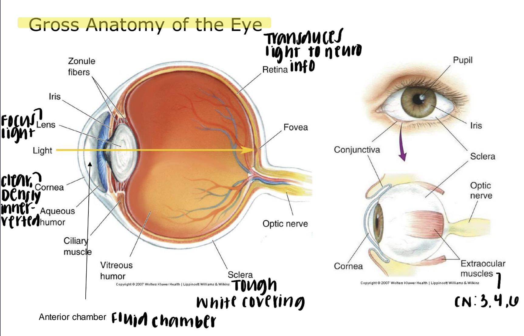
Image Formation:
Retinal images are inverted.
Upper (superior) visual field → lower (inferior) hemiretina
Lateral (temporal) visual field → nasal (medial) hemiretina
Ophthalmoscopic Appearance of the Eye:
Optic Disk
Location: where ganglion cell axons exit the eye to form the optic nerve
Does not have photoreceptors
Gives rise to a blind spot in the lateral (temporal) visual field bc. the optic disk is in the nasal (medial) hemi-retina
Blind spots are filled in by brain
Macula
Yellowish-pigmented area near the center of the retina
Responsible for high-acuity, central vision
Macular degeneration: in older adults resulting in a loss of vision in the central visual field
Pupillary Light Reflex:
Pupil controls the amount of light reaching the retina
pupil continuously adjusts to ambient light levels
Consensual b/w L & R eye: shining light in one eye influences the pupils of both eyes
Neural Circuit: the optic nerve (II) → midbrain (pretectal; allows signal to become bilateral) → midbrain (Edinger-Westphal) → oculomotor nerve (III)
Circuitry of the Retina - "Inside-Out" Organization:
Photoreceptors face the back of the eye (anterior)
Ganglion cell axons (superficial) emerge on the interior, leading to the blind spot
The octopus retina is similar to ours but flipped around (photoreceptors=interior=no blind spot)
Circuitry of the Retina - Vertical Pathway:
Photoreceptors (accept light) → Bipolar cells → Ganglion cells (project to forebrain)
Circuitry of the Retina - Horizontal (Lateral) Connections:
Amacrine cells - spike
Receive input from bipolar cells
Synapse with ganglion cells, bipolar cells, and other amacrine cells
Horizontal cells - no spike
Receive input from photoreceptors
Synapse with other photoreceptors and bipolar cells
Only ganglion cells and amacrine cells produce action potentials
Photorecep., bipolar cells, horizontal cells = non-spiking
Photoreceptors:
Photoreceptors convert light to neural signals
Regions:
Outer seg = photosensitive region
Inner seg= cellular support region
Synaptic terminal = neurotransmitter release
2 main types of photoreceptors
Rods = night vision (grey tones)
All rods respond to the same wavelength of light
Lots of photopigment bc. more membranous disks = more sensitive
Cones = color vision
Blue, Green, Red
Fewer photopigment
Distribution of Photoreceptors:
Distribution of rods and cones varies from the fovea to the retinal periphery
Peripheral Retina - good for detecting faint objects
more rods than cones
more photoreceptors than ganglion cells,
more sensitive to light
less sensitive to color (low acuity)
Fovea
pit in the retina where inner layers are pushed aside
maximizing visual acuity
Central Fovea
contains only cones
h1:1 ratio with ganglion cells
area of highest visual acuity (high acuity)
Phototransduction in Rods:
Light-activated G-protein-coupled biochemical cascade
Rods: Rhodopsin
Cones: Phosphodiesterase
Modulates cGMP-gated Na+ channels
Na+ channels open (dark) → photorecep. depol.
Na+ channels close (light) → photorecep. hyper.
Phototransduction - Photopigment:
Light interacts with photopigment to produce a change in membrane potential
Analogous to activity at G-protein-coupled neurotransmitter receptors but causes a decrease in second messenger by activating phosphodiesterase (cGMP → 5’GMP)
Phototransduction in Rods:
Dark Current:
Rod outer segments = depolarized due to a steady influx of Na+ ions
Light Current:
Photoreceptors hyperpolarize
Photoreceptors release more glutamate when depolarized and less when hyperpolarized
Adaptation to Light Level:
The human eye can function across a wide range of light levels (nine orders of magnitude)
In a given moment of time, eye can only sense a contrast ratio of one thousand
Photoreceptors change their "operating point" to adapt to the ambient light level
Dark Adaptation:
Takes approximately 20-30 minutes
1. Cones (color sensitive) become more sensitive → eventually plateaus
2. Rods (night vision: grey tones) become more sensitive → reach max. sens.
Receptive Field Organization:
Receptive field: region of the sensory surface that, when stimulated, changes the membrane potential of the neuron
Center-Surround RF: organization is common in many sensory systems
Receptive Field Organization in Photoreceptors (non-spiking):
Off-Center RF:
inhibited (hyper) by stimulation in the center
excited (depol) by stimulation in the surround
Produced by lateral inhibition at the level of the horizontal cell
Receptive Field Organization in Bipolar Cells (non-spiking):
Center-Surround & Off-Center RF’s
OFF-CENTER
hyperpolarize (-) = light stimulates the center
depolarize (+) = light stimulates the surround
ionotropic glutamate receptors (AMPA) = open Na+ channels
excitatory, sign-conserving synapse
increased light in center RF → hyper. voltage of photorecep. → lowers glutamate release from photorecep. → hyperpolarize bipolar cell (OFF)
ON-CENTER
depolarize (+) = light stimulates the center
hyperpolarize (-)= light stimulates the surround
metabotropic glutamate receptors (mGluR6) = open K+ channels
inhibitory, sign-inverting synapse
increased light in center RF → hyper. voltage of photorecep. → lowers glutamate release from photorecp. → depol. bipolar cell (ON)
Receptive Field Organization in Ganglion Cells (AP=spiking):
Center-surround receptive fields but produce spiking output (AP)
respond to spatial changes in illumination
implements a form of contrast enhancement
On-center and Off-center ganglion cell types
Off: good for detecting edges not illumination
Afferent Pupillary Defect:
Swinging flashlight test
One eye constricts normally w/ light
Other eye dilates w/ light