Synaptic Transmission
Synaptic transmission is the process by which an action potential arriving at the presynaptic terminal of a neuron is converted into a chemical signal through the calcium-dependent release of neurotransmitters. These neurotransmitters are released into the synaptic cleft, a narrow extracellular space between neurons, and diffuse across it to bind to specific receptors on the postsynaptic membrane.
A synapse is a ‘nerve-nerve’ junction.
A neuromuscular junction (NMJ) is a motoneurone excitatory synapse.
At Rest vs Active Presynaptic Cell:
At rest, the presynaptic cell maintains a resting membrane potential, characterized by a higher concentration of potassium ions (K+) inside the cell and sodium ions (Na+) outside.
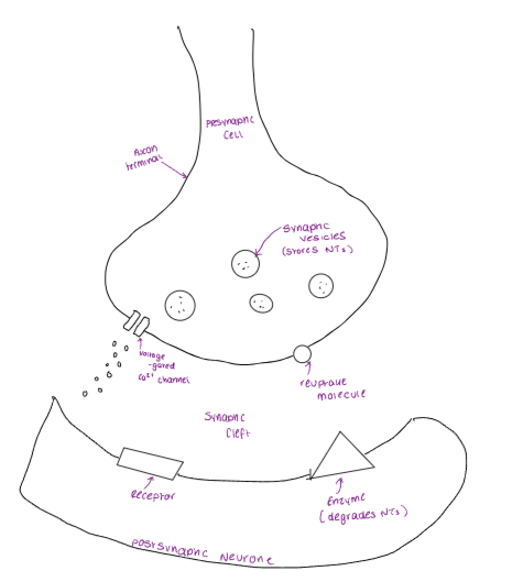
When an action potential occurs, the depolarisation of the presynaptic membrane leads to the opening of voltage-gated calcium channels, allowing Ca2+ to flow into the cell, which triggers the release of neurotransmitters into the synaptic cleft.
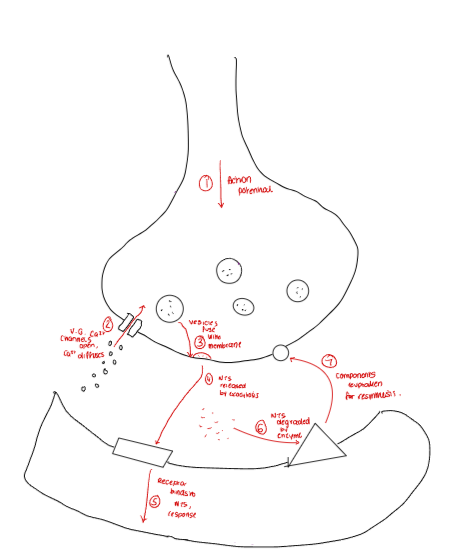
Neurotransmitters that are released from several nerve terminals elicit post-synaptic responses to affect cell body excitability.
Post-synaptic Potentials:
Local graded changes in transmembrane potential.
Not propagated and quickly decays in intensity and distance.
No refractory period.
→ Therefore, summation can occur.
EPSPs:
EPSPs are excitatory postsynaptic potentials that increase the likelihood of a neurone firing an action potential.
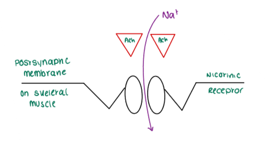
At a neuromuscular junction, acetylcholine (ACh) binds and the receptor alters shape, causing the sodium ion channels to open, leading to depolarisation.
IPSPs:
IPSPs are inhibitory postsynaptic potentials that decrease the likelihood of a neurone firing an action potential.
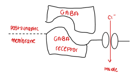
GABA (gamma aminobutyric acid) is an inhibitory neurotransmitter.
Cl- ions enter, making the internal part of the cell more negative, leading to localised hyperpolarisation.
Convergence - Summation of IPSPs and EPSPs:
Excitatory potentials are located mainly on dendrites, whereas inhibitory potentials are located mainly on the cell body. Synapses closest to the axon hillock have the greatest effect on action potentials.
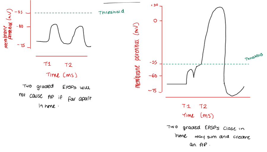
Spatial Summation is when two or more excitatory postsynaptic potentials (EPSPs) occur simultaneously at different synaptic locations on the postsynaptic membrane, their local depolarisations can spread passively through the dendritic and somatic cytoplasm and add together at the axon hillock.
→ Graded potentials individually are all below the threshold, but they sum together to create a suprathreshold signal which produces an action potential.
Temporal Summation is when multiple postsynaptic potentials (usually EPSPs) are generated at the same synapse in rapid succession by repeated stimulation from a single presynaptic neurone.
→ In order for effects to cancel out, the EPSP and IPSP must occur close enough in time that their effects overlap at the postsynaptic membrane. The hyperpolarising effect of the IPSP can reduce or neutralise the depolarising effect of the EPSP, and vice versa.
Presynaptic Inhibition:
Presynaptic Inhibition is the prevention of the release of neurotransmitters.
An action potential causes the opening of voltage-gated calcium ion channels, triggering the release of neurotransmitters.
A second action potential causes another neurotransmitter to inhibit the presynaptic calcium influx (so less of the first neurotransmitter is released).
Therefore, inhibition of presynaptic Ca2+ influx causes less neurotransmitter to be released by action potentials.
Cholinergic Synapses:
A cholinergic synapse is a type of synapse in which the neurotransmitter acetylcholine (ACh) is released from the presynaptic neurone and binds to cholinergic receptors on the postsynaptic neurone, facilitating the transmission of signals and muscle contraction.
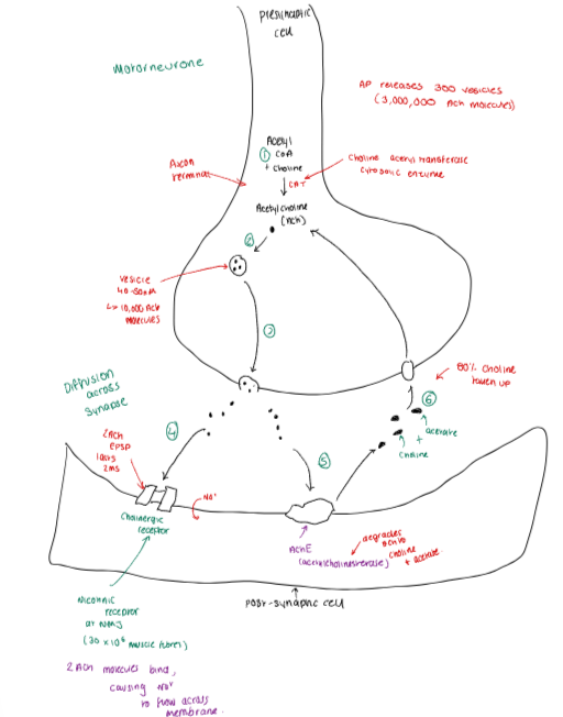
Acetyl-coenzyme A and choline react to form acetylcholine, catalysed by choline acetyl transferase cytosolic enzyme.
Acetylcholine (ACh) is packaged into vesicles (roughly 40-50nm in size), with around 10,000 ACh molecules per vesicle.
An action potential arrives at the presynaptic terminal, depolarising the membrane. This opens voltage-gated Ca²⁺ channels, and the Ca²⁺ influx triggers vesicle fusion to the outer membrane via the SNARE complex.
Ca2+ ions bind to synaptotagmin, which is associated with the synaptic vesicle. This causes a conformational change, allowing it to interact with the SNARE complex.
The interaction between synaptotagmin and the SNARE proteins leads to tightening of the SNARE complex, bringing the synaptic vesicle closer to the presynaptic membrane.
The SNARE proteins continue to pull the vesicle and membrane closer together until they fuse.
ACh-filled vesicles fuse with the presynaptic membrane and release their contents into the synaptic cleft via exocytosis.
ACh diffuses across the synaptic cleft and binds to cholinergic receptors on the postsynaptic membrane:
Nicotinic receptors (nAChRs) – ligand-gated ion channels (e.g. at the neuromuscular junction).
Muscarinic receptors (mAChRs) – G-protein-coupled receptors (e.g. in the CNS and autonomic nervous system).
Binding leads to ion flow or second-messenger activation, generating a postsynaptic response (e.g. depolarisation).
ACh is rapidly broken down in the synaptic cleft by acetylcholinesterase (AChE) into choline and acetate, with over 80% of choline molecules being re-uptaken back into the presynaptic cell for reuse in ACh synthesis, and acetate is used for other metabolic purposes.
Classification of Neurotransmitters:
Amino Acids - e.g. glycine, GABA.
Classical - e.g. noradrenaline, ACh.
Peptides - e.g. TRH, substance P.
Others - e.g. ATP, Nitric Oxide.