3. PU - Retinitis
Retinitis
Definition: Inflammation of the retina, typically secondary to choroidal inflammation in posterior uveitis.
Retinal vasculitis: Inflammation of retinal vessels (arteritis, phlebitis).
Signs include sheathing of vessels (inflammatory cells around vessels appear like a white cuff).
Cotton wool spots (due to infarcts in the nerve fibre layer).
Hemorrhage
retinal and optic disc oedema.
Major etiologies:
Infectious: tuberculosis, syphilis, herpes zoster.
Inflammatory: polyarteritis nodosa, SLE, sarcoid, Behçet’s disease, multiple sclerosis (MS).
Eale’s disease
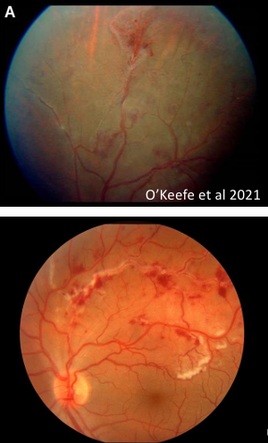
Eales’ disease
Definition: Idiopathic occlusive peripheral periphlebitis (bilateral but asymmetric).
Clinical presentation: Patients may be asymptomatic or present with floaters and blurred vision.
Epidemiology: More common in young adult men in India and the Middle East.
Disease course: Leads to recurrent retinal and vitreous haemorrhages, neovascularisation, and subsequent scarring.
Disease stages: Inflammatory, occlusive, and neovascular stages.
Eales’ disease: peripheral periphlebitis and management
Peripheral periphlebitis characterised by: sheathing of veins, retinal haemorrhages, cotton wool spots.
Complications: Branch retinal vein occlusion may occur → more haemorrhage
Ischaemia and neovascularisation.
Treatment (inflammatory stage): Periocular (around the eye), systemic, topical, and intravitreal steroids may be used.
Treatment (neovascular stage): Laser photocoagulation and anti-VEGF injections.
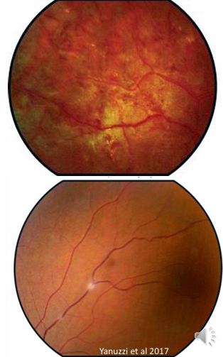
AIDS/HIV retinopathy
Prevalence: Retinal microangiopathy is the most frequent retinopathy in patients with AIDS, developing in up to of patients.
Association: Associated with declining count.
Nature: HIV retinopathy is non-infectious and often asymptomatic.
Ocular findings: Multiple cotton wool spots, haemorrhages, microaneurysms.
Course: Lesions are usually asymptomatic and may spontaneously improve over a few months.
will be symptomatic if lesions are in macular region
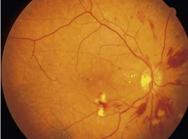
Opportunistic infections in AIDS (context for ocular disease)
opportunistic infections may occur: Toxoplasmosis, Herpes, Cytomegalovirus (CMV).
Overall incidence of opportunistic ocular infection in HIV patients has fallen since HAART (highly active antiretroviral therapy).
AIDS CMV retinitis
CMV retinitis is the most common opportunistic ocular infection in AIDS patients.
Symptoms: Frequently few or no symptoms; may have decreased vision and floaters
Natural history without treatment: Without antiviral therapy, severe visual loss is essentially inevitable. Slowly progressive haemorrhagic retinitis.
Ocular signs:
Slowly progressive, haemorrhagic retinitis.
Confluent white necrotic areas with haemorrhages along vascular arcades.
Progressive retinal atrophy.
Approximately of patients progress to retinal detachment.
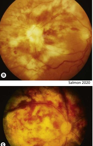
Multiple Evanescent White Dot Syndrome (MEWDS)
Description: A rare acute unilateral multifocal retinitis, usually affecting young adult women.
Preceding illness: ~ have had a preceding viral illness (e.g., upper respiratory tract infection).
Pathogenesis: Unclear; suggested roles for immunological and hormonal influences.
Presentation: Painless monocular blurring of vision (normally mild but can become severe)
Fundoscopy findings: Numerous small grey-white patches in the deep retina or retinal pigment epithelium (RPE) across the posterior pole.
Inflammatory signs: Mild vitritis may be present.
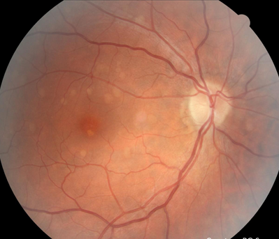
Treatment: No treatment available, referral, review and reassurance
Prognosis: Good; vision usually recovers completely in a few weeks.
Recurrence: Can recur in ~ of cases.
OCT IMAGING:
inner segment ellipsoid band (photoreceptor band in outer retina) are affected
hyper-reflective material deposits.
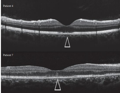
MEWDS: spectrum and differential diagnoses
MEWDS is considered part of a spectrum of overlapping conditions, including:
Acute multifocal placoid epitheliopathy (AMPPE)
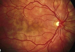
Multifocal choroiditis
Birdshot choroidopathy
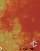
Idiopathic blind spot enlargement syndrome - only have grey patches. Disruption to outer retinal layers surrounding disc.
Acute zonal occult outer retinopathy (AZOOR) - more atrophy and pigment clumping. Some loss of peripheral vision.