(3) Cell Communication
COLOR CODE MEANINGS:
BLUE TEXT = Overall meaning of something/Need to know
PURPLE TEXT = small meanings within that something
***TYPES OF CELL SIGNALING**
Intercellular Signaling - communication between cells
Intracellular Signaling - communication within a cell
***PROCESS OF SIGNALING**
Ligands are released by signaling cells to bind to another molecule to deliver a chemical signal
Ligands bind to target cells (receptors) to deliver those chemical signals
A specific ligand will have a specific receptor that typically binds only that ligand.
1. Reception of the signal
Cells can receive a signal on the external membrane or at an intracellular receptor.
2. Transduction of the signal
A series of chemical reactions, often phosphorylation events, that activate the target molecule.
Phosphorylation Cascade - a sequence of signaling pathway events where one enzyme phosphorylates another, causing a chain reaction leading to the phosphorylation of thousands of proteins
catalyzed by protein kinases—an enzyme
causes conformational changes in the protein
Second Messenger - molecules that relay signals received at receptors on the cell surface to target molecules in the cytosol or nucleus to greatly amplify the signal. 3 major classes of 2nd messengers:
cyclic nucleotides
inositol trisphosphate and diacylglycerol
calcium ions (Ca2+)
3. Cellular response
A change in metabolism, a change in charge across the membrane, and a change in gene expression are the three possible cellular responses.
3 main responses:
Nuclear - Changes in gene expression, or the process in which information from a gene is used by the cell to produce a functional product, typically a protein.
Cytoplasmic Response - Changes in a cell's metabolic response, in which metabolic enzymes in the cell become more or less active.
Change in Membrane Potential - Ligands open ion channels in neurons that lead to a change in electrical charge across the membrane.
***FORMS OF SIGNALING**
Paracrine Signaling - Targets nearby cells
move by diffusion through the extracellular matrix
Elicit quick responses that last only a short period of time.
Endocrine Signaling - A cell targets a distant cell through the bloodstream
Originate from endocrine cells (cells within endocrine glands like thyroid, hypothalamus, etc.)
Produce a slower response but have a longer-lasting effect.
Ligands travel through the bloodstream
Autocrine Signaling - a cell targets itself
regulates pain sensation and inflammatory responses.
signal itself to undergo programmed cell death
Cellular differentiation during embryo development
Signaling Across Gap Junctions - signaling between the plasma membranes of cells
small signaling molecules, called intracellular mediators, diffuse between the two cells.
Common in the plasmodesmata
Small molecules or ions move between the cells

***TYPES OF RECEPTORS**
All receptors undergo structural (shape) changes when the signal molecule is bound.
Internal (Cytoplasmic) Receptors - found in the cytoplasm of the cell and respond to hydrophobic ligand molecules that are able to travel across the plasma membrane and promote gene expression.
pls watch this vid cuz its hard to understand by reading it. The steps typed below this vid correlate to what internal receptors do.
Step-by-Step Mechanism of Internal Receptors (don’t have to read bc its explained in vid above)
Diffusion Across the Membrane:
Ligand characteristics: The signaling molecules (ligands) are typically hydrophobic (nonpolar) and small (e.g., steroid hormones like estrogen, testosterone, or cortisol).
Because cell membranes are made of a lipid bilayer with a hydrophobic core, these molecules diffuse directly through the plasma membrane into the cytoplasm.
Ligand-Receptor Binding:
Inside the cytoplasm, the ligand binds specifically to its intracellular receptor.
This binding induces a conformational change (a shift in the 3D shape of the receptor protein).
Exposure of DNA-Binding Domains:
The conformational change exposes a specific DNA-binding domain on the receptor that was previously hidden or inactive.
It also often exposes a nuclear localization signal that allows the complex to enter the nucleus.
Translocation to the Nucleus:
The newly formed ligand-receptor complex moves through nuclear pores into the cell nucleus.
Direct Regulation of Transcription (Gene Expression):
Inside the nucleus, the complex functions as a transcription factor.
It binds directly to specific sequences of DNA (promoters or regulatory regions) near target genes.
This binding either stimulates (promotes) or represses the copying of DNA into messenger RNA (mRNA).
Protein Synthesis:
The resulting mRNA molecule exits the nucleus into the cytoplasm.
Ribosomes read the mRNA transcript and link amino acids together in the specified sequence (translation) to form new functional proteins.
Cell-Surface (Plasma Membrane) Receptors - membrane-anchored (integral) proteins that bind to external ligand molecules.
Perform signal transduction, through which an extracellular signal is converted into an intracellular signal.
Specific to individual cell types.
Cell-surface receptor proteins are fundamental to normal cell functioning, so a malfunction in any one of these proteins could have severe consequences
Three main components that make it up:
an external ligand-binding domain called the extracellular domain
hydrophobic membrane-spanning region called a transmembrane domain
an intracellular domain inside the cell (the region of the protein that extends into the cytoplasm of the cell)
Three categories of cell-surface receptors:
Ion Channel-Linked Receptors - bind a ligand and open a channel through the membrane that allows specific ions to pass through.
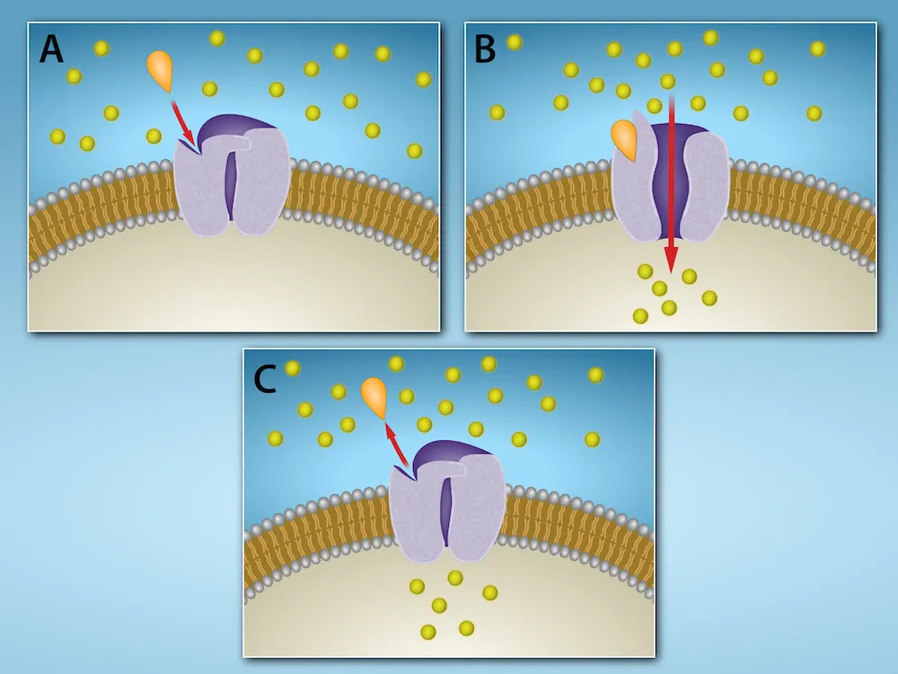
Enzyme-Linked Receptors - bind an extracellular ligand and directly or indirectly activate an enzymatic activity on their intracellular side
pls watch this vid cuz too its hard to understand by reading it. The steps typed below this vid correlate to what internal receptors do.
G Protein-Linked Receptors - translate extracellular signals into intracellular biological responses
pls watch this vid cuz too its hard to understand by reading it. The steps typed below this vid correlate to what internal receptors do.
Step-by-Step Signaling Cycle of G Protein-Linked Receptors (don’t have to read bc its explained in vid above)
Resting (Inactive) State:
The G-protein is bound to guanosine diphosphate (GDP\text{GDP}GDP).
In this form, the G-protein is inactive and consists of a trimer: an α (alpha) subunit bound to a βγ (beta-gamma) dimer.
Ligand Binding & Activation:
A signaling molecule (ligand) binds to the extracellular side of the receptor.
This induces a conformational (shape) change in the receptor that spreads to its intracellular side.
The receptor acts as a guanine nucleotide exchange factor (GEF), causing the α\alphaα subunit of the G-protein to release GDP and bind guanosine triphosphate (GTP).
Subunit Dissociation & Effector Activation:
Binding of GTP causes the G-protein to dissociate into two active signaling units:
The α-GTP subunit
The βγ dimer
Either or both fragments diffuse along the membrane to interact with and regulate target effector proteins, such as:
Enzymes (e.g., adenylyl cyclase, phospholipase C) to produce second messengers.
cyclic AMP (cAMP)
inositol trisphosphate
Ion channels to alter membrane potential or ion concentrations.
Inactivation & Resetting:
The α subunit has intrinsic GTPase activity, meaning it acts as its own timer by hydrolyzing GTP back to GDP and inorganic phosphate (Pi):
GTP→ GDP + Pi
Once GTP is converted back to GDP, the α subunit becomes inactive.
The inactive α-GDP subunit reassociates with the βγ dimer to reform the inactive trimeric G-protein, resetting the system for the next signal.
***TYPES OF RECEPTORS**
Signaling Molecules - Basically, they are just ligands that act as chemical signals that travel to the target cells to coordinate responses.
Small Hydrophobic Ligands - It’s right in the name, and they diffuse through the plasma membrane and interact with internal receptors.
Primarily like steroid hormones
Water-Soluble Ligands - Sometimes too large to pass through + are polar.
Most water-soluble ligands bind to the extracellular domain of cell-surface receptors.
***HORMONES**
Steroid Hormones - small hydrophobic ligands.
Estradiol and testosteron aid in gene expression
Estradiol
Testosterone
Cholesterol
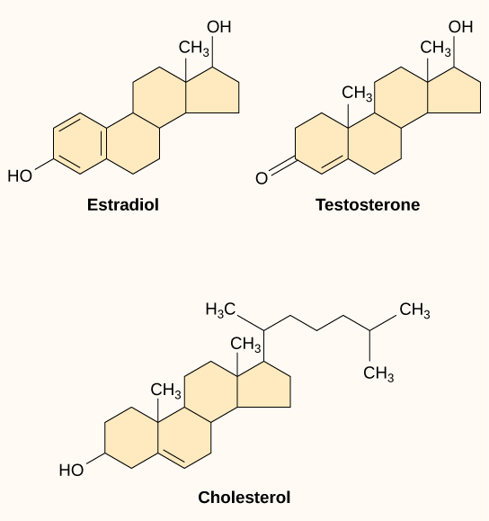
Peptide Hormones - hormones are polar, water-soluble ligands and therefore cannot pass through the plasma membrane unaided
bind to cell-surface receptors