Week 9
Cardiovascular System
Topic 1: The Heart and Cardiac Cycle
Briefly describe the structure of the heart and trace the pathway of blood through the heart
Describe the structure and function of the valves of the heart
Name the components of the conduction system of the heart and trace the conduction pathway
Identify the waves of a normal electrocardiograph trace
Describe the timing and events of the cardiac cycle
Describe how stoke volume, cardiac output and heart rate are regulated
Topic 2: Blood vessels and Blood pressure
Compare and contrast the structure and function of arteries, veins, and capillaries
Examine how blood flow, blood pressure and resistance relate to blood circulation
Describe the mechanisms involved in regulation of blood pressure
Outline the factors involved in capillary exchange and bulk flow
Heart structure and blood flow
The heart
The heart is a muscular organ located in the chest. It often described as a ‘pump’, but actually the heart is two pumps side by side, which are responsible for receiving deoxygenated blood, recycling it through the lungs and supplying oxygenated blood to the body. About the size of your fist, it beats hundreds or thousands of times a day, pumping blood around blood vessel networks throughout the body to deliver oxygen and nutrients to body cells and at the same time remove carbon dioxide and other waste products. Blood is pumped from the lungs, through the heart and to the tissues in the body. From there it returns via the veins to the heart and the cycle begins again:


Heart structure
The heart is a muscle that works like a pump to send blood around the body. It has a right side and left side, separated by the septum.
It has 4 chambers:
Right side | Left side |
|---|---|
Right atrium | Left atrium |
Right ventricle | Left ventricle |
It also has 4 valves that keep blood moving in the correct direction:
Valve | Location |
|---|---|
Tricuspid valve | Right atrium → right ventricle |
Pulmonary valve | Right ventricle → pulmonary artery |
Mitral/bicuspid valve | Left atrium → left ventricle |
Aortic valve | Left ventricle → aorta |
Blood flow through the heart
1. Deoxygenated blood returns from the body
Blood low in oxygen comes back to the heart through the superior and inferior vena cava and enters the right atrium.
2. Right atrium contracts
The tricuspid valve opens, and blood moves into the right ventricle.
This is part of atrial systole.
3. Right ventricle contracts
The tricuspid valve closes to stop backflow.
The pulmonary valve opens, and blood is pumped into the pulmonary artery toward the lungs.
This is ventricular systole.
4. Blood becomes oxygenated in the lungs
Oxygen-rich blood returns from the lungs through the pulmonary veins and enters the left atrium.
5. Left atrium contracts
The mitral valve opens, and blood moves into the left ventricle.
6. Left ventricle contracts
The mitral valve closes to stop backflow.
The aortic valve opens, and oxygen-rich blood is pumped into the aorta and then to the rest of the body.
Key idea
The right side of the heart pumps blood to the lungs.
The left side of the heart pumps blood to the body.
Heart valves and sounds
Heart valves
The four valves in the heart ensure blood flows in the correct direction through the chambers of the heart. Each valve is like a one-way door that keeps the blood flowing in the same direction. If we take a cross-section of the heart, you can see these four valves, each containing two or three leaflets.

Heart sounds
The heart sounds you hear using a stethoscope are associated with the closing of these valves. The first heart sound that you hear is the closing of the tricuspid and mitral valves, also known collectively as the atrioventricular valves. This sound is called the LUB.
The second heart sound is the closing of the pulmonary and aortic valves, also known as semilunar valves. This is called the DUB.
The LUB-DUB sounds you can hear are due to the leaflets of the valves slapping against one another while closing.

Heart's intrinsic conduction system
The conducting system of the heart consists of cardiac muscle cells and conducting fibres that initiate the normal cardiac cycle and coordinate the contractions of cardiac chambers.
In order for the heart to pump efficiently and the systemic and pulmonary circulations to operate correctly, the events in the cardiac cycle must be synchronised by the structures of the conduction system. Both atria contract together, as do the ventricles, but atrial contraction occurs first.
This conducting system provides the heart its automatic rhythmic beat.

Sinoatrial node (or SA node)
This is the natural pacemaker of the heart. It initiates all heart beats and determines heart rate. Electrical impulses from the SA node spread throughout both atria and stimulate them to contract.
Located in the right atrium near the entrance of the superior vena cava.
Atrioventricular node (or AV node)
The AV node serves as an electrical gateway to the ventricles. It delays the passage of electrical impulses to the ventricles. The delay is to ensure that the atria have ejected all the blood into the ventricles before the ventricles contract.
Located on the opposite side of the right atrium, near the tricuspid valve.
Atrioventricular bundle
Also referred to as AV bundle or bundle of His. The AV node receives signals from the SA node and passes them here.
Left and right bundle branches
The AV bundle is divided into left and right bundle branches, which conduct the impulses towards the apex of the heart.
Purkinje fibres
The signals are passed here from left and right bundle branches, turning upwards and spreading throughout the ventricular myocardium and stimulating them to contract.
These signals stimulate contraction first of the right and left atrium, and then the right and left ventricles in sequence, allowing efficient pumping of blood throughout the body.
Intrinsic conduction system
The independent but coordinated activity of the heart is a function of gap junctions and the activity of the heart’s own conduction system. The electric currents generated in and transmitted through the heart spread throughout the body and can be detected with a device called anelectrocardiograph. An electrocardiogram (ECG) is a graphic record of heart activity.


WATCH THIS:
🫀 Cardiac Cycle = One Complete Heartbeat
Systole = contraction | Diastole = relaxation
Phase | Valves | What happens |
|---|---|---|
🟦 Atrial diastole | AV valves initially closed | Atria relax and fill with blood from the vena cava and pulmonary veins |
🟩 Atrial systole | AV valves open | SA node fires → atria contract → blood moves into ventricles |
🟨 Ventricular diastole | Both valves closed, then AV valves open | Ventricles relax → pressure falls → ventricles fill |
🟥 Ventricular systole | AV valves close, then semilunar valves open | Ventricles contract → blood is ejected into pulmonary artery and aorta |
🟦 Atrial Diastole: Atria Fill
The atria are relaxed and blood enters them.
Right atrium | Left atrium |
|---|---|
Receives deoxygenated blood from superior + inferior vena cava | Receives oxygenated blood from pulmonary veins |
At first, the AV valves are closed, so blood briefly pools in the atria.
🟩 Atrial Systole: Atria Contract
The SA node sends an electrical signal across the atria. This causes the atria to contract. Atrial pressure becomes higher than ventricular pressure, so the AV valves open.
Blood moves: atria → ventricles
AV valves:
Right side | Left side |
|---|---|
Tricuspid valve | Mitral/bicuspid valve |
🟨 Ventricular Diastole: Ventricles Relax and Fill
At the start, both the AV valves and semilunar valves are closed. The ventricles relax, pressure falls, but volume stays the same.
This is called isovolumetric relaxation.
Iso = same
Volumetric = volume
So it means same volume while relaxing.
Then ventricular pressure drops below atrial pressure, so the AV valves open and blood flows into the ventricles.
At the end of this phase, the ventricles are full. This is called:
End-diastolic volume = preload
🟥 Ventricular Systole: Ventricles Contract and Eject Blood
The electrical signal travels:
SA node → AV node → Bundle of His → right + left bundle branches → ventricles
The AV node delay gives the atria time to finish emptying before the ventricles contract.
As ventricles contract, ventricular pressure becomes higher than atrial pressure, so the AV valves close.
“Lub” = AV valves closing
At first, all valves are closed and pressure rises without blood leaving. This is called isovolumetric contraction.
Then ventricular pressure becomes high enough to open the semilunar valves.
Right ventricle ejects blood into | Left ventricle ejects blood into |
|---|---|
Pulmonary artery | Aorta |
This is the ejection phase.
⭐ Key Volume Terms
Term | Meaning |
|---|---|
End-diastolic volume / preload | Blood in ventricle before contraction |
End-systolic volume | Blood left in ventricle after contraction |
Stroke volume | Blood actually pumped out per beat |
Stroke volume = end-diastolic volume − end-systolic volume
🌈 Full Sequence to Memorize
Atria fill → atria contract → ventricles fill → ventricles contract → blood ejects → ventricles relax → repeat
Or:
Atrial diastole → atrial systole → ventricular systole → ventricular diastole
Cardiac output, Heart rate and Stroke volume
Cardiac output
The cardiac output represents the volume of blood that is delivered to the body. Its an important measure in determining the effectiveness of the heart to deliver blood to the rest of body. It is defined as the amount of blood pumped from each ventricle each minute (L/min).
Two important variables contribute to cardiac output: the heart rate and stroke volume. The equation representing this relationship is:
Cardiac output (L/min) = Heart rate (beats/minute) × Stroke volume (L/beat)
Heart rate
The heart rate is the number of heart beats per minute. The resting heart rate is determined by the pacemaker cells at the SA node. The rate at which the SA node generates electrical impulses (60 to 100 times per minute) determines the heart rate. However, the autonomic nervous system and hormones (like adrenaline) exert control over SA node activity, thus significantly influence heart rate.
🫀 Stroke Volume
Stroke volume = the volume of blood pumped out by one ventricle with each beat.
Cardiac output = heart rate × stroke volume
So, if stroke volume increases, cardiac output also increases.
🟦 Main factors that determine stroke volume
🟥 1. Contractility = strength of contraction
Contractility refers to the intrinsic strength of cardiac muscle contraction.
When contractility increases:
stronger heart contraction → more blood pumped out → larger stroke volume → increased cardiac output
🟩 2. End-diastolic volume / Preload = how full and stretched the ventricle is
End-diastolic volume is the amount of blood in the ventricles at the end of diastole, when the ventricles are full.
Preload is the degree of stretch of the cardiac muscle fibres at the end of diastole.
When EDV/preload increases:
more blood in ventricle → more stretch → more blood pumped out → increased stroke volume → increased cardiac output
Preload depends on two factors:
🟢 Duration of ventricular diastole
The longer the heart is at rest/relaxed, the more time the ventricle has to fill with blood.🔵 Venous return
The amount of blood returning to the heart through the veins.🟧 3. Afterload = pressure the heart must overcome
Afterload is the pressure the heart must overcome before the semilunar valve can open and eject blood.
For the left ventricle, ejection begins when:
left ventricular pressure > aortic pressure
At that point:
higher ventricular pressure → aortic valve opens → blood is ejected into the aorta

Venous return
Venous return refers to the process of blood flowing back to the heart through the veins. Venous return mechanisms help ensure blood returns to the heart efficiently, despite the challenge of low venous pressure, especially from lower parts of the body against gravity. Stroke volume increases when the venous return increases, because when more blood returns to the heart, more blood can be pumped per beat.
Three key mechanisms assist in venous return include:
1. Vein valves
Veins contain one-way valves that prevent blood from flowing backward.
These valves ensure unidirectional blood flow toward the heart, even against gravity.
2. Skeletal muscle pump
Contraction of skeletal muscles compresses nearby veins.
The compression propels blood toward the heart due to the presence of one-way valves in veins, which prevent backflow.

Human Anatomy and Physiology, Marieb and Hoehn, Global edition (2018)
3. Respiratory Pump
During inhalation, the diaphragm moves downward, increasing abdominal pressure and decreasing thoracic pressure.
This pressure gradient helps push blood from the abdominal veins into the thoracic veins and then into the heart.
These mechanisms work together to maintain effective circulation and ensure adequate blood return to the heart, crucial for maintaining blood pressure and cardiac output.
Blood vessels
Completion requirements
Having learned about the heart, we now move onto blood vessels. Blood vessels form a closed delivery system that begins and ends at the heart. They are like ‘pipes’that circulate blood throughout the body. However, unlike rigid pipes, blood vessels are dynamic structures that pulsate, constrict and relax. There are three main types of blood vessels: arteries, capillaries and veins.
Blood vessel structures
There are three main types of blood vessels: arteries, veins and capillaries. The walls of the arteries and veins have three layers:
The lining consists of endothelium, a single layer of cells.
The middle layer consists of muscles and elastic fibres.
The outer layer is made of connective tissue with elastic fibres.
Capillaries have a single layer of endothelial cells to allow oxygen and nutrients to readily pass through.

Arteries
Arteries are large, thick-walled vessels that carry oxygenated blood away from the heart to various parts of the body.
The largest artery, the aorta, emerges from the left ventricle of the heart and branches into smaller arteries, which further divide into arterioles that delivers blood to capillaries
Arteries are characterised by their strong and elastic walls, allowing them to withstand the high pressure generated by the pumping action of the heart.
Predominantly consisting of smooth muscle cells and elastic fibers for blood flow regulation and maintenance of pressure.
Veins
Veins are vessels that carry deoxygenated blood back to the heart from various parts of the body.
Veins generally have thinner walls compared to arteries and are less elastic.
Veins have valves that prevent the backward flow of blood.
Smaller veins called venules merge into larger veins, ultimately leading to the superior and inferior vena cava, which return blood to the right atrium of the heart.
Pressure is much lower in veins.
Capillaries
Capillaries are tiny, thin-walled vessels that connect arteries and veins. They form an extensive network throughout tissues and organs.
Capillaries are the site of exchange between the blood and surrounding tissues. Nutrients and oxygen diffuse from the blood into the tissues, while waste products move from the tissues into the blood.
The walls of capillaries are so thin that molecules can pass through them easily - Single endothelial cell layer.
Connect arterioles and venules.
Mean arterial blood pressure
The cardiovascular system must maintain adequate blood pressure in order for the blood to be delivered to all the organs and tissues. If the pressure drops too low, the organs will not receive an adequate perfusion of nourishing blood. If the pressure rises too high, it could damage the delicate inner lining of the blood vessels and eventually lead to heart disease or stroke.
Blood leaves the left ventricle under relatively high pressure of about 100 to 140 mmHg. With each beat, the blood gets pushed out into the arteries against the artery walls. These walls are normally quite elastic and flexible and allow the blood to flow with little resistance.
Blood pressure is the force of blood pressing against the walls of the blood vessels, or arteries, each cardiac cycle. Pressure fluctuates with each heartbeat during the cardiac cycle. Thus, we calculate mean arterial pressure (MAP), or the average arterial blood pressure during a single cardiac cycle.
MAP is calculated using two measurements: systolic and diastolic pressure (see figure below). Hence, blood pressure is reported as one number ‘over’ another; for example, 120 (systolic) over 80 (diastolic), or 120/80. Note that MAP is roughly one-third of the way between diastolic and systolic pressures, as the heart spends more time resting than contracting.

There are main cardiovascular variables that affect MAP:

Blood pressure gradient
Completion requirements
The pressure difference between the arteries and veins is the driver of blood flow through the circulatory system. This pressure gradient is maintained by the heart's pumping action and the resistance offered by the blood vessels.
The arterial system is a high-pressure system because it receives blood directly from the heart, which ejects blood into the arteries during systole (ventricular contraction). Arteries are referred to as pressure reservoirs because of their elastic and muscular walls which allow them to store and maintain blood pressure generated by the heart. As blood moves through the arterial system (arteries → arterioles → capillaries), pressure gradually decreases due to friction and the resistance of the vessels.
The venous system is a low-pressure system because blood has lost much of its initial energy as it passed through the resistance of the capillaries. Veins are referred to as volume reservoirs because of their thin and compliant (stretchable) walls which allow them to store a large volume of blood at relatively low pressure.

Blood pressure regulation (Baroreceptor reflex and Hormonal control)


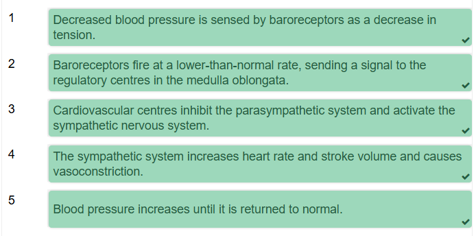
Indirect renal mechanism
Blood pressure regulation is complex and involves a variety of mechanisms involving both short- and long-term responses by the kidneys, as well as other systems of the body. This section will focus on the long-term mechanisms of blood pressure regulation via the renin-angiotensin-aldosterone system, or RAS, which indirectly restores blood pressure homeostasis by regulating blood volume.

Although blood volume varies with age, gender and body size, renal mechanisms usually keep blood volume close to around five litres. The renin-angiotensin mechanism influences both blood volume and blood pressure by regulating the release of aldosterone, and, therefore, sodium and water reabsorption by the kidneys. The effects of this system are slow (hours to days).
Let’s look at the steps in this process one at a time, tracking the change from a decreased blood pressure to an increase in blood pressure back to normal levels.
Renin
If we have a decrease in blood pressure, there will be a decrease in the ‘stretch’ of the wall of the aorta and carotid bodies. The baroreceptor reflex will detect this change and compensate by triggering the release of an enzyme called renin in the juxtaglomerular cells of the nephron in the kidneys. This is the first step in the process.

Angiotensin
Once renin is in the bloodstream, it will enzymatically cleave angiotensinogen, a plasma protein made by the liver, into angiotensin I. In turn, angiotensin I enters the blood vessels, where it is converted into angiotensin II as it passes by the lungs. This is done by an enzyme called the angiotensin converting enzyme, or ACE.

Angiotensin II
Angiotensin II has a few important roles which, themselves, increase mean arterial pressure:
It is a potent vasoconstrictor. This results in an increase in peripheral resistance and a decrease in size of our blood vessels, which will increase blood pressure.
It will also increase a hormone called antidiuretic hormone, which is released by the posterior pituitary. This, together with an increase in thirst, increases water reabsorption by the kidneys. As a result, there is an increase in blood volume and, therefore, increasing blood pressure.
Aldosterone
Angiotensin II also stimulates the adrenal cortex, causing the adrenal glands to secrete aldosterone. Aldosterone promotes sodium reabsorption in the kidneys which, together with the ADH release from the angiotensin II, results in an increase in water reabsorption. Again, this leads to an increase in blood volume and thus arterial pressure.
Summary
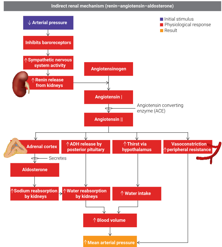
We can now zoom out to see the process as a whole, from when a drop in arterial pressure is first detected by the baroreceptors, through to an increase in mean arterial pressure.

Capillary exchange
Capillaries are the smallest blood vessels. They have thin walls with tiny pores that allow water and small solutes to pass, but do not allow larger protein molecules to pass. This makes capillaries ideally suited for their role to enable the exchange of fluid, gases, nutrients and hormones between blood and cells of the body. This process is called capillary exchange. Two forces control the movement of fluid and its dissolved contents into and out of capillaries: hydrostatic pressure and oncotic pressure.


Hydrostatic pressure is greater than oncotic pressure:


