Neurobio Structures Networks LECTURE
Lecture: 02/09
Hypofrontality
prefrontal cortex and frontal circuitry have been the long-standing focus of inquiry into the pathology of schizophrenia
hypofrontality - reduced activation of the frontal cortex in schizophrenia, most consistently observed during executive-function tasks
particularly the dorsolateral prefrontal cortex (DLPFC)
early neuroimagining evidence (1970’s - 1980’s)
regional cerebral blood flow (rCBF) studies suggested reduced frontal perfusion
PET studies later showed reduced frontal glucose metabolism
fMRI findings (1990’s onward)
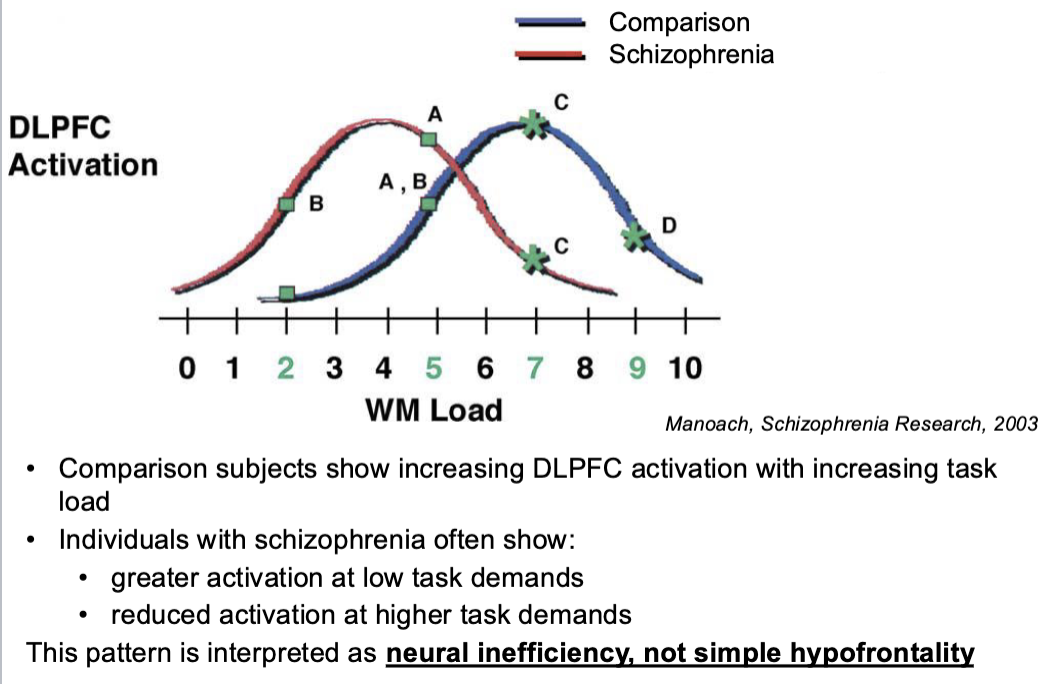
many studies showed reduced DLPFC activation during working-memory tasks
however, some studies reported increased or inefficient PFC activation, depending on task demands and performance level
current interpretation
hypofrontality reflects inefficient recruitment of prefrontal control networks, rather than simply “less frontal activity”
why did the field move beyond the hypofrontality hypothesis?
these early brain imaging studies did not fully account for important confounds
early functional imaging methods has limited spatial andn temporal resolution and small sample sizes
task performance differences (attention, IQ, working-memory capacity)
negative symptoms and motivation
medication side effects (i.e., the effect of antipsychotic drugs on the vascular system)
abnormalities in non-frontal regions that influence frontal activity (e.g., parietal cortex functioning)
in summary:
people living with schizophrenia often show impaired performance on tasks engaging the prefrontal cortex, especially working memory and executive control
early reports of reduced frontal metabolism and blood flow were influential but later found to be context-dependent and methodologically confounded
current models emphasize prefrontal network dysfunction, rather than simple hypo- or hyperactivity
Hippocampal Abnormalities
early neuroimaging studies in the 1990’s reported increased resting cerebral blood flow and metabolism in the hippocampus in schizophrenia
later task-based fMRI studies also showed increased hippocampal activation, particularly during memory-related tasks
there findings contributed ot the idea that hippocampal hyperactivity may be a core feature of schizophrenia, in contrast to early models focused primarily on prefrontal dysfunction
schizophrenia is associated with impaired hippocampal habituation, meaning reduced ability to downregulate neural responses after repeated presentation of emotionally salient stimuli
during repeated viewing of facial expressions, individuals with schizophrenia show sustained hippocampal activation, whereas comparison participants show the expected decrease in activation over time (Holt et al., 2005)
post-mortem studies reveal:
normal total number of hippocampal neurons
selective reduction of inhibitory interneurons (e.g., PV+ interneurons)
MRI studies reveal:
reduced hippocampal volume
Hippocampal Structure
the majority of hippocampal neurons (~90%) are glutamatergic pyramidal cells, which provide long-range excitatory projections outside the hippocampus
a smaller population (~10%) consists of GABAergic interneurons
interneurons play a critical role in:
synchronizing pyramidal neuron firing
maintaining excitation-inhibition balance
regulate information flow through hippocampal circuits
Do structural changes in the hippocampus predict the onset of psychosis?
individuals at Clinical High Risk (CHR) for psychosis participated in a longitudinal MRI study
CHR participants includes individuals with
attenuated psychotic symptoms
brief intermittent psychotic symptoms
familial risk plus recent functional decline
participants were followed over time to determine who transitioned to psychosis
the key question: do hippocampal structural changes predict later conversion to psychosis?
participants were followed at monthly intervals to complete a structural clinical symptom rating scale for the first year, at 3 month intervals during the second , and the third year and annually thereafter until transition to frank psychosis or until end of follow-up period (5 years)
those who converted tro psychotic disorder were scanned again after conversion, those who did not convert were scanned at the end of the study period
all patients were antipsychotic naive at the time of baseline
total of 18 patients completed the study (8 converted to psychosis, 10 did not)
at the time of follow up scan, 5 of the converters were treated with antipsychotics
a significant main effect of time: hippocampal volume decreased over time in all subjects
no difference between converters and non-converters
Hippocampal abnormalities in schizophrenia
increased hippocampal activity in schizophrenia, possibly reflecting impaired habituation or regulation of hippocampal responses
no reduction in total hippocampal neuron number, but selective loss of GABAergic interneurons
reduced hippocampal volume is frequently observed, though not specific to schizophrenia and not predictive of psychosis onset
environmental risk factors are also at play
for instance, cannabis use is high in psychotic disorders
the early and heavy use of cannabis alters hippocampal structure (Cousijn et al., 2012)
dopamine hypothesis of schizophrenia has been linked to hippocampal hyperactivity
Effects of cannabis use on hippocampal volume
they found reduced hippocampal volumes in cannabis dependence, not in non-dependent cannabis users
THC use and cannabis dependence are associated with increased risk for psychosis, particularly in individuals with existing vulnerability
Findings suggest hippocampal circuitry may be especially vulnerable across multiple pathways linked to psychosis risk
Cerebellar abnormalities in schizophrenia
once thought to primarily support motor coordination, the cerebellum is now known to contribute to cognitive and emotional regulation
the cerebellum participates in distributed circuits linking cortex → cerebellum → thalamus → cortex (CCTCC)
post-mortem studies suggest reduced Purkinje cells desity in the cerebellum
neuroimaging studies show reduced cerebellar activation across multiple cognitive tasks
these findings contributed to Dr. Andreasen’s “cognitive dysmetria” hypothesis of schizophrenia
The hyper and hypo connectivity pattern of the thalamus with the cortex has recently been replicated for striato-cortical and cerebello-cortical connectivity in an MRI study of 159 chronic schizophrenia patients and 162 matched controls.
These results highlight that the cortico-cerebellar-thalamic-cortical circuit (CCTCC) is widely affected in schizophrenia
It’s brain wide alterations rather than exclusively focal functional disturbances that are altered in schizophrenia.
Thalamic abnormalities in schizophrenia
intrinsic brain networks revealed with resting-state functional connectivity MRI
thalamus → sensory-motor cortex hyperconnectivity
thalamus → prefrontal cortex hypoconnectivity
also observed in CHR & predicts conversion
Communication between the thalamus and the cortex is modulated by the thalamic reticular nucleus (TRN).
TRN is a very small structure that receives collaterals from thalamocortical and corticothalamic projections
TRN is made up almost entirely of GABAergic interneurons
Abnormal wiring of the thalamocortical circuitry in psychotic disorders.
It predicts conversion to psychosis in clinical high risk.
Not unique to schizophrenia.
Possibly due to loss of GABAergic interneurons in the thalamic reticular nucleus
Lecture: 2/11
Can E/I imbalance explain schizophrenia?
Global alteration of excitation inhibition balance in schizophrenia
BUT- the extend to which different brain functions are affected by schizophrenia vary greatly:
severe alterations of belief, thought, and higher-order executive cognitive function.
mild alterations in lower-level sensory and motor functions
If these neural changes are pervasive, how do we explain the presence of deficit “hotspots”?
Can brain abnormalities predict the emergence of psychosis?
An important goal in our field is to identify structural and functional changes in the brain that can reliably predict who will develop a psychotic disorder.
This is typically done by recruiting a large group of participants who are at heightened risk for developing psychosis and either:
obtain brain imaging data longitudinally (e.g. every 6 months) to identify progressive changes in brain structure and volume
Obtain brain images at a single time point but monitor participants clinically to determine diagnostic outcomes, then compare brain imaging at baseline between high-risk converters and high-risk non-converters
North American Prodrome Longitudinal Study (NAPLS3).
Determining whether brain grey matter structure changes prior to conversion to a psychotic disorder
Whether gray matter changes can elucidate illness-related neurobiological processes
If yes, can we target the prevention of these brain changes?
One possible target could be interventions that are geared towards protecting excitation/inhibition balance