Anatomy and Physiology Final Exam 2024 Review v2
General Concepts
anatomy = structure, physiology = function
levels of organization from simplest to most complex
molecule
cell
tissue
organ
organ system
organism
Anatomical Directions (see textbook Table 1.1)
superior v. inferior
Above v Below
anterior v. posterior
Front v back
medial v. lateral
Closer to midline v farther from midline
proximal v. distal
Closer to point of attachment (trunk) v farther from point of attachment (trunk)
Anatomical Planes
sagittal (or longitudinal)
slicing body vertically separating it from left and right
frontal (or coronal)
slicing body from front and back
transverse
slicing body horizontally separating it from top and bottom
Homeostasis
general definition
The process of maintaining balance in the both in a changing environment
receptor
receives info from environment
effector
responds to commands from control center/enacts change
control center
processes info
Nervous System
Functions and Classifications
sensory v. motor
Sensory bring signals in CNS
Motor bring signals out CNS
CNS v. PNS
CNS= Control center
PNS= communication line
somatic v. autonomic
Somatic=voluntary
Autonomic=involuntary
parasympathetic v. sympathetic
Parasympathetic=relaxed
Sympathetic=fight or flight response
afferent v. efferent
(mnemonic: afferent ARRIVES in brain, efferent EXITS)
Neuron Structure and Function
locate on a neuron diagram and know the function of the following
dendrite
Receives signals
cell body
nucleus
axon
sends signals away
node of Ranvier
speeds up axons, has lil gaps
Schwann cell
makes myelin in PNS
axon terminal
Releases neurotransmitters
myelin sheath
a layer around Schawan
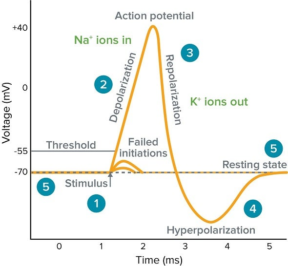
Action Potential
purpose of the action potential is to conduct nerve impulses and send information
neuron starts positive on outside, negative on inside
sodium (Na+) rushes into neuron, depolarizing the interior, changing to positive charge inside
action potential moves down the neuron as more ion channels open and let sodium in
after action potential passes, cell resets to resting potential
or
Threshold
depolarization
re-polarization
hyper polarization
resting phase
Synapses
general function - chemically link one neuron to another, or to a muscle
neurotransmitter - chemical messenger released by axon into synaptic cleft
ion channel - flow regulation
synaptic cleft - space in between two neurons
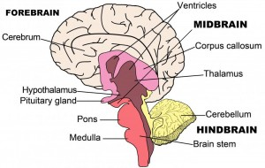
Brain Structure and Function
general locations and functions (see text Table 7.1)
cerebral cortex - control voluntary skeletal muscle, intellectual and emotional processing
thalamus - relays impulses to cerebral cortex
hypothalamus - regulation of autonomic nervous functions, regulates temp, water balance, food
limbic system - mediates emotional response
medulla oblongata - relays sensory input, controls HR, respiratory rate
cerebellum - smooths skeletal muscle movements, regulates balance and posture
Protection of CNS
three layers of meninges
Outside to inside: Dura mater, Arachnoid mater, Pia mater
functions of cerebrospinal fluid (CSF)
Cushions the CNS from shock, provides nutrients, and removes waste.
Brain and Spinal Cord Dysfunction
concussion
A mild traumatic brain injury that affects brain function. Effects are often short term and can include headaches and trouble with concentration, memory, balance, mood and sleep.
stroke (CVA)
something blocks blood supply to part of the brain or when a blood vessel in the brain bursts. Parts of the brain become damaged or die.
transient ischemic attack (TIA)
Caused by a brief blockage of blood flow to the brain. A TIA usually lasts only a few minutes and doesn't cause long-term damage.
Muscular System
General Functions
movement
maintain posture
stabilizing joints
generating heat
control/regulation
Muscle Types
Skeletal | Cardiac | Smooth | |
where found in the body? | everywhere | heart | organs |
appearance? | striated | striated | not striated (smooth) |
control of contraction? | voluntary | involuntary | involuntary |
Muscle Movements
origin
The point where a muscle starts and stays still during movement
insertion
The point where a muscle ends and moves towards the origin during movement
Flexion
Bending a joint to decrease the angle between two body parts
extension
Straightening a joint to increase the angle between two body parts
abduction
Moving a body part away from the midline of the body
adduction
Moving a body part towards the midline of the body
Muscle Interactions
agonist (prime mover)
The main muscle responsible for a movement
antagonist
The muscle that opposes or reverses a prime mover
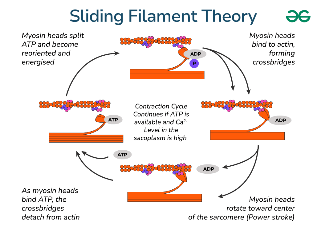
Sliding Filament Model - model of muscle contraction
calcium - activates binding sites on actin filament
myosin - protein with head that attaches to actin and performs power stroke (thick filament)
actin - protein that myosin head pulls in power stroke, causing muscle to shorten (thin filament)
sarcomere - basic unit of muscle contraction, shortens as actin filaments are pulled toward center
Muscle Use and Exercise
ATP Generation Methods
Aerobic | Anaerobic | |
amount of ATP produced | 32 - 36 ATP per glucose | 2 ATP per glucose |
oxygen required? | yes | no |
advantages | more energy | available when oxygen level is low |
disadvantages | not available when oxygen level is low | can only last for a minute or so, produces lactic acid in muscle |
isometric - muscle tenses up but does not change length (ex. plank)
isotonic - muscle changes length (ex. push ups)
Gross Anatomy - location and type of movement associated with each
head and neck
masseter-Closing the jaw
trapezius-Shoulder elevation (shrugging), scapular retraction (pulling the shoulder blades together)
chest, shoulder and arm
deltroid-Shoulder abduction (raising the arm away from the body), flexion, and extension
pectoralis major-Shoulder flexion, adduction (bringing the arm across the chest), and medial rotation
biceps brachii-Elbow flexion (bending the arm)
triceps brachii-Elbow extension (straightening the arm)
latissimus dorsi-houlder adduction (bringing the arm down and towards the body), extension, and medial rotation
abdomen
rectus abdominis-Trunk flexion (bending forward)
external oblique-Trunk flexion, lateral trunk flexion (side bending), and trunk rotation
pelvis, hip and thigh
gluteus maximus-Hip extension (straightening the hip), hip abduction (moving the leg away from the midline)
biceps femoris-Hip extension, knee flexion (bending the knee)
rectus femoris-Knee extension (straightening the knee), hip flexion
lower leg
gastrocnemius-Plantar flexion (pointing the foot downward), knee flexion
soleus-Plantar flexion of the foot, stabilizing the ankle joint’
Cardiovascular System
Introduction
basic functions of the cardiovascular system
Transport oxygen, nutrients, and waste throughout the body, help regulate temperature and pH, support the immune system, and maintain fluid balance.
Heart Anatomy
heart location within the chest
Center slightly to the left
Pericardium
The pericardium is a protective sac around the heart.
Chambers and Great Vessels
atria v. ventricles
Atria are smaller, upper chambers that receive blood
Ventricles are larger, lower chambers that pump blood out.
pulmonary v. systemic circulation
Pulmonary circulation carries blood to the lungs for oxygenation
Systemic circulation delivers oxygenated blood to the body.
The right side of the heart drives pulmonary circulation
The left side drives systemic circulation.
path of blood thru heart and lungs
Blood flows from the right atrium to the right ventricle, then to the lungs for oxygenation, returning to the left atrium, then to the left ventricle, and finally to the body.
Heart Valves
total number and names of valves
There are four valves in the heart: tricuspid valve, pulmonary valve, mitral valve (bicuspid), and aortic valve.
function of AV valves
AV valves (tricuspid and mitral) prevent backflow of blood into the atria during ventricular contraction. They are open during heart relaxation (diastole) and closed during ventricular contraction (systole).
function of semilunar valves
Semilunar valves (pulmonary and aortic) prevent blood from flowing back into the ventricles after contraction. They are closed during diastole and open during ventricular contraction (systole).
Physiology of the Heart
function of the sinoatrial (SA) node
The SA node initiates the heartbeat and is located in the right atrium.
function of the atrioventricular (AV) node
The AV node delays the electrical impulse to allow atria to contract before ventricles and is located in the lower part of the right atrium.
function of the atrioventricular (AV) bundle
The AV bundle conducts the impulse from the AV node to the ventricles and is located in the interventricular septum.
function of Purkinje fibers
Purkinje fibers distribute the impulse throughout the ventricles and are located in the walls of the ventricles.
systole v. diastole
Systole is the contraction phase, diastole is the relaxation phase.
Blood Vessels
arteries v. veins - number of layers, presence of valves, mass of muscle tissue, direction of flow
Three layers in arteries and veins
Arteries have teacher muscle tissue than veins
Artiers flow blood away from heart
Veins flow blodd towards the heart
Physiology of Circulation
systolic pressure v. diastolic pressure
Systolic pressure is the pressure during ventricular contraction
Diastolic pressure is the pressure during ventricular relaxation.
vasoconstriction v. vasodilation
Vasoconstriction narrows blood vessels, increasing blood pressure.
Vasodilation widens blood vessels, decreasing blood pressure.
things that increase/decrease BP
Factors like stress, obesity, and high salt intake can increase blood pressure.
Factors like dehydration, bleeding, and shock can decrease blood pressure.
Homeostatic Imbalances (describe/define each)
myocardial infarction
Death of heart muscle due to blocked blood flow.
fibrillation
Abnormal heart rhythm causing rapid, irregular contractions.
tachycardia
Abnormally fast heart rate.
bradycardia
Abnormally slow heart rate.
hypertension
High blood pressure.
Hypotension
Low blood pressure.