Neuroanatomy
Cerebral Spinal Fluid and Associated Organs
Q: What are the four ventricles responsible for circulating cerebral spinal fluid (CSF) around the brain?
A: Left + Right Lateral Ventricles, Third Ventricle (connect the lateral ventricles and the cerebral aqueduct), Fourth Ventricle (connects to the cerebral aqueduct and sends CSF to the subarachnoid layer of the meninges)
Q: Where is CSF created?
A: By cells called the choroid plexus in the fourth ventricle
Q: What are the meninges and what layers is it made of?
A: The meninges are thin membranes that surround the central nervous system and are made up of the Dura Mater (Tough Mother), Arachnoid Mater (Spider Mother), Subarachnoid Layer, Pia Mater (Little Mother) (Moving from the inside to outside).
Q: What functions do the Ventricles and Meninges serve in the Central Nervous System (CNS)?
A: It cushions the brain from injury by dispersing the impact, it provides buoyancy to the brain so that it doesn’t get crushed under its own weight, and it allows the circulation of CSF which provides nutrition to the brain
Blood Brain Barrier
Q: What molecules can pass through the Blood Brain Barrier (BBB) without the aid of Transport Proteins)?
A: Small, uncharged (Not ions), Lipophilic (Dissolves in lipids/fats) molecules like oxygen or carbon dioxide
Q: What major molecules can only pass the BBB with the aid of Active Transporter Proteins?
A: Mostly important nutrients like Glucose and Amino Acids
Q: What major molecules cannot pass through the BBB?
A: The pathogens and other larger hydrophilic (Dissolves in water) molecules
Peripheral Nervous System
Q: How many pairs of nerves does each of the following sections spinal cord have?
Section | Number of Pairs |
Cranial | 12 |
Cervical | 8 |
Thoracic | 12 |
Lumbar | 5 |
Sacral | 5 |
Coccygeal | 1 |
Somatic Nervous System
Q: What is the Somatic Nervous System responsible for?
A: Communicating voluntary motor signals and sensory information to and from the CNS
Q: What routes (ventral vs. dorsal) send sensory information to the CNS?
A: Dorsal
Q: What routes (ventral vs. dorsal) send motor information from the CNS to the muscles?
A: Ventral
Autonomous Nervous System
Sympathetic Nervous System
Q: What is the function of the Sympathetic Nervous System
A: To prepare the body for vigorous activity by controlling the function of certain organs
Q: What section of the spinal cord do the nerves of the Sympathetic Nervous System extend from?
A: The Thoracic and Lumbar sections
Q: Does the Sympathetic Nervous System have long pre-ganglionic or post-ganglionic nerves?
A: Long post-ganglionic cells
Parasympathetic Nervous System
Q: What is the function of the Parasympathetic Nervous System?
A: Facilitate nonemergency responses such as digestion
Q: What section of the spinal cord do the nerves of the Parasympathetic Nervous System extend from?
A: Cranial and Sacral
Q: Does the Sympathetic Nervous System have long pre-ganglionic or post-ganglionic nerves?
A: Long pre-ganglionic nerves
Central Nervous System
Cerebral Cortex
Frontal Lobe
Q: What are the functions of each of the following regions of the Frontal Lobe
Region | Function |
Primary Motor Cortex | Most important region of the brain responsible for movement |
Premotor Cortex | Helps prepare for movement by integrating movement intentions with information about the body’s current position (p.g. 242 of textbook). Also, contains mirror neurons which makes it important for learning motor movements through imitation. |
Supplementary Motor Cortex | Important for inhibiting habitual motor movements and planning of more complex movements |
Prefrontal Cortex | Made of multiple cortices responsible for functions such as self-control, emotion regulation and language |
Broca’s Area | Important for coordinating the motor responses necessary for communicating language (e.g. mouth movements in speaking or hand movement in writing) |
Q: What word is used to describe the way that the Primary Motor and Primary Somatosensory Cortices are arranged and what does it mean?
A: Somatotopic, regions of the cortex that are adjacent usually correspond to regions of the body that are adjacent
Temporal Lobe
Q: What are the functions of the following regions of the Temporal Lobe?
Region | Function |
Primary Auditory Cortex | Responsible for processing auditory stimulus |
Medial Temporal Lobe | AKA The hippocampus (see below) |
Wernicke’s Area | Responsible for functions related to language comprehension |
Q: What word is used to describe the way that the Primary Auditory Cortex is arranged and what does it mean?
A: Tonotopic, regions of the cortex responsible for processing similar frequencies are usually adjacent.
Parietal Lobe
Q: What are the major functions of the Primary Somatosensory Cortex?
A: Integrating sensory information including pertaining to somatosensation and proprioception.
Occipital Lobe
Q: What is the major function of the Occipital Lobe?
A: Processing of visual information
Limbic System
Q: What are the major functions of the following regions of the limbic system?
Region | Function |
Amygdala | Processing of emotions (particular fear) and fear learning |
Hippocampus | Responsible for the encoding of declarative memory |
Cingulate Gyrus | Processing physical and emotional pain |
Thalamus | Acts as a ‘relay station’ for sensory information before it is sent to the corresponding cortex for processing |
Hypothalamus | Maintains homeostasis for the body by communicating with the Pituitary Gland |
Basal Ganglia | Responsible for reward learning and habit formation |
Basal Ganglia and Other Structures Important for Reward Learning
Q: What structures make up the Basal Ganglia?
A: Globus Pallidus and the Striatum (with is made of the Caudate Nucleus and the Putamen)
Q: In which structure of the Basal Ganglia will you find the Nucleus Accumbens?
A: The Putamen
Q: What are the two pathways between the striatum and thalamus, and what is the function of each?
A: The Direct pathway (Striatum -> Thalamus) is implicated in simple excitatory movement signals, while the Indirect pathway (Striatum -> Globus Pallidus -> Thalamus) is responsible for inhibiting maladaptive movements
Q: Which structure in the midbrain makes connections to the Basal ganglia that are important for reward learning?
A: The Ventral Tegmental Area
Q: What are the two dopaminergic pathways, what regions of the brain do they connect, and what are each of them responsible for?
Pathway | Regions | Function |
Mesocorticolimbic | VTA -> Nucleus Accumbens VTA -> Frontal Cortex | Reward Learning |
Nigrostriatal | Substantia Nigra -> Striatum | Movement |
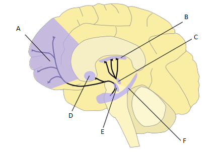
Q: Identify the regions of the brain in the picture below
Region | Name |
A | Frontal Cortex |
B | Striatum |
C | Substantia Nigra |
D | Nucleus Accumbens |
E | Ventral Tegmental Area |
F | Hippocampus
|