PERIPHERAL NERVOUS SYSTEM
Peripheral Nerves:
Consist of parallel bundles of axons surrounded by 3 connective tissue sheaths:
1. Endoneurium: Separates individual axons
2. Perineurium: Surrounds fascicles
3. Epineurium: Encloses the entire nerve trunk
Peripheral nerves Supplies either the viscera or somatic structures
Somatic peripheral nerves are usually mixed and include the sensory, autonomic, and motor axons
Cutaneous branches supply the skin and subcutaneous tissues; muscular branches supply the muscles, tendons, and joints
Peripheral axons are classified according to speed of conduction and diameter
Junctions of anterior rami form 4 nerve plexuses: Cervical, Brachial, Lumbar, Sacral
Cervical plexus: Provides cutaneous sensory information from the posterior scalp to the clavicle Innervates the anterior neck muscles and diaphragm
Brachial plexus: Entire upper limb is innervated by the brachial plexus branches
Lumbar plexus: Branches innervate the skin and muscles of the anterior and medial thigh
Sacral plexus: Innervates the posterior thigh and most of the leg and foot; contains parasympathetic axons
MOVEMENT AND NERVE HEALTH
Movement optimizes the health of nerves by promoting blood flow throughout the nerves and the flow of axoplasm through the axons. Movement causes axoplasm to thin and flow more easily
NEUROMUSCULAR JUNCTION
Motor axons synapse with muscle fibers at neuromuscular junctions. Unlike neuron-neuron synapses, no summation of action potentials is required to depolarize the postsynaptic membrane
In a normal motor unit, every depolarization of the motor axon releases sufficient acetylcholine (ACh) to initiate action potentials in the innervated muscle fibers
DYSFUNCTION OF PERIPHERAL NERVES
Signs of peripheral nerve damage include the following changes: Sensory, Autonomic, Motor
All signs are in a peripheral nerve distribution
SENSORY CHANGES Include decreased or lost sensation, Abnormal sensations include the following: Hyperalgesia, Dysesthesia, Paresthesia, Allodyni
AUTONOMIC CHANGES
Signs depend on the pattern of axonal dysfunction
Single nerve: Signs are usually observed only if the nerve is completely severed
Many nerves: Problems may include difficulty regulating blood pressure, heart rate, sweating, bowel and bladder functions, and impotence
MOTOR CHANGES
Signs of peripheral nerve damage include paresis or paralysis
If the muscle is denervated, electromyogram (EMG) recordings show no activity for approximately 1 week after the injury
Muscle atrophy progresses rapidly
Muscle fibers begin to develop a generalized sensitivity to ACh along the muscle membrane; fibrillation ensues
Fibrillation is not diagnostic of a specific lesion but always pathological
DENERVATION:
TROPHIC CHANGES: When the nerve supply is interrupted, trophic changes begin in the denervated tissues
Muscles atrophy, skin becomes shiny, nails become brittle, and subcutaneous tissues thicken
Ulceration of cutaneous and subcutaneous tissues, poor healing of wounds and infections, and neurogenic joint damage are common, secondary to blood supply changes, loss of sensation, and lack of movement
CLASSIFICATION OF NEUROPATHIES
Peripheral neuropathy can involve a single nerve, several nerves, or many nerves
Mononeuropathy: Involves a single nerve and is considered a focal dysfunction
Multiple mononeuropathy: Involves several nerves and is multifocal (i.e., asymmetrically involves individual nerves)
Polyneuropathy: Involves many nerves and is a generalized disorder that typically has a distal and symmetrical presentation
TRAUMATIC INJURY TO A PERIPHERAL NERVE: MONONEUROPATHY
Various types of trauma may injure peripheral nerves
Depending on the severity of damage, traumatic injuries to peripheral nerves are classified into 3 categories: Traumatic myelinopathy, Traumatic axonopathy, Severance
TRAUMATIC MYELINOPATHY Refers to the loss of myelin limited to the site of injury. Peripheral myelinopathies interfere with the function of large-diameter axons. Focal compression of a peripheral nerve causes traumatic myelinopathy. Repeated mechanical stimuli may cause focal compression
Carpal tunnel syndrome is a common compression injury of median nerve in the space between carpal bones and the flexor retinaculum
Carpal tunnel syndrome is more prevalent in people whose occupations require working on a cold environment or gripping of vibrating tools
TRAUMATIC AXONOPATHY Disrupts axons and Wallerian degeneration; occurs distal to the lesion
Axonopathies affect all sizes of axons; reflexes, somatosensation, and motor functions are significantly reduced or absent. Regenerating axons are able to reinnervate appropriate targets because myelin and connective tissues remain intact
SEVERANCE Occurs when nerves are physically divided by excessive stretching or a laceration. Axons and connective tissue are completely interrupted, causing immediate loss of sensation and/or muscle paralysis in the area supplied. If proximal and distal nerve stumps are apposed and scarring does not interfere, some sprouts enter the distal stump and are guided to their target tissue in the periphery
MULTIPLE MONONEUROPATHY Involves 2 or more nerves in different parts of the body. Vasculitis may cause multiple mononeuropathy. If vasculitis is suspected, an urgent referral should be made for an electrodiagnostic evaluation. Individual nerves are affected, producing a random, asymmetrical presentation of signs
POLYNEUROPATHY Hallmark signs include symmetrical involvement of sensory, motor, and autonomic fibers, often progressing from distal to proximal. Symptoms typically begin in the feet and then appear in the hands and areas of the body supplied by the longest axons. Polyneuropathies are not the result of trauma or ischemia Cause can be toxic, metabolic, or autoimmune. Most common causes include diabetes, nutritional deficiencies secondary to alcoholism, and autoimmune diseases. Variety of therapeutic drugs, industrial and agricultural toxins, and nutritional disorders can cause polyneuropathy
DIABETIC POLYNEUROPATHY Axons and myelin are damaged. Decreased sensation along with pain, paresthesias, and dysesthesias. Balance and strength training reduce the risk for falls in people with diabetic neuropathy. Gait and balance improve with exercise
One-third of patients with Guillain-Barré syndrome require a ventilator
DYSFUNCTIONS OF THE NEUROMUSCULAR JUNCTION
Myasthenia gravis is an autoimmune disease that damages ACh receptors at the neuromuscular junction. Repeated use of a muscle leads to increasing weakness
Botulism causes interference with the release of ACh from the motor axon. Produces acute, progressive weakness, with loss of stretch reflexes; sensation remains intact
IDIOPATHIC POLYNEUROPATHY Older people without diabetes also develop peripheral neuropathy. Among people over 60 with polyneuropathy.
HEREDITARY MOTOR AND SENSORY NEUROPATHY, Also known as Charcot-Marie-Tooth disease. Usually causes paresis of muscles distal to the knee, with resulting foot drop, a steppage gait, frequent tripping, and muscle atrophy. Significant numbness is unusual. Therapy involves strengthening, stretching, conditioning, and joint, muscle, and skin protection.
MYOPATHY Are disorders unique to muscles. Sensation and autonomic function remain intact because the nervous system is not affected by myopathy. Coordination, muscle tone, and reflexes are unaffected until muscle atrophy becomes so severe that muscle activity cannot be elicited
Dysfunction of peripheral nerves and the muscles they innervate can be evaluated by electrodiagnostic studies
Recording electrical activity from nerves and muscles by nerve conduction and EMG studies reveal the pathologic location and are often diagnostic
EMG differentiates between nerve and muscle disorders (neuropathy from myopathy)
Nerve conduction studies can be used to differentiate among the following: Primarily myelinopathy (or demyelinating) and axonopathy processes, Upper motor neuron (motor tract) and lower motor neuron (motor neuron) paresis, Mononeuropathy and polyneuropathy, Local conduction block and Wallerian degeneration
3 clinical signs of peripheral neuropathy are revealed in patients 50 years of age and older; presence of two or three signs correlate highly with electrodiagnostic evidence of peripheral neuropathy: Absence of ankle jerk reflex despite facilitation, Impaired vibration, Impaired position sense of the great toe
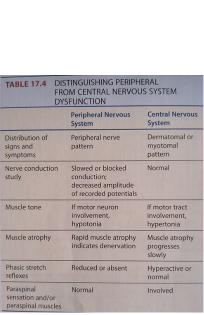
Spinal Region
Spinal region includes all neural structures contained within the vertebrae, Spinal cord, Dorsal and ventral roots, Spinal nerves, Meninges
VENTRAL AND DORSAL ROOTS
Rootlets: Are small groups of axons sending information to the periphery
Ventral root: Is the coalescence of ventral rootlets
Dorsal root: Contains sensory axons bringing information into the spinal cord; it enters the posterolateral spinal cord via rootlets
SEGMENTS OF THE SPINAL CORD
Segmental organization is a significant feature of the spinal cord. Each segment of cord is connected to a specific region of the body by axons traveling through a pair of spinal nerves. Connections of nerve rootlets to the exterior of the cord indicate segments.
SPINAL NERVES AND RAMI
Spinal nerves carry all motor and sensory axons of a single spinal segment. In the cervical region, spinal nerves are found above the corresponding vertebrae (except the eighth spinal nerve). In the remainder of the cord, spinal nerves lie below the corresponding vertebrae.
Spinal cord ends at level L1-2 intervertebral space
INTERNAL STRUCTURE OF THE SPINAL CORD
Dorsal horn processes sensory information.
Lateral horn processes autonomic information.
Ventral horn processes motor information.
Much of the gray matter is composed of spinal interneurons.
Spinal interneurons include cells that remain entirely within the gray matter and also cells whose axons travel in white matter to different levels of the cord.
MENINGES Are layers of connective tissue that surround the spinal cord. Are continuous with the meninges surrounding the brain.
Pia mater closely adheres to the spinal cord surface.
Arachnoid is separated from the pia by cerebrospinal fluid in the subarachnoid space.
Dura is the tough, outer layer
Blood is supplied to spinal cord by three spinal arteries running vertically along the cord: One in the anterior midline, Two are posterior, on either side of midline but medial to dorsal roots.
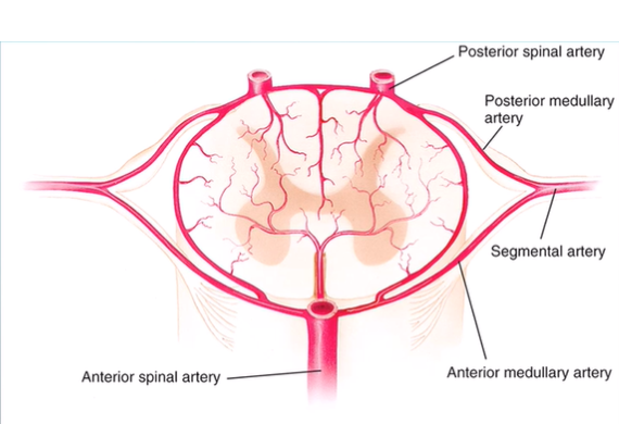
MOVEMENTS OF THE SPINAL CORD
Hip flexion produces anterior movement of the cauda equina, stretching the lumbosacral roots.
Physiologic motions do not significantly change the vertebral canal space in people with normal vertebral canals.
Extending and/or lateral bending of the neck increases the intervertebral foramen pressure at all cervical levels.
Neck extension increases cervical nerve root signs and symptom
Nerve roots and spinal nerves are protected from excessive mechanical loads by. Occupying 23% to 50% of available space within the intervertebral foramina, cushioning by fat. ligaments that maintain the spinal nerve within the intervertebral space and relieve traction on spinal nerve
FUNCTIONS OF THE SPINAL CORD
Segments of the spinal cord exchange information with other spinal cord segments, with peripheral nerves, and with the brain. Information conveyed by a motor tract to a motor neuron is only one of many influences on that motor neuron.
Afferent and descending information converges on the same spinal interneurons.
Reflexes and voluntary control act together to produce goal-oriented movements.
Reflexes are not hardwired but depend on the environmental context and the task.
By integrating volleys of peripheral, ascending, and descending inputs, spinal circuitry provides the following: Modulation of sensory information, Coordination of movement patterns, Autonomic regulation
Intraneuronal circuits integrate the activity from all sources and adjust output of motor neurons.
Interneurons coordinate activity in all the muscles when a limb moves.
What determines whether a single alpha motor neuron will fire? Summation of activity at 20,000 to 50,000 synapses determines whether an alpha motor neuron will fire.
STEPPING PATTERN GENERATORS Are adaptable neural networks that produce rhythmic output. Contribute to stepping by activating motor neurons, eliciting alternating flexion and extension at the hips and knees. Are normally activated when the person voluntarily sends signals from the brain to the stepping pattern generators in the spinal cord to initiate walking
When walking, electrical stimulation to a single point on the foot produces different responses, depending on the phase of the gait cycle. If stimulus occurs at the onset of swing phase, then tibialis anterior activity increases. If stimulus occurs at the end of swing phase, then tibialis anterior activity decreases and antagonist muscle activity increases.
REFLEXES
Spinal reflexes (except monosynaptic phasic stretch reflex) involve interneurons.
Withdrawal reflex is the capacity of interneuronal circuits to generate complex movements.
Afferent information from skin, muscles, and/or joints can elicit withdrawal movements.
Specificity of the movement pattern is referred to as a local sign indicating that the response depends on the site of the stimulation.
Sometimes information is relayed to other cord segments by collaterals of the primary afferent and by interneurons.
Crossed extension reflex example: If a person is standing when a lower limb is abruptly withdrawn, another interneuronal circuit quickly adjusts the muscle activity in the stance limb to prevent the person from falling.
INHIBITORY CIRCUITS
Interneurons in inhibitory circuits contribute to spinal cord motor coordination. Inhibitory interneurons provide: reciprocal inhibition, recurrent inhibition.
RECURRENT INHIBITION Effects opposite to reciprocal inhibition: Inhibition of agonists and synergists, disinhibition of antagonists
Renshaw cells: Interneurons that produce recurrent inhibition. Are stimulated by a recurrent collateral branch from the alpha motor neuron. Inhibit the same alpha motor neuron that gives rise to the collateral and also inhibit alpha motor neurons of synergists. Focus motor activity
Loss of descending influence on Renshaw cell activity may cause difficulty in achieving fine- motor control.
<<SPINAL CONTROL OF PELVIC ORGAN FUNCTION<<
Bowel control is similar to bladder control.
Signal to empty the bowels is stimulation of stretch receptors in the wall of the rectum. Lower spinal cord is vital for sexual function.
Erection of the penis or clitoris is controlled by parasympathetic fibers from S2 to S4 spinal cord levels; ejaculation elicited by sympathetic nerves originating in L1-L2 and the pudendal nerve with cell bodies in S2-S4.
EFFECTS OF SEGMENTAL AND TRACT LESIONS IN THE SPINAL REGION
Lesion in the spinal region may interfere with the following: Segmental function, Affects the function of a spinal cord segment, Segmental lesions interfere with neural function only at the level of the lesion.
Vertical tract function: Conveys ascending and descending information. Lesions interrupting vertical tracts result in a loss of function below the level of the lesion. Both segmental and vertical tract function
SIGNS OF SEGMENTAL DYSFUNCTION
A lesion affecting a single level of the spinal cord causes segmental signs at that level
Segmental signs include abnormal or lost sensation in a dermatomal distribution and/or motor neuron signs in a myotomal distribution
A lesion of the ventral horn, ventral root, or spinal nerve interferes with motor neuron function
SIGNS OF VERTICAL TRACT DYSFUNCTION
Lesions interrupting vertical tracts result in loss of communication to and/or from the spinal levels below the lesion
Ascending tract signs include problems with the regulation of blood pressure (BP), sweating, and bladder and bowel control
Descending tract signs include paralysis, spasticity, and muscle hypertonia; if the lateral corticospinal tract is interrupted, Babinski’s sign is present
All signs of damage to vertical tracts occur below the level of the lesion
Spinal region lesions may cause both segmental and tract signs
DIFFERENTIATING SPINAL REGION FROM PERIPHERAL REGION LESIONS
Peripheral region lesions produce deficits in the distribution of a peripheral nerve
Peripheral nerve lesions cause: altered or lost sensation in a peripheral nerve distribution, decrease or loss of muscle power in a peripheral nerve distribution, no vertical tract signs, decreased or lost phasic stretch reflex
Spinal region segmental signs occur when nerve roots and/or spinal nerves are compromised
Segmental signs include: altered or lost sensation in a dermatome, decreased or lost muscle power in a myotome, decreased or lost phasic stretch reflex
Spinal region vertical tract signs include: altered or lost sensation below the level of the lesion, altered or lost descending control of BP, pelvic viscera, and thermoregulation
motor tract signs including decrease or loss of muscle power, spasticity, muscle hypertonia, and if the lateral corticospinal tract is involved, positive Babinksi’s sign and clonus
SPINAL REGION SYNDROMES Syndrome is a collection of signs and symptoms that do not indicate a specific cause
Syndromes resulting from tumors or trauma
Anterior cord syndrome: Interferes with pain and temperature sensation and motor control
Central cord syndrome: In a small lesion, loss of pain and temperature occurs at the level of the lesion. In a large lesion, the upper limb motor function is impaired
Brown-Séquard syndrome: Below the level of lesion, voluntary motor control, conscious proprioception, and light touch are lost ipsilaterally; pain and temperature sensation are lost contralaterally
Cauda equina syndrome: Causes sensory impairment and flaccid paresis or paralysis of lower limb muscles, bladder, and bowels
Tethered cord syndrome: Causes low back and lower limb pain, difficulty walking, excessive lordosis, scoliosis, problems with bowel and/or bladder control, and foot deformities
EFFECTS OF SPINAL REGION DYSFUNCTION ON PELVIC ORGAN FUNCTION
Effects of spinal region lesions on bladder, bowel, and sexual function depend on the level of cord damage
The effect of spinal cord lesions on bowel control and sexual organ function is similar to the effect of SCI on bladder function
Sexual function is a significant issue for many following SC
TRAUMATIC SPINAL CORD INJURY
Is usually caused by motor vehicle accidents, sports injuries, falls, or penetrating wounds
First three types of injuries typically do not sever the spinal cord; damage is due to crush, hemorrhage, edema, or infarction
Penetrating wounds, by a knife or a bullet, directly sever neurons in the cord
Immediately after traumatic injury to spinal cord, cord functions below the lesion are depressed or lost (spinal shock). Is due to the interruption of descending tracts that supply tonic facilitation to the spinal cord neurons
During spinal shock, the following are lost or impaired: Somatic reflexes, including stretch reflexes, withdrawal reflexes, and crossed extension reflexes
Autonomic reflexes, including smooth muscle tone and reflexive emptying of the bladder and bowels
Autonomic regulation of BP, resulting in hypotension. Control of sweating and piloerection is lost
Several weeks after the injury most people experience some recovery of cord function, which leads to a return of the reflex activity below the lesion
In some people, spinal neurons become excessively excitable, resulting in stretch reflex hyperreflexia
Hyperreflexia develops as neuroplasticity produces new synapses in the reflex pathway
ABNORMAL INTERNEURON ACTIVITY IN CHRONIC SPINAL CORD INJURIES
Chronic SCIs: Is the period after recovery from spinal shock when the neurologic deficit is stable. The period can last for decades
Additional changes secondary to SCIs: Loss of motor neurons, changes in mechanical properties of muscle fibers (atrophy of muscle fibers, fibrosis, alteration of contractile properties toward tonic muscle characteristics
Two abnormalities occur in interneuron activity below the level of the lesion: Inhibitory interneuron response to type Ia afferent activity is diminished (correlates with hyperreflexia), Transmission from cutaneous afferents to motor neurons is facilitated (occurs because of the loss of descending inhibition)
CLASSIFICATION OF SPINAL CORD INJURIES
Whether the injury is complete or incomplete
Complete injury: Lack of sensory and motor function in the lowest sacral segment
Incomplete injury: Preservation of sensory and/or motor function in the lowest sacral segment
Neurologic level of the injury: Most caudal level with normal sensory and motor function bilaterally
American Spinal Injury Association (ASIA) developed standardized assessment for evaluating neurologic level in SCI
Key sensory points tested with safety pin to determine the ability to distinguish sharp from dull; light touches with cotton to determine ability to localize light touch
AUTONOMIC DYSFUNCTION IN SPINAL CORD INJURIES
During spinal shock, neural control of the pelvic organs is depressed. Bladder and bowel walls are atonic, allowing overfilling of these viscera, and overflow leaking occurs. Overfilling and overflow leaking can be avoided by establishing a regular bladder and bowel-emptying routine
Complete lesions at higher levels of the spinal cord cause more serious abnormalities of autonomic regulation
Loss of descending sympathetic control as a result of lesions above T6 results in three dysfunctions: Autonomic dysreflexia, Poor thermoregulation (body temperature regulation), Orthostatic hypotension
AUTONOMIC DYSREFLEXIA Medical emergency that can affect people with SCIs above T6
Stroke is the immediate life-threatening concern when someone experiences autonomic dysreflexia. Occurs when an unperceived noxious stimulus below the level of injury elicits uncoordinated autonomic control responses that cause Elevated BP, Pallor below the lesion, Sweating and flushing below the lesion, Reduced heart rate
POOR THERMOREGULATION May interfere with the ability to maintain homeostasis
Compensation: Excessive sweating may occur above the level of the lesion
People with complete lesions above the T6 level should avoid exposure to high ambient temperatures because of the risk of heat stroke
Signs of heat stroke include high body temperature; rapid pulse; and dry, flushed skin
Signs of hypothermia include irritability, mental confusion, hallucinations, lethargy, clumsiness, slow respiration, and slowing of the heartbeat
Orthostatic hypotension is a 20 mm Hg or greater fall in systolic BP or a 10 mm Hg or greater fall in diastolic BP on assuming an upright position
In people with SCI, orthostatic hypotension results from the loss of sympathetic vasoconstriction, combined with the loss of muscle-pumping action for blood return
PROGNOSIS AND TREATMENT IN SPINAL CORD INJURY
Severed axons in the adult spinal cord fail to functionally regenerate
Barriers to regeneration are inhibitory molecules on oligodendrocytes, impenetrable glial scars, and decreased rate of growth in mature neurons
Some functional losses after SCI are not due to the original trauma; rather, they are due to secondary changes such as bleeding, edema, ischemia, pain, and inflammation
Typical complications after SCI include urinary tract infection, spasticity, chills and fever, decubiti, autonomic dysreflexia, contractures, heterotropic ossification, pneumonia
Upright posture can provide some protection against urinary tract infection and pneumonia; mobility can help avoid contractures and decubiti
MENINGOMYELOCELE Is a developmental defect arising from the failure of the inferior neuropore to close. Outcomes are roughly equivalent to SCI in later life
SPASTIC CEREBRAL PALSY Is a motor disorder that develops in utero or during infancy. Is characterized by excessive muscle contraction and phasic stretch hyperreflexia
LESIONS OF DORSAL AND VENTRAL NERVE ROOTS
Radiculopathy is a lesion of a nerve root; the term also often used clinically to refer to damage of a spinal nerve. Mechanical irritation or infection produces pain in the innervated dermatome and in muscles innervated by the spinal cord segment
Erb’s paralysis is paralysis as a result of forceful separation of the head and shoulder. Birth trauma and motorcycle accidents are often causes of Erb’s paralysis
Klumpke’s paralysis is the result of avulsion of the motor roots of C8 and T1. Precipitating injury is traction on the abducted arm. Complete severance of the ventral root deprives the muscles in its myotome of motor innervation, resulting in muscle atrophy and fibrillation
LESIONS OF DORSAL ROOT GANGLIA
Dorsal root ganglia (DRG) are more sensitive to mechanical damage than the proximal or distal axons of primary nociceptive afferents
DRG compression induces alterations in the production of neuropeptides, receptors, and ion channels in primary nociceptive afferents
Sciatica: Pain radiating from low back and down the lower limb along the path of the sciatic nerve
MULTIPLE SCLEROSIS Is characterized by random, multifocal demyelination limited to the central nervous system. Signs and symptoms are exceptionally variable because the demyelination can occur in a wide variety of locations; the extent of lesion also varies. Sensory complaints may include numbness, paresthesia, and Lhermitte’s sign. Frequently produces asymmetrical weakness and ataxia of the lower limbs
TRANSVERSE MYELITIS Is a rare immune disorder that damages a limited part of the spinal cord. Signs and symptoms include myotomal weakness, dermatomal sensory loss, loss of somatosensation, bladder, bowel, and sexual dysfunction. Resulting inflammation spreads across the width of the spinal cord, producing spinal segment losses and blocking signals travelling up and down the cord
Tumors outside the dura mater or in the subarachnoid space may compress the spinal cord, nerve roots, spinal nerve, or their blood supply
Tumors can also occur within the spinal cord. Pain, aggravated by coughing or sneezing, is the most common initial symptom
VERTEBRAL CANAL STENOSIS is a narrowing of the vertebral canal, compressing the neural and vascular structures. Is usually a degenerative disorder caused by bone growth, facet hypertrophy, bulging disks, and hypertrophy of the ligamentum flavum
CERVICAL STENOSIS Signs and symptoms vary. Narrowing of intervertebral foramina compresses spinal nerves, resulting in a dermatomal distribution of abnormal sensations, pain, and numbness, along with myotomal distribution of weakness and atrophy in the upper limb. Compression of vertical tracts affects both the upper and lower limbs
LUMBAR STENOSIS Produces lower limb and lower back pain that may be aggravated by walking and improved with rest. If stenosis is severe, compression of spinal nerve roots and/or cauda equina causes additional signs and symptoms
SYRINGOMYELIA Is a rare progressive disorder most frequently occurring in people 35 to 45 years of age. Segmental signs occur in the upper limbs, including loss of sensitivity to pain and temperature stimuli, as a result of an interruption of axons crossing the midline in the anterior white commissure; paresis; and muscle atrophy. Sensory loss is often distributed similar to a cape draped over the shoulders
Signs and symptoms that indicate a spinal cord lesion are: bilateral alteration or loss of somatosensation, incoordination
motor tract signs: Decreased muscle power, spasticity, muscle hypertonia, Babinski’s sign, and clonus
Signs and symptoms that indicate a possible cauda equina lesion are: difficulty with urination and/or defecation, decreased or lost sensation in the saddle area, low back pain, unilateral or bilateral sciatica, lower limb paresis and sensory deficits, decreased or lost lower limb reflexes.
Signs and symptoms that indicate intermittent claudication are: pain in the buttock, posterior lower limb, and/or foot while walking or exercising that disappears after brief rest, decreased pulse in the lower limb, cyanosis
Brainstem Region
Brainstem is superior to the spinal cord and inferior to the cerebrum.
From inferior to superior: Medulla, Pons, Midbrain
Connections of CNs follow 2-4-3 rule: two CNs (3 and 4) connect with midbrain; four CNs (5 through 8) connect with pons; remaining three CNs (9, 10, and 12) connect with medulla
VERTICAL TRACTS IN THE BRAINSTEM
Sensory, autonomic, and motor vertical tracts travel through the brainstem. Some tracts continue through the brainstem without alteration. Brainstem acts as a conduit
LONGITUDINAL SECTIONS OF THE BRAINSTEM
Brainstem is divided longitudinally into 2 sections: basilar and tegmentum.
Midbrain has an additional longitudinal section, posterior to the tegmentum, called the tectum
Tectum includes structures involved in reflexive control of intrinsic and extrinsic eye muscles and in movements of the head: Pretectal area, Superior and inferior colliculi
Basilar section is located anteriorly and contains predominantly motor system structures: Descending axons from the cerebral cortex: corticospinal, corticobrainstem, corticopontine, corticoreticular tracts. Motor nuclei: substantia nigra, pontine nuclei, inferior olive. Pontocerebellar axons
RETICULAR FORMATION Is a complex neural network that includes the reticular nuclei, their connections, and ascending and descending reticular pathways. Integrates sensory and cortical information. Regulates somatic motor activity, autonomic function, and consciousness. Modulates nociceptive (pain) information
RETICULAR NUCLEI AND THEIR NEUROTRANSMITTERS/NEUROMODULATORS: Regulate neural activity throughout the central nervous system (CNS)
Major reticular nuclei are as follows: Ventral tegmental area (VTA), Pedunculopontine nucleus (PPN), Raphe nuclei, Locus ceruleus and medial reticular area
VENTRAL TEGMENTAL AREA: DOPAMINE Most neurons that produce dopamine are located in the midbrain
Two production areas of dopamine: Ventral tegmental: part of reticular formation, Substantia nigra: part of basal ganglia circuit
Activation of VTA affects the ventral striatum, producing feelings of pleasure and reward. Morphine is habit forming because it inhibits inhibitory inputs to the VTA.
PEDUNCULOPONTINE NUCLEUS: ACETYLCHOLINE: Is located in the caudal midbrain
Ascending axons from the PPN project to the inferior part of the frontal cerebral cortex and intralaminar nuclei of the thalamus
PPN influences movement via the following connections: Globus pallidus and subthalamic nucleus, Emotion system, Reticular areas that give rise to reticulospinal tracts
RAPHE NUCLEI: SEROTONIN Most cells that produce serotonin are found along the midline of the brainstem.
The pontine raphe nuclei modulate neural activity throughout the brainstem and in the cerebellum.
Raphespinal endings in the anterior horn provide nonspecific activation of interneurons and motor neurons.
LOCUS COERULEUS AND MEDIAL RETICULAR ZONE: NOREPINEPHRINE
Locus coeruleus and the medial reticular zone are sources of most norepinephrine in the CNS.
Axons from locus coeruleus project throughout the brain and spinal cord.
Medial reticular zone produces both norepinephrine and epinephrine. Regulates autonomic functions through projections to the hypothalamus, brainstem nuclei, and lateral horn of the spinal cord
REGULATION OF CONSCIOUSNESS BY THE ASCENDING RETICULAR ACTIVATING SYSTEM
Consciousness: awareness of self and surroundings
Consciousness system: governs alertness, sleep, and attention
Brainstem components: reticular formation and ascending reticular activating system (ARAS)
EXTERNAL ANATOMY OF THE MEDULLA Has two vertical bulges called pyramids, Lateral to the pyramids are two small oval lumps, called olives
Cranial nerve 12 connects with the medulla between the pyramid and the olive
INFERIOR MEDULLA Inferior half of the medulla contains a central canal that is continuous with the central canal of the spinal cord. Anteriorly, the descending axons of the corticospinal tract form the pyramids
Lower medulla contains cranial nerve structure
In the upper half of the medulla, the central canal widens to form part of the fourth ventricle
Tracts in the rostral medulla maintain approximately the same positions as in the caudal medulla, except that the medial longitudinal fasciculus is located more posteriorly
Most cranial nerve nuclei in the rostral medulla are clustered in the dorsal section
FUNCTIONS OF THE MEDULLA Contributes to: Controlling eye and head movements, Coordinating swallowing, Helping regulate cardiovascular, respiratory, and visceral activity
Pons processes motor information from the cerebral cortex and forwards the information to the cerebellum
Pontine cranial nerve nuclei process sensory information from the face (CN 5)
Controls contraction of muscles involved in: Processing sensation from the face (CN 5), Lateral movement of the eye (CN 6), Chewing (CN 5), Facial expression (CN 7), Also conveys info about sound, head position and head movements (CN 8)
MIDBRAIN Is the uppermost part of the brainstem. Connects the diencephalon and the pons, Is divided into 3 regions from anterior to posterior: 1. Basis pedunculi 2. Tegmentum 3. Tectum
Cerebral aqueduct, a small canal through the midbrain, joins the third and fourth ventricles
BASIS PEDUNCULI Is formed by the cerebral peduncles and the substantia nigra
Substantia nigra is one of the nuclei in the basal ganglia circuit
Other basal ganglia nuclei are the caudate, putamen, globus pallidus, PPN, and subthalamic nucleus
TEGMENTUM Contains vertical sensory tracts, superior cerebellar peduncle, red nucleus, PPN, and the nuclei of cranial nerves 3 and 4
Most vertical tracts occupy similar positions as in the pons, The spinothalamic tract and medial lemniscus are located more laterally in the midbrain
TECTUM Contains the pretectal area and the colliculi
Pretectal area is involved in the pupillary, consensual, and accommodation reflexes of the eye
Inferior colliculi relay auditory information from the cochlear nuclei to the superior colliculus and to the medial geniculate body of the thalamus
Superior colliculi are involved in reflexive eye and head movements
CEREBELLUM Function is entirely dependent on input and output connections with the brainstem
Cerebellum and brainstem share the tightly confined space of the posterior fossa
Summary of cerebellar functions: Coordination of movement, Motor planning, Cognitive functions, including rapid shifts of attention
ARTERIAL SUPPLY TO THE BRAINSTEM AND CEREBELLUM Branches of the vertebral arteries and branches of the basilar artery supply the brainstem and the cerebellum.
Near pontomedullary junction, the vertebral arteries join to form the basilar artery.
At junction of the pons and midbrain, the basilar artery divides to become the posterior cerebral arteries.
BRAINSTEM RULE OF FOUR Method for recalling brainstem anatomy and arterial supply. Developed by Gates.
In the rule of four, there are four rules 1. There are four structures near the midline that begin with M 2. There are four structures to the side that begin with S 3. There are four CNs in the medulla, four in the pons, and two in the midbrain 4. The four motor nuclei in the brainstem are numbers that divide equally into 12
BRAINSTEM RULE OF FOUR There are four structures near the midline (medial) that begin with M. Motor nuclei of CNs that innervate muscles that move the eyes (CNs 3, 4 and 6) or move the tongue (CN 12). Motor tract = corticospinal tract 3. Medial longitudinal fasciculus: neurons within the brainstem that coordinate eye and head movements. Medial lemniscus: part of the DCML pathway
There are four structures to the side (lateral) that begin with S. Sympathetic tract: raise eyelid and pupillary dilator (efferents). Spinothalamic tract (fast nociception and temperature). Sensory tract of trigeminal nerve: trigeminal lemniscus (fast nociception and temperature). Spinocerebellar tract (unconscious proprioception)
There are four CNs in the medulla, four in the pons, and two in the midbrain
Medulla = 8, 9, 10, 12
Pons = 5, 6, 7, 8
Midbrain = 3, 4
DISORDERS IN THE BRAINSTEM REGION
Evaluating function of cranial nerves and vertical tracts can localize lesions within the brainstem
Single brainstem lesion may cause a mix of ipsilateral and contralateral signs
Occur because cranial nerves supply the ipsilateral face and neck; many of the vertical tracts cross midline in the brainstem to supply the contralateral body
Lesions in the brainstem may also interfere with vital functions and consciousness
VERTICAL TRACT SIGNS
Lateral corticospinal, dorsal column medial lemniscus, and spinothalamic tracts connect the spinal cord with the contralateral cerebrum
Lesions of lateral corticospinal and dorsal column tracts in the brainstem usually cause contralateral signs because these tracts cross the midline in the inferior medulla
CORTICOBRAINSTEM LESIONS
Convey motor signals from cerebral cortex to CN nuclei in brainstem. Neurons with axons in the corticobrainstem tract serve as motor tract neurons to the motor neurons in CNs 5, 7, 9, 10, 11, and 12
Motor tract lesions are associated with muscle hypertonia
Motor neuron lesions are associated with hyporeflexia and muscle flaccidity
CONTRALATERAL AND IPSILATERAL SIGNS
Single lesion in the upper anteromedial medulla on left side can cause: Paralysis of the right hand and foot Loss of discriminative touch and proprioceptive information on the right side of the body, Paresis of the left side of the tongue.
DISORDERS OF VITAL FUNCTIONS Disruption of vital functions secondary to brainstem damage may cause: Heart to stop beating, Blood pressure to fluctuate, Breathing to cease, Areas in the medulla and pons regulate vital functions
FOUR DS OF BRAINSTEM REGION DYSFUNCTION Cardinal signs of brainstem dysfunction:
Dysphagia: difficulty in swallowing
Dysarthria: difficulty in speaking
Diplopia: double vision
Dysmetria: inability to control the distance of movement
DISORDERS OF CONSCIOUSNESS States of altered consciousness may occur with lesions to either the brainstem or the cerebrum. Brainstem damage affecting reticular formation and/or axons of the ARA system interfere with consciousness.
Damage to cerebrum interfering with hypothalamic/thalamic activating areas or with the function of the entire cerebral cortex may also impair consciousness
Tumors within cerebellum or brainstem cause increased intracranial pressure
Pressure may cause headache, nausea, vomiting, cranial nerve disorders, or hydrocephalus
If the tumor is within the cerebellum, ataxia commonly occurs
Damage caused by a benign tumor may be extensive because the unyielding bone and dura prevent brain tissue from moving away from the pressure
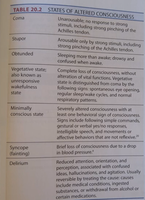
Ischemia in the brainstem produces abrupt onset of neurologic symptoms including dizziness, visual disorders, weakness, incoordination, and somatosensory disorders
Vertebrobasilar artery insufficiency produces transient symptoms of brainstem region ischemia when the neck is extended and rotated
Cranial Nerves
Cranial Nerves Exchange information between the peripheral nervous system (PNS) and the central nervous system (CNS). Serve sensory, motor, and autonomic functions. Differ from spinal nerves in specialization; some are only motor, others are only sensory, and some are both sensory and motor
Cranial nerves have 4 functions: Supply motor innervation to the muscles of the face, eyes, tongue, jaw, and two neck muscles. Transmit somatosensory information from the skin and muscles of the face and from the temporomandibular joint (TMJ). Transmit special sensory information related to visual, auditory, vestibular, gustatory, olfactory, and visceral sensations. Provide parasympathetic regulation of pupil size, curvature of the lens of the eye, heart rate (HR), blood pressure (BP), breathing, and digestion
Olfactory nerve (1) is sensory Sense of smell is dependent on olfactory nerve function. Much of the information attributed to taste is olfactory in origin because the information from taste buds is limited to chemoreceptors for salty, sweet, sour, umami (i.e. “savory”), and bitter
Optic nerve (2) is sensory. Retina is the inner layer of the posterior eye; light striking the retina is converted into neural signals by the photosensitive cells. Visual signals sent to the midbrain are involved in reflexive responses of the pupil, awareness of light and dark, and orienting the head and eyes.
Oculomotor (3), trochlear (4), and Abducens (6) nerves are primarily motor. Contain motor neuron axons innervating the six extraocular muscles that move the eye and control reflexive constriction of the pupil
PUPILLARY, CONSENSUAL, AND ACCOMMODATION REFLEXES Involve the optic and oculomotor nerves
Pupillary and consensual reflexes are elicited by the same stimulus (e.g., shining a bright light into one eye).
Pupillary reflex is the constriction of the pupil in the eye directly stimulated by the bright light. Consensual reflex is the constriction of the pupil in the other eye
Trigeminal nerve (5): mixed nerve containing both sensory and motor fibers. Sensory fibers transmit information from the face and TMJ. Trigeminal nerve named for its 3 branches: Ophthalmic, Maxillary, Mandibular
Facial (7): Innervates muscles of facial expression and most glands in the head; also conveys sensory information from the posterior ear canal and taste from the anterior tongue. Carries efferent signals for the corneal reflex. Signals to and from CN VII are processed in the nuclei located in the pons, medulla, and upper spinal cord
Vestibulocochlear (8): Sensory nerve with 2 distinct branches: Vestibular branch transmits information related to head position and head movement, Cochlear branch transmits information related to hearing. Peripheral receptors for these functions are located in the inner ear (the labyrinth)
Labyrinth consists of the vestibular apparatus and the cochlea.
Sound is converted to neural signals by a sequence of mechanical actions. When sound waves enter the external ear, vibration moves the ossicles
Ossicles vibrate the membrane at the opening of the upper chamber, moving the fluid contained in the upper chamber, Fluid moves inside the cochlea, vibrating the basilar membrane and its attached hair cells
Auditory information Orients the head and eyes toward sounds. Increases the activity level throughout the CNS. Provides conscious awareness and recognition of sounds
From the cochlear nuclei, auditory information is transmitted to 3 structures:
Reticular formation: Connections account for the activating effect of sounds on the CNS.
Inferior colliculus: Directly and via the superior olive, auditory information is integrated from both ears to detect the location of sounds.
Medial geniculate body: Serves as a thalamic relay station for auditory information to the primary auditory cortex.
3 cortical areas are dedicated to processing auditory information:
Primary auditory cortex is the site of conscious awareness of the intensity of sounds
Auditory association cortex compares sounds with memories of other sounds, then categorizes the sounds as language, music, or noise
Wernicke’s area is where comprehension of spoken language occurs
GLOSSOPHARYNGEAL (9): Mixed nerve containing both sensory and motor fibers. Sensory fibers transmit somatosensation from the soft palate and pharynx and information from taste receptors in the posterior tongue. Motor component innervates a pharyngeal muscle and the parotid salivary gland.
Vagus Nerve (10): Provides afferent and efferent innervation of the larynx, pharynx, and viscera. Far-reaching connections allow the vagus to decrease HR, constrict the bronchi, affect speech production, and increase digestive activity. Motor function can be tested by eliciting the gag reflex
Accessory (11): Provides innervation to the trapezius and sternocleidomastoid muscles. Originates in the spinal accessory nucleus in the upper cervical cord, travels upward through the foramen magnum, and then leaves the skull through the jugular foramen. Cell bodies are in the ventral horn at levels C1 to C4
Hypoglossal (12): Provides innervation to the intrinsic and extrinsic muscles of the ipsilateral tongue. Cell bodies are located in the hypoglossal nucleus of the medulla. Activity of the hypoglossal nerve is controlled by both voluntary and reflexive neural circuits.
Swallowing: Involves 3 stages, Oral, Pharyngeal and laryngeal, Esophageal
SPEAKING Requires cortical control At the CN level, sounds generated by the larynx (CN 10) are articulated by the soft palate (CN 10), lips (CN 7), jaws (CN 5), and tongue (CN 12)
SYSTEMS CONTROLLING CRANIAL NERVE MOTOR NEURONS CNs 3-7 and 9-12 contain motor neuron fibers. Activity of the motor fibers in CNs are controlled through descending inputs from both voluntary and limbic structures of the brainstem and cerebrum and also via local reflex mechanism
DESCENDING CONTROL OF MOTOR CRANIAL NERVES CN efferents receive descending regulation by the corticofugal tracts and the memory system. Descending limbic pathways are separate from corticobulbar tracts. Eye movements can be voluntarily controlled, or the eyes may be automatically drawn toward or avoid disturbing sights. Speaking is mostly voluntary but can occur automatically in highly emotional contexts
In instances in which brain damage interferes with voluntary speech, the ability of the memory system to produce emotionally charged words, such as profanity, may be preserved
Extreme emotions, by activating limbic pathways that influence motor activity, can interfere with the ability to eat and speak
Lesions of olfactory nerve can result in an inability to smell. Smoking or excessive nasal mucus may also interfere with the function of the olfactory nerve
Complete interruption of the optic nerve results in ipsilateral blindness and the loss of the pupillary light reflex. Pupillary light reflex is the constriction of the pupil in response to a light shining into the eye. Loss of reflex may also occur with a lesion of CN 3 because the oculomotor nerve is the efferent limb of the reflex
Complete lesion of oculomotor nerve causes: Ptosis, Ipsilateral eye looks outward and down, Diplopia, Deficits in moving the ipsilateral eye medially, downward, and upward
Loss of pupillary reflex and consensual response to light. Loss of constriction of the pupil in response to focusing on a near object.
Lesion of trochlear nerve prevents activation of the superior oblique muscle; consequently, the ipsilateral eye cannot look downward and inward. Symptoms include double vision, difficulty reading, and visual problems when descending stairs. Other possible causes of eye movement asymmetry must be ruled out
Complete severance of any branch of the trigeminal nerve results in anesthesia of the area supplied by the ophthalmic, maxillary, or mandibular branch. If the ophthalmic division is affected, afferent limb of the blink reflex will be interrupted, preventing blinking in response to touch stimulation of the cornea. If the mandibular branch is severed, the jaw will deviate toward the involved side when the mouth is opened; the masseter reflex will be lost.
Trigeminal Neuralgia: Dysfunction of the trigeminal nerve that produces severe, sharp, stabbing pain in the distribution of one or more branches of the trigeminal nerve. In most cases, pressure of a blood vessel on the nerve causes local demyelination and ectopic foci that sensitize the trigeminal nerve root and the trigeminal nerve nucleus.
Complete lesion of the abducens nerve will cause the eye to look inward; paralysis of the lateral rectus muscle leaves the pull of the medial rectus muscle unopposed. Person with this lesion will be unable to voluntarily abduct the eye and will have double vision. Other causes of asymmetrical eye movements must be ruled out
FACIAL NERVE Lesion of the facial nerve causes paralysis or paresis of the ipsilateral muscles of facial expression. Unilateral facial palsy can result from a lesion of the CN 7 nucleus or from a lesion of the axons of CN 7
RAMSAY HUNT SYNDROME Facial and vestibulocochlear nerves are both affected. Caused by varicella zoster infection. Usually consists of acute facial paralysis accompanied by ear pain and blisters on the external ear
VESTIBULOCOCHLEAR NERVE AND DISORDERS OF THE AUDITORY SYSTEM
Loss of hearing in one ear interferes with the ability to locate sounds; normally, the timing of input from each ear is compared with the location of the sounds in space Deafness as a result of peripheral disorders is classified as either conductive or sensorineural
Conductive deafness occurs when transmission of vibrations is prevented in the outer or middle ear. Common causes include excessive wax in the outer ear canal or otitis media
Sensorineural deafness is due to damage of the receptor cells or the cochlear nerve and is less common than conductive deafness. Common causes include acoustic trauma, ototoxic drugs, Ménière's disease, and acoustic neuroma
Tinnitus in the form of infrequent, mild, and high-pitched “ringing” that lasts for seconds to minutes is normal, particularly in quiet environments. May be caused by medications, stimulation of receptors in the ear, or central sensitization after deafferentation
Treatment for tinnitus includes masking sounds provided by a hearing aid, medication, habituation techniques, and/or transcranial magnetic stimulation of the central auditory system
Glossopharyngeal nerve Complete lesion interrupts the afferent limb of both the gag reflex and the swallowing reflex. Salivation is also decreased
Complete lesion of vagus nerve results in difficulty speaking and swallowing, poor digestion, asymmetrical elevation of the palate, and hoarseness
Complete lesion of accessory nerve causes paralyzes the ipsilateral sternocleidomastoid and trapezius muscles. Motor tract (upper motor neuron) lesions cause paresis rather than paralysis because cortical innervation is bilateral, and the muscles become hypertonic rather than hypotonic.
Complete lesion of hypoglossal nerve causes atrophy of the ipsilateral tongue. When a person with this lesion is asked to stick out the tongue, the tongue protrudes ipsilaterally rather than in the midline. Problems with tongue control result in difficulty speaking and swallowing
Dysphagia is difficulty with swallowing. Frequent choking, lack of awareness of food in one side of the mouth, or food coming out of the nose may indicate dysfunctions of CNs 5, 7, 9, 10 or 12. Motor tract lesions may also cause swallowing dysfunctions
Dysarthria is poor control of the speech muscles. Only vocal speech is affected; people with dysarthria can understand the spoken language and can write and read
Causes of dysarthria: Motor neuron (lower motor neuron) involvement of CNs 5, 7, 10 or 12. Motor tract (upper motor neuron) lesions or muscle dysfunction
Sensory: olfactory (I), optic (II), vestibulocochlear (VIII)
Motor (with some sensory fibers): oculomotor (III), trochlear (IV), abducens (VI), accessory (XI), hypoglossal (XII)
Mixed (both sensory and motor): trigeminal (V), facial (VII), glossopharyngeal (IX), vagus (X)
VISUAL SYSTEM
SIGHT: INFORMATION CONVEYED FROM THE RETINA TO THE CORTEX
Visual pathway begins with cells in the retina that convert light into neural signals. Signals are processed within the retina and are conveyed to the retinal output cells. Retinal output is conveyed by the axons that travel in the optic nerve, optic chiasm, and optic tract; synapses in the lateral geniculate nucleus of the thalamus
Optic nerve is the bundle of axons that pass from the retina to the optic chiasm. Nerves merge at the optic chiasm, where some axons cross the midline. Optic tract conveys visual information from chiasm to the lateral geniculate
PROCESSING OF VISUAL INFORMATION
Information reaching the primary visual cortex stimulates neurons that discriminate the shape, size, or texture of objects
Information conveyed to the visual association cortex is analyzed for colors and motion
Action stream: Is a stream of visual information that flows dorsally and used to direct movement
Perception stream: Is a stream of visual information that flows ventrally and used to recognize visual objects
DISORDERS OF THE VISUAL SYSTEM: Consequences of damage along the retinogeniculocortical pathway vary according to the location of the lesion. Visual losses are described by referring to the visual field deficit
OPTIC NERVE LESIONS Complete interruption of the optic nerve results in ipsilateral blindness; loss of direct pupillary light reflex. Optic nerve is entirely myelinated by oligodendroglia and is frequently affected by multiple sclerosis
Visual loss is described by referring to the visual field deficit
Complete lesion of the retina or of the optic nerve results in total loss of vision in ipsilateral eye.
Bitemporal hemianopia: Loss of information in both temporal visual fields
Homonymous hemianopia: Loss of visual information from the same visual field, right or left, in both eyes
Cortically blind: The person has no awareness of any visual information due to a lesion in the brain
Blindsight: The ability of a cortically blind individual to orient to, point to, or detect movements of visual objects
(Oculomotor) CN 3 has parasympathetic neurons that innervate the intrinsic muscles of the eye: the pupillary sphincter and the ciliary muscle
Accommodation: increases refraction of light rays so that the focal point will be maintained on the retina
THE PUPILLARY LIGHT REFLEX Elicited by shining a bright light into one eye
Shining light into one eye causes pupil constriction in the eye directly stimulated by bright light. Pathway consists of neurons that sequentially connect: the retina to the pretectal nucleus in the midbrain, the pretectal nucleus to the parasympathetic nuclei of the oculomotor nerve, parasympathetic nuclei of the oculomotor nerve to the ciliary ganglion, ciliary ganglion to the pupillary sphincter muscle
THE NEAR TRIAD Consists of adjustments to view a near object: 1. the pupils constrict 2. the eyes converge 3. the lens become more convex.
The accommodation reflex requires activation of the visual cortex and an area in the frontal lobe of the cerebral cortex (frontal eye field)
Oculomotor, trochlear, and abducens nerves are primarily motor, containing motor neurons innervating the six extraocular muscles that move the eye
Extraocular muscles include four straight (rectus) muscles and two oblique muscles, Two oblique muscles attach to the posterior half of the eyeball
When eye is adducted, the superior oblique muscle depresses and the inferior oblique muscle elevates the eye
CN 3 (oculomotor nerve): Controls contraction of the superior, inferior, and medial rectus
CN 4 (trochlear nerve): Controls the superior oblique muscle, which rotates the eye, or, if the eye is adducted, depresses the eye
CN 6 (abducens nerve): Controls the lateral rectus muscle, which moves the eye laterally
Coordination of the two eyes is maintained via synergistic action of the extraocular muscles
Signals conveyed by the Medial Longitudinal Fasciculus (MLF) coordinate head and eye movements by providing bilateral connections among vestibular and ocular motor nuclei in the brainstem and spinal accessory nerve nuclei in the spinal cord
Lesions affecting the CNs (3, 4, and 6) that innervate extraocular muscles or the MLF cause misalignment of the eyes. With suppression of vision from one eye, the person will lose depth perception
Complete lesion of the oculomotor nerve causes: Severe ptosis (drooping of eyelid), Ipsilateral eye is aimed outward and down, Diplopia (double vision), Deficits in moving ipsilateral eye medially, downward, and upward, Loss of direct (ipsilateral) pupillary light reflex, Loss of constriction of pupil in response to focusing on a near object
Lesion of trochlear nerve prevents activation of superior oblique muscle. People with lesions of trochlear nerve complain of double vision, difficulty reading, and visual problems when descending stairs
Complete lesion of abducens nerve will cause eye to deviate inward. A person with this lesion will be unable to voluntarily abduct the eye and will have double vision
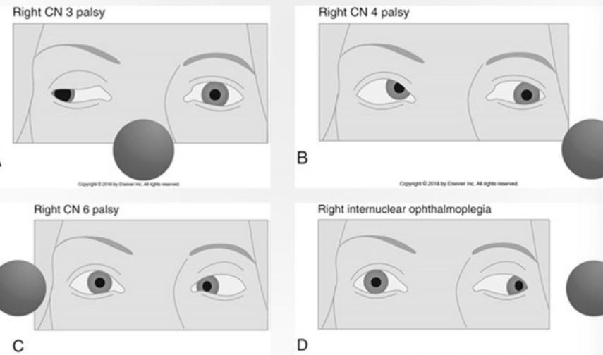 A lesion affecting the MLF produces internuclear ophthalmoplegia by interrupting signals from abducens nucleus to the oculomotor nerve. When connection between the abducens nucleus and the oculomotor nucleus is interrupted, the eye contralateral to the lesion moves normally, but the eye ipsilateral to the lesion cannot adduct past midline when contralateral eye moves laterally
Superior colliculus coordinates reflexive orienting movements of the eyes and head via the MLF
Two objectives of eye movement: Keeping position of the eyes stable during head movements, Directing the gaze at visual targets
Gaze stabilization (aka visual fixation) during head movements is achieved by the vestibulo-ocular reflex (VOR) optokinetic nystagmus
Direction of gaze is accomplished by: saccades, smooth pursuits, vergence movements
VESTIBULO-OCULAR REFLEXES Stabilize visual images during head movements.
Vestibular receptors for the VOR are in three fluid-filled tubes inside each inner ear called semicircular canals. Stimulating a pair of semicircular canals induces eye movements in roughly the same plane as the canals
OPTOKINETIC NYSTAGMUS Adjusts eye position during slow head movements. When a person is walking, the head moves relative to objects in the environment. Is elicited by moving visual stimuli. Allows the eyes to follow large objects in the visual field
Physiologic nystagmus: Normal response that can be elicited in an intact nervous system by optokinetic stimulation, rotational or temperature stimulation of the semicircular canals, or by moving eyes to extreme horizontal position
Pathologic nystagmus: Sign of nervous system abnormality
Adjusting relative levels of activity of the paramedian pontine reticular formation and midbrain reticular formation controls diagonal saccades
Saccades can be generated voluntarily or elicited by a variety of stimuli (e.g., visual, tactile, auditory, nociceptive)
Smooth pursuit eye movements are used to follow a moving object. Example: Watching someone walk across a room.
Moving visual stimulus is essential for the production of smooth pursuit movements.
Convergence of eye movement: During reading and other activities in which the visual object is near eyes; eyes are aimed toward midline to allow image to fall on corresponding areas of retinas
Trophia: Deviation of one eye from forward gaze when both eyes are open
Phoria: Deviation from forward gaze apparent only when person is looking forward with one eye and other eye is covered
Binocular fusion: Blending of the image from each eye to become a single image
Abnormalities of eye movement occur with lesions involving: CNs that control extraocular muscles, neuromuscular junction or extraocular muscles, MLF, vestibular system, cerebellum, eye fields in the cerebral cortex
MOTION SICKNESS Is nausea, headache, anxiety, and vomiting experienced in moving vehicles, May be caused by a conflict between different types of sensory information or by postural instability. Seasickness may be caused by a conflict between visual and vestibular information
Vestibular
Vestibular information is essential for postural control and for the control of eye movements.
Vestibular apparatus contains sensory receptors that respond to the position of the head relative to gravity and to head movements
Information is converted into neural signals that are conveyed by the vestibular nerve to the vestibular nuclei
Projections from the vestibular nuclei contribute to: sensory information about head movement and head position relative to gravity, gaze stabilization (i.e., control of eye movements when the head moves), postural adjustments, autonomic function and consciousness
VESTIBULAR APPARATUS Consists of bony and membranous labyrinths and hair cells, Receptors inside the membranous labyrinth are hair cells, Bending of the hairs determines the frequency of signals conveyed by the vestibular nerve
Receptors in semicircular canals detect movement of the head (rotation, angular, not affected by gravity) by sensing the motion of endolymph Is made up of three hollow rings arranged perpendicular to each other. Each opens at both ends into the utricle; has an ampulla that contains a crista
Crista: Is a body of supporting cells and sensory hair cells. Hair cells are embedded in a gelatinous mass called the cupola
Each canal in a pair produces reciprocal signals; increased signals from one canal occur simultaneously with decreased signals from its partner. Reciprocal signals are essential for normal vestibular function. If signals from a pair of semicircular canals are not reciprocal, then difficulties will result with the control of posture, abnormal eye movements, and nausea
Utricle and saccule: Both are otolithic organs; membranous sacs within the vestibular apparatus. Are not sensitive to rotation but respond to head position relative to gravity and to linear acceleration and deceleration. Macula are hair cells enclosed by a gelatinous mass topped by calcium carbonate crystals located within the utricle and saccule
Otoconia are calcium carbonite crystals that are more dense than the surrounding fluid and gelatinous support
Changing head position tilts the macula; the weight of the otoconia displaces the gelatinous mass, bending the embedded hairs. Bending hairs stimulates or inhibits the hair cells, depending on the direction of the bend, which determines the frequency of firing of neurons in the vestibular nerve
VESTIBULAR NERVE Transmits information from semicircular canals and otolithic organs to the vestibular nuclei in the medulla and pons and to the flocculonodular node of the cerebellum. Peripheral part of vestibular system consists of vestibular apparatus and peripheral part of vestibular nerve
Most common symptom of vestibular system dysfunction is vertigo. Pathologic vertigo occurs with both peripheral and central disorders and arises from disturbance of spatial orientation in the vestibular cortex
Vestibular disorders may also cause pathologic nystagmus, unsteadiness, ataxia, nausea, vomiting
PERIPHERAL VESTIBULAR DISORDERS Typically cause recurring periods of vertigo, accompanied by moderate to severe nausea. Nystagmus almost always accompanies peripheral vertigo. Certain drugs may also cause peripheral vestibular damage
BENIGN PAROXYSMAL POSITIONAL VERTIGO Inner ear disorders that cause the acute onset of vertigo and nystagmus are
Benign: Is not malignant
Paroxysmal: Has a sudden onset of a symptom or disease
Positional: Denotes head position as the provoking stimulus. Rapid change of head position, resulting in vertigo and nystagmus, will subside in less than 2 minutes, even if the provoking head position is sustained
Activities that frequently provoke benign paroxysmal positional vertigo (BPPV) Getting into or out of bed, Bending over to look under a bed, Reaching up to retrieve something from a high shelf, Turning over in bed Are caused by the displacement of otoconia from the macula into a semicircular canal
The attachments of otoliths to the cupula cause atypical BPPV. Characterized by more intense dizziness and longer duration than typical BPPV, no latency before onset, and prolonged persistence of dizziness and nystagmus when provoking position is maintained
VESTIBULAR NEURITIS Is an inflammation of the vestibular nerve and is usually caused by a virus. Symptoms include disequilibrium, spontaneous nystagmus, nausea, and severe vertigo (for up to 3 days). Hearing is unaffected, Caloric testing shows decreased or absent response on the involved side. During the acute phase, medication may be used to suppress nausea, vertigo, and vomiting
MÉNIÈRE’S DISEASE Causes a sensation of fullness in the ear, tinnitus, severe acute vertigo, nausea, vomiting, and hearing loss. Is associated with abnormal fluid pressure in the inner ear; authorities are not certain if this is the cause or an effect. Drugs that suppress vertigo are useful during acute attacks; in the extreme, the vestibular nerve may be surgically severed to relieve symptoms
BILATERAL LESIONS OF THE VESTIBULAR NERVE Interfere with reflexive eye movements in response to head movement; the initial complaint is usually oscillopsia
Oscillopsia is the subjective sensation of visual objects bouncing when the head is moving. The world seems to bounce up and down as the person walks because normal reflexive adjustments for head movement are decreased. Over time, the nervous system adapts to the change; people report less difficulty with disorienting movements of the visual field
CENTRAL VESTIBULAR SYSTEM Comprised of 4 nuclei, 6 pathways, the vestibulocerebellum and the vestibular cortex
Vestibular system has two roles in motor control Gaze stabilization, Postural adjustment
PERCEPTION: INHIBITORY VISUAL-VESTIBULAR INTERACTION IN THE CEREBRAL CORTEX Activity in the visual cortex and the vestibular cortex is reciprocally inhibitor. Visual inhibition can be experienced by comparing visual details you see when turning your head and eyes slowly from one side of the room to the other side. When vestibular activity increases, visual details are suppressed
CENTRAL VESTIBULAR DISORDERS Are the result of damage to the vestibular nuclei or to the connections within the brain. Typically produce milder symptoms than peripheral disorders. Are most commonly the result of ischemia or tumors in the brainstem/cerebellar region, cerebellar degeneration, multiple sclerosis, or Arnold-Chiari malformation
LESIONS OF THE VESTIBULOTHALAMOCORTICAL PATHWAY Create abnormal perception of vertical without vertigo. No dizziness occurs because the signals in the vestibular nuclei are symmetric. People with lesions that affect the vestibular system superior to the vestibular nuclei experience head tilt, misidentification of vertical, and lateropulsion
VESTIBULAR MIGRAINE Migraine may cause vestibular dysfunction. Diagnosis of vestibular migraine based on dizziness symptoms that do not fit other syndromes, plus a history of migraine, family history of migraine, and susceptibility to motion sickness. Vestibular rehabilitation decreases imbalance and the severity of dizziness in people with migrainous vertigo
PERSISTENT POSTURAL-PERCEPTUAL DIZZINESS Dizziness and unsteadiness that persists for more than 3 months, Worst with upright posture. Aggravated by motion of person, environment, or visual demands.
Events that frequently precede persistent postural-perceptual dizziness (3PD): Vestibular disorders, Mild traumatic brain injury, Anxiety disorders, Depression, Medical problems or medications that cause dizziness/unsteadiness
__UNILATERAL VESTIBULAR LOS__S Causes problems with posture, eye movement control, and nausea. Signals from the damaged side are not correctly balanced with signals from the intact side
Peripheral lesion interfering with otolithic function on one side causes imbalance; information from otoliths on the normal side are not balanced by information from lesioned side. Affects the vestibulospinal system, producing a tendency to fall toward the side of the lesion
BILATERAL VESTIBULAR LOSS Bilateral loss of otolith input eliminates a person’s internal sense of gravity. Person must rely on visual and proprioceptive cues for spatial orientation • Creates difficulty walking in the dark and walking on uneven surfaces • No asymmetry of the vestibular information equals no vertigo
EVALUATING THE VESTIBULAR SYSTEM Patients reporting dizziness are often describing different experiences. Clinicians must distinguish among Vertigo: Illusion of movement, Near syncope: Feeling of impending faint, Disequilibrium: Loss of balance, Light-headedness: Inability to concentrate
SENSORY TESTING Results can be used to localize a vestibular lesion. Hearing, proprioception, and vibration are tested. Because impaired proprioception can cause unsteadiness, proprioception and vibration tests are used to distinguish between lesions of the conscious proprioception pathways and vestibular lesions
SPECIALTY CLINIC TESTING OF VESTIBULO-OCULAR REFLEXES The gain of the vestibulo-ocular reflex (VOR) depends on the frequency of the stimulus. VOR may be tested in five ways:
- By passive, rapid head turns
- By testing dynamic visual acuity
- By use of rotating chair
- By caloric testing
- By electronystagmography
Rehabilitation does not directly affect central dysfunctions of the vestibular system and is ineffective for active Ménière's disease. Rehabilitation is effective for BPPV, unilateral vestibular loss or dysfunction and bilateral vestibular loss, and central vestibular disorders that benefit from movement retraining
Exercises are designed to promote movement retraining, habituation, or substitution to improve function.
Movement retraining: Practicing and modifying movements
Habituation: Exposure to positions or movements that produce symptoms, followed by relaxation until the symptoms abate
Substitution: Using alternative sensory inputs or motor responses or using predictive or anticipatory strategies