Obstetrics!
important medical HX… conditions….
cystic fibrosis
sickle cell disease
type 2 diabetes = close relatives w/it = gestational diabetes more likely
pre-eclampsia = if maternal mother or sister was affected, increased risk
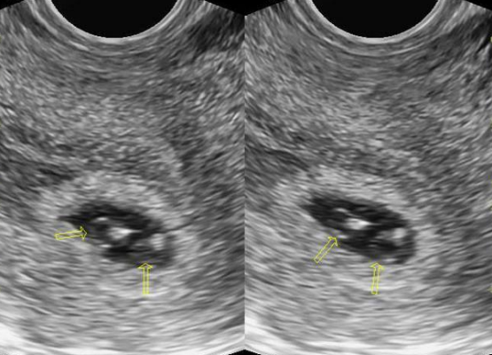
4-5 wks = gestational sac, first sign
5.5 wks = yolk sac, Important role in human embryonic development. Transfer of nutrients to the developing embryo in the third and fourth weeks
6 wks = embryo pole, diamond ring, lil dot (embryo)
7-8 wks = amnion, Embryo is attached to yolk sac via vitelline duct but is within the Amnion. The YS is seen out of the Amnion. By 16 weeks the amnion will grow to fill the chorionic cavity completely
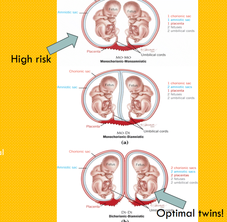
chorion (CORD/PLACENTA) vs amniotic sac
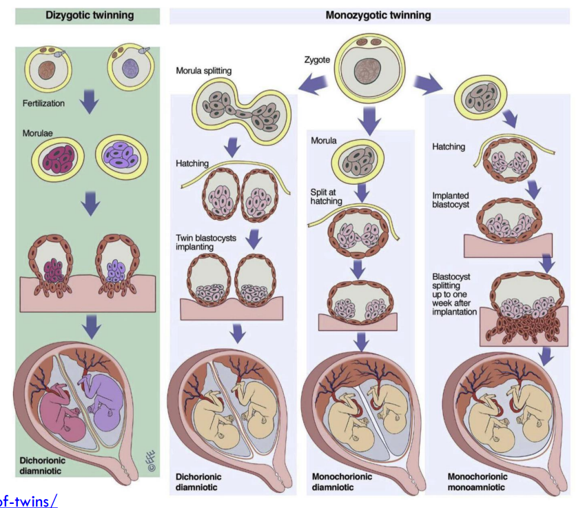
MO-MO = monochorionic + monoamniotic = usually separate during implantation
MO-DI = monochorionic + diamniotic = separate before implantation around hatching
DI-DI = dichorionic + diamniotic = usually split at the morula
Viability exam - first trimester
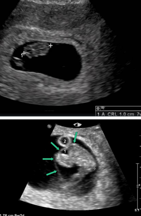
Fetal number
Location of gestational sac (GS)
Measure Yolk sac
Measure GS
Crown rump length (CRL)
Fetal cardiac activity (m-mode)
Visualize the ovaries
Posterior cul de sac→ pouch of Douglas!
SCREENING → trisomy 21, 13, 18
maternal age
gestational age
nuchal translucency
maternal serum markers
additional things that are examined
EDD = estimated due date
fetal HR
brain structures & measurements
abdomen
placenta location
bladder
extremities
nasal bone
2nd trimester
Cervical assessment
Placental assessment
Fetal Cardiac activity using → M Mode
Detailed imaging of Fetal organs..What is included?
Fetal situs (position of organs - heart/bowels)
Amniotic fluid
3rd trimester
Cervical assessment <32 weeks
Placental Localization
Fetal Presentation Breech vs cephalic
Quick assessment of Fetal Biometry (BPD-biparietal diameter, HC- head circumference, AC-abdominal circumference, FL- femur length)
Amniotic fluid assessment (Polyhydramnios vs. Oligohydramnios)
Doppler assessment
MRI → may be used if additional imaging is necessary in pregnancy (cleft lip, club foot, heart defects
Amniocentesis
Prenatal diagnosis of chromosome defects as well as metabolic disorders
Family history or previous child with a genetic disease or metabolic disorder, such as Down syndrome
Risk of open neural tube defects, such as spina bifida
Maternal age over 35 years by the pregnancy due date
Abnormal maternal screening tests
Chorionic villus sampling = 10-12 wks, done as early as 8wks and as late as 16wks, examines placental cells for fetal abnormalities
Cordocentesis = done after 18wks quick results
Fetoscopic laser photocoagulation
allows the OB to see the fetus and intrauterine environment during pregnancy. Used to help diagnose anomalies that may be difficult to assess by US ex: NTD or to treat certain conditions ex: TTTS, fetal biopsies, spina bifida
TTTS = twin-to-twin transfusion syndrome, shared placenta between twins, uneven blood supply, one is more nourished and is larger