Let's Get Pumping! (Lesson 7.01)
Overview of Blood
Blood is not merely a liquid; it is a specialized tissue composed of cells and dissolved chemicals that serve critical functions in the body.
Blood maintains homeostasis through the circulation of its components in relation to the heart and lungs.
Blood Tissue
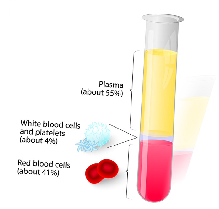
Blood consists of:
Fluid: Plasma
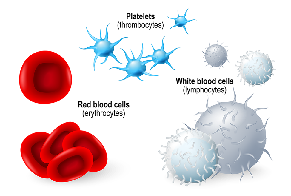
Cells: Red blood cells (RBCs), white blood cells (WBCs), platelets, and cell fragments.
Plasma:
Described as a straw-colored fluid, consisting of 90% water and 10% solutes.
Solutes in plasma include:
Proteins (e.g., albumin, fibrinogen, globulins, prothrombin)
Nutrients, waste products, gases, and substances that help in homeostasis.
Makes up 55% of blood volume.
Functions of Plasma Proteins
Clotting: Essential for hemostasis (the process of blood clotting).
Immune System: Essential for mounting an immune response.
Viscosity and Volume: Maintain the consistency of blood viscosity and overall blood volume.
Osmotic Pressure: Regulates fluid balance within and outside of cells, facilitating homeostasis across body systems.
Impact of Liver Disease on Blood
Liver disorder leads to:
Impaired immunity due to reduced plasma protein production.
Impaired blood clotting functions.
Potential disruptions in electrolyte and fluid balance.
Percentages in Blood Composition
Plasma and other solutes account for 55% of blood, while the remaining 45% is made up of formed elements.
In a typical drop of blood:
Approximately 5 million RBCs.
7,500 WBCs.
300,000 platelets.
Formed Elements: Types of Blood Cells
Hematopoiesis: The process of creating new blood cells, predominantly occurring in the red bone marrow (myeloid tissue).
Lymphocytes: Produced in lymphoid tissues such as lymph nodes, spleen, and thymus gland.
Red Blood Cells (Erythrocytes)
Lifespan: 105 to 120 days.
Characteristics:
Concave disc shape and lack a nucleus.
Packed with hemoglobin, crucial for oxygen transport and acid-base homeostasis.
Functions:
Transport inhaled oxygen from lungs to tissues.
Carry about 24% of carbon dioxide waste back to lungs for exhalation.
Flexible structure aids in navigating narrow capillaries.
Platelets
Description: Small, non-nucleated cell fragments.
Lifespan: 8 to 11 days.
Functions: Key role in blood clotting (hemostasis).
Synthesis stimulated by thrombopoietin, released by kidneys.
Approx. one-third stored in the spleen for emergencies.
White Blood Cells (Leukocytes)
Comprise 1% of formed elements yet are critical for immune response.
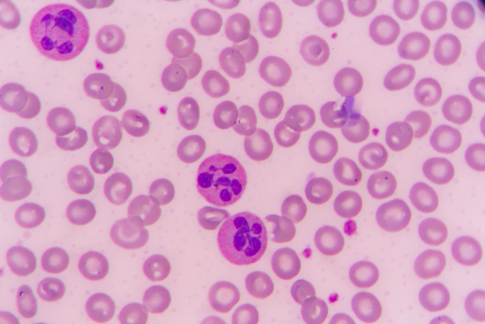
Characteristics: Sphere-shaped with variably shaped nuclei; color differentiation when stained.
Types of Leukocytes
Neutrophils
Function: Protect against bacterial and viral infections through phagocytosis.
Lifespan: From hours to 3 days.
Appearance: Pink-purple when stained.
Lymphocytes
Structure: Single-lobed nucleus, minimal cytoplasm.
Function: Secretion of antibodies; involved in immune response against antigens (viruses, pollen, bacteria).
Lifespan: Days to years; stained appearance is purple.
Monocytes
Characteristics: Kidney bean-shaped nucleus; large cytoplasm.
Function: Perform phagocytosis; destroy bacteria and waste products.
Lifespan: Months; stained appearance is steel blue.
Eosinophils
Structure: Single-lobed nucleus, abundant cytoplasm.
Function: Regulate allergic reactions, perform phagocytosis, combat parasitic infections.
Lifespan: 10 to 12 days; orange-red appearance when stained.
Basophils
Function: Secretion of heparin (anticoagulant) and histamine (inflammatory response).
Lifespan: Hours to 3 days; stained light purple.
Hemostasis: The Process of Blood Clotting
Hemostasis comprises several stages:
Vasoconstriction: Smooth muscle spasms in damaged vessel walls reduce blood loss.
Platelet Plug Formation: Platelets adhere to damaged areas, forming a temporary plug, akin to sealing a leak.
Coagulation (Clotting Phase): Sequential chemical reactions (clotting cascade) release substances (thrombin and fibrin) to promote optimized clotting mechanisms.
Hemostasis and Tissue Repair
The hemostatic process also involves tissue repair through a structured sequence:
Hemostasis: Initial bleeding and clot formation.
Inflammation: Immune cells target foreign materials and bacteria at the injury site.
Proliferation: New cells support epidermis growth; new blood vessels form.
Remodeling: Post-healing adjustments where excess cells break down and skin normalizes.