Unit 2 - Cell Structure and Function
Subcellular components & Cell Structure and Function
All cells contain a plasma membrane, cytoplasm (cytosol), chromosomes, and ribosomes.
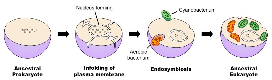
Endosymbiont theory:
EVERY CELL HAS A CELL MEMBRANE
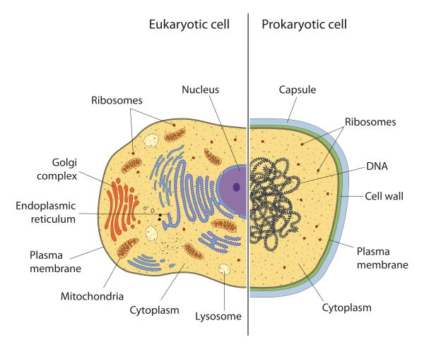
Prokaryotes:
Domains are archaea and bacteria
Lack a nucleus and membrane-bound organelles
Have free ribosomes and a plasma membrane
Contain DNA in the nucleoid
Eukaryotes:
Domains are protists (unicellular lil guys 😃), plants, animals, fungi
Much larger than prokaryotic cells
Double layer phospholipid membrane (hydrophilic region faces outwards, hydrophobic inwards)
Plant cell specific organelles:
Chloroplasts → photosynthetic organelle that converts sunlight into chemical energy stored in sugar molecules, contain stroma (fluid) and thylakoids (small green sacs within a granum stack)
Central vacuole → used as storage, breaks down waste products, hydrolysis of macromolecules, maintains turgor pressure, stores ions
Cell wall → outer layer that gives structure, made of cellulose in plant cells (chitin in fungi, peptidoglycan in prokaryotes) + other polysaccharides + protein
Plasmodesmata → channels through cell walls that connect the cytoplasm of adjacent cells
Animal cell specific organelles:
Lysosomes → digestive organelle where macromolecules are hydrolyzed (hydrolysis)
Centrosomes with centrioles → Organizes cell’s microtubules and contains a pair of centrioles
Flagella → Motility structure composed of microtubules within an extension of the plasma membrane
Cell junctions - adhere, interact, communication of animal cells
Tight Junctions - Plasma membranes of each cell are pressed together tightly to prevent leakage of extracellular fluid
Gap junctions - Membrane proteins surround a pore through which sugars, amino acids, etc. pass, necessary for cell communication
Desmosomes - Intermediate filaments anchor desmosomes into cytoplasm, fasten cells together
Organelles and their function:
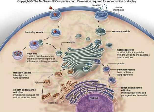
ENDOPLASMIC RETICULUMS
Smooth endoplasmic reticulum - synthesizes lipids, metabolizes carbohydrates, detoxifies drugs and poisons, stores calcium ions (secreted during muscle movement), abundant in liver cells
Rough endoplasmic reticulum - studded ribosomes secrete glycoproteins, distributes transport vesicles, serves as a “membrane factory”, makes proteins for transport
NUCLEUS - Center of DNA
Nuclear envelope - Double membrane enclosing nucleus, perforated and continuous with endoplasmic reticulum
Nucleolus - Produces ribosomes, one or more in a cell
Chromatin - Material consisting of DNA and proteins, visible in a divided cell as chromosomes
Plasma membrane - Regulates material moving in and out of cell (see plasma membrane section)
Ribosomes - made in nucleolus (dark, metabolically active spot on nucleus), protein + RNA, synthesize proteins (amino acid chains, polypeptides)
Golgi apparatus - sorts, modifies, ships proteins
Lysosome - Breaks down macromolecules using hydrolytic enzymes (digestion and recycle of cell’s organic material), involved in apoptosis (cell self destruct)
Vesicle - Transport membranes, merges with lysosomes to deliver macromolecules to be digested for recycle, merges with cell membrane to secrete proteins (path: RER → golgi app. → cell membrane)
Mitochondrion - Break down macromolecules to produce ATP energy (Krebs cycle), double membrane provides compartments for different metabolic reactions, folding of membrane increases surface area which produces more ATP
Peroxisome - metabolic compartment that produce hydrogen peroxide and convert it to H2O (oxidization)
Microvilli - Increase cell’s surface area
CYTOSKELETON - Maintenance of cell shape
Microfilaments - Changes in cell shape, division of animal cells
(Smallest)
Intermediate filaments - Maintenance of cell shape, serve as anchorage (Medium)
Microtubules - Cell motility, chromosome and organelle movement (Biggest)
Centrosome - Region where cell’s microtubules are initiated; contains a pair of centrioles
Flagellum - Motility structure present in SOME animal cells, cluster of microtubules within extension of plasma membrane
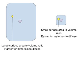
Cell Size
Cells must be smaller to compensate for volume growth (to facilitate the entrance and exit of nutrients and waste)
The more available surface area, the faster diffusion of nutrients into cell can occur (huge door vs doggy door)
Smaller cells are less metabolically needy with a large surface area to rapidly exchange nutrients
Cellular structures such as folds aid in a large surface area to volume ratio
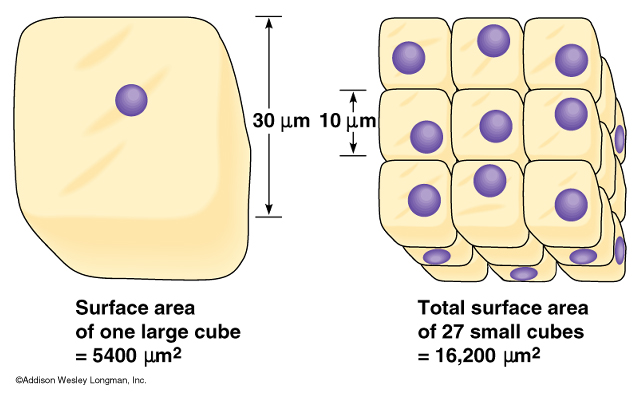
Surface area grows by r², volume grows by r³
Cells must maintain a larger surface area relative to volume (many small cubes has more surface area than one large cube
As organisms increase in size, their SA/V ratio decreases
Summary:
Surface area grows at a slower rate than volume, so cells must compensate for the metabolic need of the cell volume by increasing surface area. A larger surface area in relativity to the cell volume better facilitates the necessary exchange of nutrients into the cell to metabolically function. Smaller cells aid in rapid exchange of nutrients because smaller cells are less metabolically needy (surface area can increase while maintaining volume, bigger organisms have more cells not bigger cells). In combination with a larger surface area compared to volume, diffusion occurs much quicker so that the cell can function more efficiently. In conclusion, smaller cells with more surface area = more efficient and rapid metabolic function.
Plasma Membranes & Membrane Permeability
Amphipathic nature of hydrophilic heads and hydrophobic tails of phospholipid bilayer creates selective permeability
Fluid mosaic model = membrane held together by weak interactions (fluid) + phospholipids, proteins, carbs (mosaic)
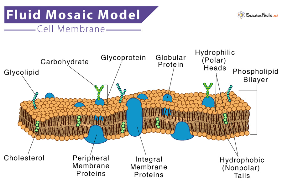
Cholesterol - regulates fluidity, limits fluidity at high temps, hinder close packing at low temps
Carbohydrates - cell to cell recognition
Glycoproteins - one or more carbohydrate attached to a membrane protein
Glycolipids - lipid with one or more carbohydrate attached
Permeability
Small, nonpolar molecules pass easily (Nitrogen, oxygen, carbon dioxide)
Hydrophilic substances and large polar molecules cannot pass membrane easily
Channel proteins - hydrophilic tunnel that allows specific target molecules to pass through
Carrier proteins - Change shape to move a target molecule across membrane
Membrane Transport
Concentration gradient - One solute is more concentrated than the other (high → low)
PASSIVE TRANSPORT
Net movement of molecules from high to low concentration without ATP
Diffusion - directly across membrane (small, nonpolar) movement of molecules
Facilitated diffusion - movement of molecules from high to low concentration through transport proteins
ACTIVE TRANSPORT
Requires direct input of ATP to move molecules against their concentration gradient
Protein pump (carrier protein) → requires ATP (active transport)
Establish and maintain concentration gradients
Cotransport - Secondary active transport that uses the energy from an electrochemical gradient to transport two different ions across the membrane through a protein (Symport - 2 different ions are transported in same direction, antiport - 2 different ions are transported in opposite directions
ENDOCYTOSIS
Phagocytosis - cell takes in large particles
Pinocytosis - cell takes in extracellular fluid containing dissolved substances
Receptor-mediated - Receptor proteins are used to capture specific target molecules through a vesicle
EXOCYTOSIS
Internal vesicles use energy to fuse with the plasma membrane and secrete large macromolecules (hormones, waste)
Facilitated Diffusion
movement of molecules from high to low concentration through transport proteins (large molecules and small polar molecules)
Water channel protein - Aquaporin
Sodium, potassium require channel proteins
Glucose is transported through facilitated diffusion
Tonicity and Osmoregulation
Tonicity - The relative concentration of solutes inside and outside of a cell
Osmoregulation - the maintenance of constant osmotic pressure in the fluids of a cell by the control of water and solute concentrations
Hypotonic - more water, less solute > all relative to the cell
Hypertonic - less water, more solute > all relative to the cell
Isotonic - equal ratios of solute and water relative to cell
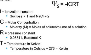
Water potential - tendency of water to move by osmosis
Ψ = ΨS + ΨP (unit of pressure is bars)
More negative water potential → more water movement
Water potential of pure water = 0
Increasing amount of solute in water → Increase in solute potential, decrease in water potential
Increasing water potential will cause an increase in pressure potential
Decreasing pressure potential will cause a decrease in pressure potential
In an open system, pressure = 0
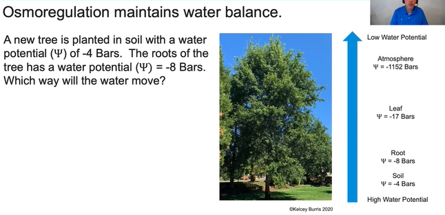
Water will move from soil to the roots because the soil has a higher water potential that the roots!
Mechanisms of Transport & Compartmentalization
Compartmentalization allows for a variety of metabolic processes and enzymatic reactions to occur simultaneously, increasing the efficiency of the cell.
Membranes minimize competing interactions
The hydrolytic enzymes of the lysosome function at an acidic environment (contained in lysosome membrane)
Folding of mitochondria maximizes amount of metabolic actions that can occur, allowing the production of MORE ATP
Thylakoids are highly folded to increase efficiency of the light dependent reactions
Origins of cell compartmentalization
Endosymbiont theory - (mitochondria and chloroplasts) A free-living aerobic prokaryote was engulfed by the engulfing cell by endocytosis and was not digested, developed symbiotic relationship, eventually became mitochondria (same for chloroplasts)
Evidence of endosymbiotic cells
Double membranes to regulate material passage
Have their own circular DNA, like prokaryotes
Contain their own ribosomes and synthesis proteins