Chapter 16 The Neurological Exam
Ch 16.1 Overview of the Neurological Exam
The neurological exam is a clinical assessment tool used to identify which specific parts of the Central Nervous System (CNS) are affected by damage or disease.
It can be performed in a short time—sometimes as quickly as 5 minutes—to establish neurological function.
In emergency departments, this rapid assessment is critical for determining the proper course of treatment and the potential extent of recovery.
The exam is organized into five major sections, each targeting specific regions and functions of the CNS:
Mental Status Exam
Focus: Assesses higher cognitive functions such as memory, orientation, and language.
CNS Correlation: Cerebrum, primarily the cerebral cortex.
Cranial Nerve Exam
Focus: Tests the sensory and motor functions of the 12 cranial nerves.
CNS Correlation: Diencephalon and brain stem (including olfactory connections to the forebrain).
Sensory Exam
Focus: Tests sensory functions associated with spinal nerves.
CNS Correlation: Spinal cord and its connections through spinal nerves.
Motor Exam
Focus: Tests motor functions associated with spinal nerves.
CNS Correlation: Spinal cord and its connections through spinal nerves.
Coordination Exam
Focus: Tests the ability to perform complex and coordinated movements.
CNS Correlation: Cerebellum.
Gait Exam: Frequently cited as a sixth major component, it specifically assesses the motor function of walking and is considered part of the coordination exam because walking requires coordinated movement.
Neuroanatomy and the Neurological Exam
Localization of function is the concept that circumscribed locations are responsible for specific functions.
The neurological exam highlights this relationship by mapping clinical deficits to localized brain regions.
Language Localization: In most individuals, language function is localized to the left hemisphere between the superior temporal lobe and the posterior frontal lobe, including the intervening connections through the inferior parietal lobe.
Clinical Application and Rapid Assessment:
The power of the neurological exam lies in the link between anatomical structure and physiological function, allowing for an accurate estimation of where the nervous system is damaged.
A rapid assessment (taking less than 5 minutes) often includes:
Cranial Nerve Tests: Asking the patient to smile, raise eyebrows, stick out tongue, and shrug shoulders, and following a pen tip through the visual field.
Motor Tests: Assessing muscular strength by providing resistance against the arms and legs.
Sensory Tests: Having the patient indicate when they feel a sensation (e.g., the tip of a pen) with their eyes closed.
Observational Mental Status: A formal mental status exam may not be required if no deficits are apparent during normal interaction; deeper testing is pursued only if cognitive or language deficits become evident.
Localization Example: A patient reporting "pins and needles" or a lack of sensation in limbs suggests a problem within the sensory systems between the spinal cord and the brain, providing a lead for localization before imaging (like a CT scan) is performed.
Clinical Management: If a blockage is suspected, a patient might be put on aspirin therapy to limit the formation of blood clots in the event of an embolus—an obstruction, such as a blood clot, that blocks the flow of blood in an artery or vein.
Causes of Neurological Deficits
Damage to the nervous system can be localized to individual structures or distributed across broad areas of the brain and spinal cord. Localized, limited injury is most often the result of circulatory problems.
Oxygen Sensitivity: Neurons are extremely sensitive to oxygen deprivation; deterioration begins within 1 or 2 minutes, and permanent damage (cell death) can result within a few hours.
Stroke (Cerebrovascular Accident or CVA): The loss of blood flow to part of the brain.
Main Types of Stroke:
Ischemic Stroke:
Occurs when blood flow is lost because vessels are blocked or narrowed.
Often caused by an embolus (blood clot or fat deposit), thickening of the blood vessel wall,
or hypovolemia (a drop in blood volume in the brain).
Transient Ischemic Attack (TIA):
Similar to a stroke, but symptoms are resolved within a 24-hour period because adequate blood flow is restored.
By diagnostic definition, stroke effects must last at least 24 hours.
Hemorrhagic Stroke:
Bleeding into the brain resulting from a damaged blood vessel.
Accumulated blood fills a region of the cranial vault and exerts physical pressure on brain tissue,
which can lead to loss of function and the squeezing of local arteries.
This damage can cause the blood-brain barrier to break down, allowing additional fluid to accumulate, a condition known as edema.
While focal injuries like strokes result in localized deficits, other events can cause widespread damage or disruptions to neurological function.
Widespread and Traumatic Damage:
Infectious Diseases: Can lead to loss of function throughout the CNS. Nervous tissue components, specifically astrocytes and microglia, react to the disease, potentially altering function.
Blunt Force Trauma: Physical damage to the CNS, such as that resulting from a motor vehicle accident, can cause broad or localized injury.
Neurodegenerative Diseases:
A class of disorders characterized by progressive loss of nervous tissue.
Examples: Alzheimer’s disease, Parkinson’s disease, Huntington’s disease, amyotrophic lateral sclerosis (ALS), Creutzfeldt–Jakob disease, and multiple sclerosis (MS).
Mechanisms:
Neuron Death: Occurs in diseases like Alzheimer’s, Parkinson’s, and ALS.
Myelin Damage: Specifically characterizes Multiple Sclerosis (MS).
Impact on Neurological Exam: These disorders often present with broad effects, such as memory deficits (compromising the mental status exam) or movement deficits (affecting the motor, coordination, or cranial nerve exams).
Etiology: Causes vary from genetics (Huntington’s) and autoimmunity (MS) to those not entirely understood (Alzheimer’s and Parkinson’s).
Developmental Disorders:
Result from genetic factors or environmental influences during development that disrupt the normal growth of the CNS.
Autism Spectrum Disorder (ASD): A primary example where the cause is complex and often involves multiple factors.
Connectivity vs. Localization:
In stroke-induced prosopagnosia (inability to recognize faces), there is clear cell death in the fusiform gyrus.
In ASD, the relevant brain regions may be physically present, but they are not connected properly to other regions, leading to similar functional deficits.
Differentiating Acute vs. Global Signs:
The neurological exam helps differentiate between an acute event (like a stroke) and a chronic or global condition (like trauma or infection).
Discrete vs. Global Loss: A discrete loss of one function (e.g., being unable to say specific words) suggests an acute, localized event. A global loss of function (e.g., loss of language in all its aspects) is more likely a global or widespread event.
Ch 16.2 The Mental Status Exam
In a clinical setting, the mental status exam is used to understand the relationship between the brain and the body by assessing behavior.
Behavioral Indicators: Observations of tremors related to intentional movements, incoordination, or the neglect of one side of the body can indicate failures in the connections of the cerebrum, either within the hemispheres or to other portions of the nervous system and musculature.
Eliciting Behavior: Assessment can be as simple as asking a patient for their name. This verifies:
Orientation and Awareness: Ensuring the patient is capable of interacting and understands reality.
Speech Function: Identifying potential problems in understanding or producing speech.
Functions of the Cerebral Cortex
The cerebrum serves as the seat of higher mental functions, including memory, learning, language, and conscious perception.
Physical Structure:
The cerebral cortex is a thin layer of gray matter on the outside of the cerebrum.
It is approximately 2.55 mm thick on average.
It is highly folded to increase surface area within the limited space of the cranial vault.
Localization and Cytoarchitecture:
Cytoarchitecture refers to the study of the brain’s regions based on its cellular composition.
Brodmann’s Areas: Developed by Korbinian Brodmann, this system identifies approximately 50 separate regions of the cortex based on the size, shape, and number of neurons.
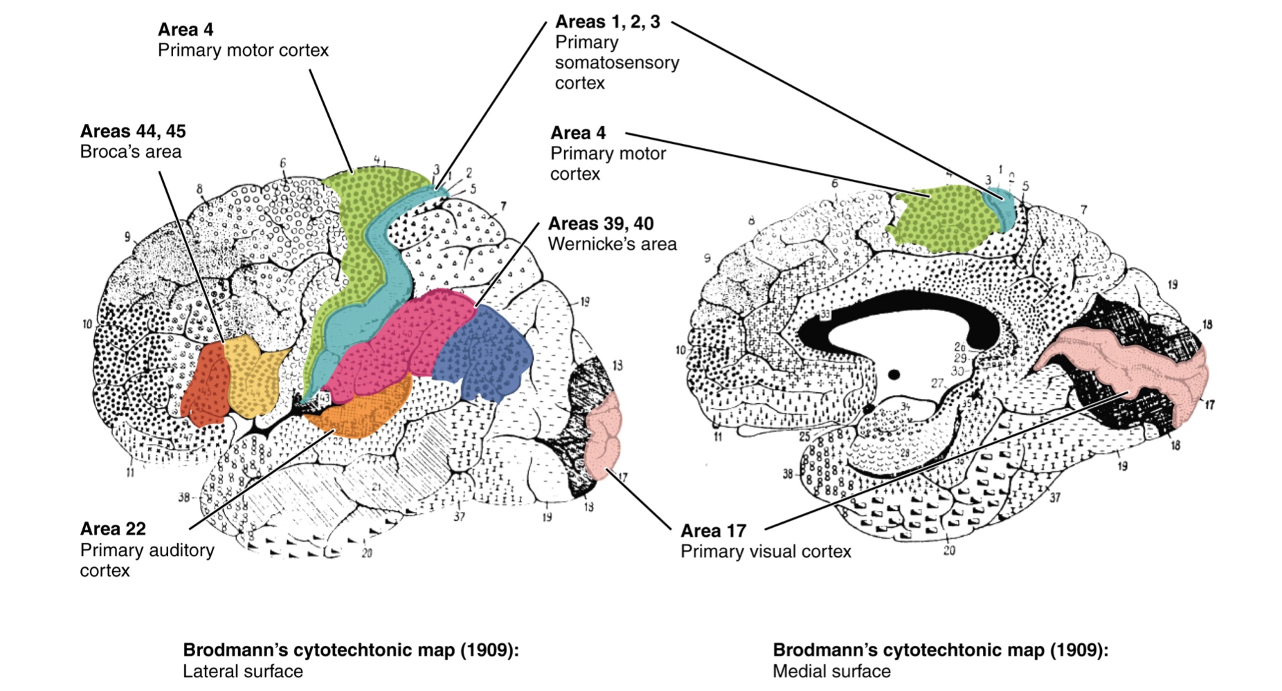
Current neurological practice still references these areas due to the strong correlation between anatomical structure and physiological function.
Specific Functional Areas:
Primary Somatosensory Cortex (Areas 1, 2, 3): Located in the postcentral gyrus; encompasses sensory submodalities like touch, vibration, and proprioception.
Primary Visual Cortex (Area 17): Handles initial visual processing, while adjacent Areas 18 and 19 constitute subsequent regions of visual processing.
Primary Auditory Cortex (Area 22): Handles initial auditory info, followed by Area 23 for further processing.
Primary Motor Cortex (Area 4): Located in the precentral gyrus; is the source of descending motor commands.
Premotor Cortex (Area 6): Suggests specialization for functional processing and motor planning.
Classification of Cortical Areas:
Primary Cortical Areas (1,2,3,4,17,22):
Where sensory information is initially received from the thalamus for conscious perception.
In the case of the motor cortex, where descending commands are sent down to the brain stem or spinal cord to execute movements.
Association Areas:
Adjoining regions to primary areas that further process modality-specific input.
Integrative Areas:
Located in the spaces between sensory or motor domains; they process multisensory information or process data in more complex ways.
Multimodal Integration: Found where modality-specific regions meet; can process multiple modalities together (e.g., spatial processing in vision and somatosensation).
Example - Posterior Parietal Cortex: Lies between the somatosensory and visual cortex. It coordinates visual and motor functions, such as reaching for a glass, by integrating visual data with proprioceptive feedback regarding the weight and position of the limb.
Cognitive Abilities
Assessment of cerebral functions is directed at cognitive abilities, which are separated into four major groups during the mental status exam:
Orientation and Memory
Language and Speech
Sensorium
Judgment and Abstract Reasoning
Orientation and Memory
Orientation: Patient’s awareness of their immediate circumstances.
Time: Awareness of the date and current clock time.
Place: Awareness of where they are and why they are there.
Identity: Recognition of personal identity and the ability to relate to the examiner.
Testing: Questions include "What is the date?", "Where are you?", and questions addressing remote memory like "Who is the President?".
Memory Assessment:
Three-word recall test: The patient is given three words (e.g. book, clock, shovel) to recall after a short interval.
Secondary Tasks: Reciting months in reverse, spelling words backwards, or reciting number sequences back to the examiner.
Neuroanatomy of Memory:
Primarily a function of the temporal lobe, hippocampus, and amygdala (medial temporal lobe).
The Case of Patient HM (Henry Molaison):
Following a bilateral lobectomy of the medial temporal lobes, HM developed anterograde amnesia (inability to form new memories) and some retrograde amnesia (loss of memories prior to surgery).
Episodic Memory: HM could not form new autobiographical memories.
Procedural Memory: HM retained the ability to remember how to perform tasks (e.g. riding a bike).
Differentiation and Consolidation:
Short-term Memory (Working Memory): Localized to the prefrontal cortex.
Memory Consolidation: The function of the hippocampus and medial temporal structures is to move short-term memories from the prefrontal lobe to long-term storage in the temporal lobe.
Prefrontal Cortex Subtests:
Set Generation: Tests the ability to organize information by asking the patient to list at least 10 words starting with the same letter (excluding proper nouns) within 1 minute.
Language & Speech
Significance: Language is a fundamental human aspect of neurological function and is central to self-awareness, often relating to the philosophical concept of Cogito Ergo Sum ("I think, therefore I am").
Subtests for Language Assessment:
Understanding Language: Measures the patient's ability to follow complex verbal instructions,
such as "touch your right finger to your left elbow and then to your right knee."
Fluency and Coherency: Assessed by having the patient generate descriptions of drawings, recite specific sentences, or explain the meaning of a written passage.
Aphasia and Functional Localization:
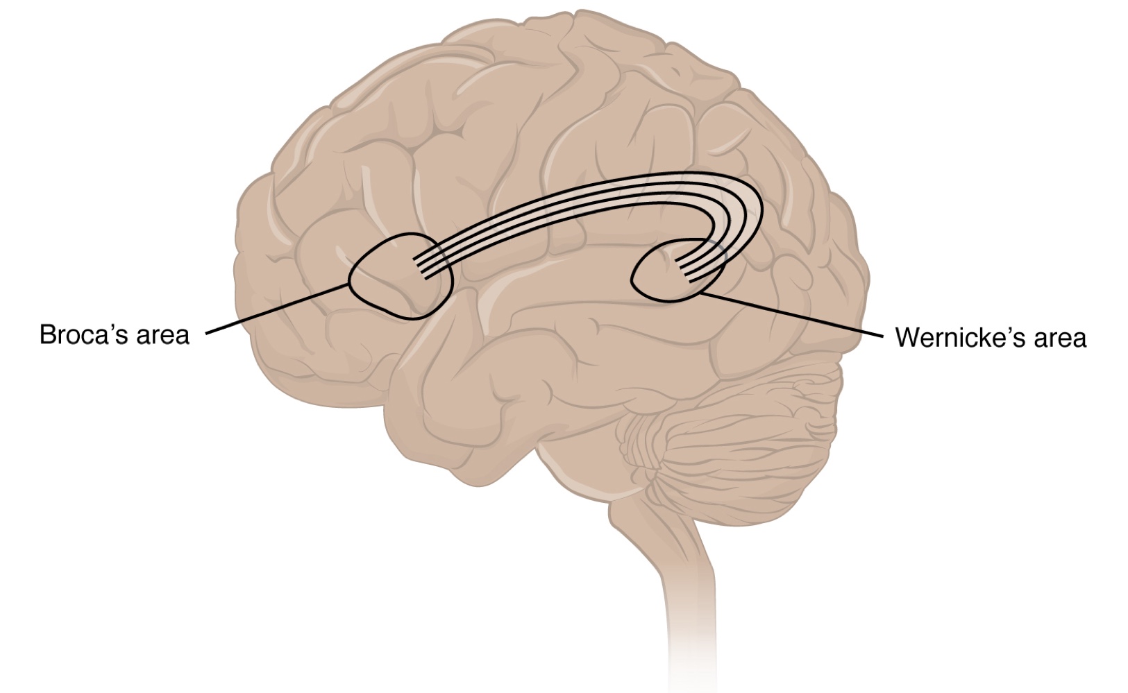
Aphasia: A loss of language and speech functions resulting from brain damage.
Broca’s Area: Located in the lateral aspect of the frontal lobe, anterior to the motor cortex region for the head and neck.
Expressive Aphasia: Characterized by compromised speech production. The speech is often non-fluent, broken, or halting, and grammar may be lost.
Wernicke’s Area: Found adjacent to the auditory association cortex at the end of the lateral sulcus.
Receptive Aphasia: Characterized by a loss of content understanding.
Patients can produce speech but cannot comprehend what is said to them or understand the meaning of their own words.
Conduction Aphasia: Results from damage to the white matter tracts connecting the posterior temporal lobe and the lateral frontal lobe.
Deficit: Primarily presents as an inability to faithfully repeat spoken language despite maintaining basic understanding and production capability.
Sensorium
Definition: Collectively refers to the parts of the brain involved in the reception and interpretation of sensory stimuli.
Role of the Cerebral Cortex: The cortex is the seat of conscious sensory perception. Information moves from primary cortical areas (somatosensory, visual, auditory, and gustatory) to association areas for processing.
Subconscious Processing: Sensory information can also be processed in deeper brain regions; for instance, the cerebellum uses proprioceptive information to maintain balance subconsciously.
Specific Subtests for Cortical Functions:
Praxis:
A practical exercise where a patient performs a task (.e.g. placing a hand palm down and flipping it 4 times) based solely on verbal instructions without any physical demonstration.
Requirements: Requires the patient to understand verbal instructions, transform them into motor movements, and use sensory feedback (visual and proprioceptive) to execute the task correctly.
Gnosis:
Refers to the naming and recognition of stimuli, involving two primary tasks:
Stereognosis: Identifying common objects (ee.g. a nickel vs. a quarter) through touch and manipulation with eyes closed, relying on sensory cues like weight, size, and edges.
Graphesthesia: Identifying numbers or letters traced onto the palm of the hand using a dull pointer, such as a pen cap.
Language and Cognitive Integration:
Praxis and gnosis are integrated with language, as functions depend on the relationship between words describing actions (verbs) or objects (nouns).
V and N Impairments:
V Impairment: A deficit in using or understanding verbs, often localized to the region where the frontal and temporal lobes meet, including the insula.
N Impairment: A deficit in using or understanding nouns, typically associated with damage to the middle and inferior temporal lobes.
Judgment and Abstract Reasoning
Overview: Planning and producing responses require the ability to make sense of the world. Judgment and reasoning in the abstract allow individuals to determine logical actions and process how specific decisions (ee.g. hitting the snooze button for 10 extra minutes) might affect their day.
Neuroanatomy:
Prefrontal Cortex: The region located anterior to the motor association areas (e.g. premotor cortex, Broca’s area, and frontal eye fields) of the frontal lobe.
Functions: It is responsible for working memory, planning, decision-making, judgment, and abstract reasoning.
It acts as the antecedent to movement by judging whether a movement should be made.
Mental Status Subtests:
Problem Solving: The examiner asks situational questions, such as "If you see a house on fire, what would you do?".
Proverb Interpretation: The patient is asked to explain the abstract meaning of common proverbs, such as "Don't look a gift horse in the mouth."
Comparison of Similarities: Asking the patient to identify commonalities between pairs of words, such as apple and orange, or lamp and cabinet.
Personality and the Prefrontal Cortex:
The prefrontal cortex is deeply linked to an individual's personality.
Phineas Gage Case: A railroad worker in the mid-1800s who survived a metal spike impaling his prefrontal cortex. The injury caused his personality to shift from dependable and quiet to raucous and irritable.
Prefrontal Lobotomy: A psychiatric procedure common in the 1940s and early 1950s where the white matter connections of the prefrontal cortex were severed. This resulted in significant changes to mood and personality and was eventually phased out in favor of antipsychotic drugs.
Ch. 16.3 The Cranial Nerve Exam
The cranial nerve exam provides directed testing of the forebrain and brainstem. The 12 cranial nerves serve the head and neck and are categorized by their specific clinical functions.
Overview of Functional Groupings:
Sensory:
3 nerves are dedicated strictly to special senses;
4 others handle a combination of special and general senses.
Motor:
Extraocular: 3 nerves control gaze by regulating the extraocular muscles.
Face/Pharynx: 4 nerves control facial expressions, chewing (mastication), swallowing, and speech.
Neck: 1 nerve assists with head and neck movements.
Autonomic:
4 nerves provide preganglionic parasympathetic fibers (e.g. the Vagus nerve, X) to control pupillary size, salivation, and thoracic/abdominal viscera.
Sensory Nerves
The olfactory (I), optic (II), and vestibulocochlear (VIII) nerves are dedicated primarily to the special senses of smell, vision, equilibrium, and hearing.
Olfactory Nerve (I)
Function: Sense of smell.
Clinical Testing: Asking the patient to identify common smells (e.g.coffee or mint) while testing one nostril at a time.
Anosmia: The clinical term for the loss of the sense of smell.
Causes include blunt force trauma to the head or the natural aging process.
Regeneration: Olfactory neurons are rare in that they regularly regenerate; however, if the brain moves within the cranium (e.g. in a motor vehicle accident), the axons can be sheared, leading to potentially permanent loss.
Significance: Olfaction is critical for the enjoyment of food; without it, food often tastes bland despite intact gustatory senses.
Optic Nerve (II)
Anatomy and Pathway:
Optic nerves enter the cranium through the optic canals and meet at the optic chiasm.
At the chiasm, fibers sort such that the two halves of the visual field are processed by the opposite sides of the brain.
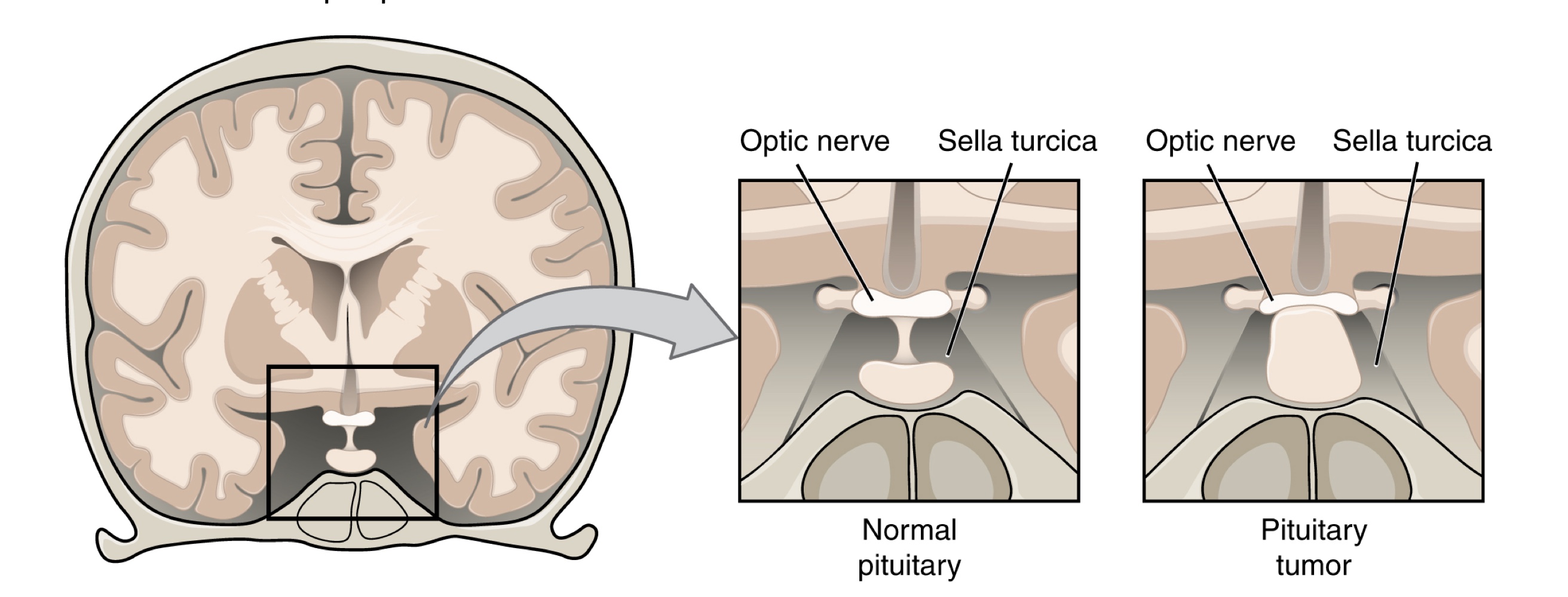
Decussation: The axons that decussate (cross) in the chiasm are from the medial retinae of either eye, which carry information from the peripheral visual field.
Visual Acuity: Assessed with a Snellen chart, which presents standard Roman letters in varying sizes.
Results are expressed as a ratio (e.g.20/60), where the first number is the testing distance (20 feet) and the second is the distance from which a person with normal acuity could read that same line.
Visual Field Testing: Used to establish the boundaries of peripheral vision.
Clinical Deficits: Deficits in visual field perception often suggest damage along the length of the optic pathway between the orbit and the diencephalon.
Pituitary Tumors: The pituitary gland is seated in the sella turcica of the sphenoid bone, directly inferior to the optic chiasm. A tumor pressing on the chiasm typically results in the loss of peripheral vision.
Ophthalmic Inspection: An ophthalmoscope is used to physically inspect the optic disk, the location where the optic nerve emerges from the eye.
Vestibulocochlear Nerve (VIII)
Function: Carries equilibrium and auditory sensations from the inner ear to the medulla.
Anatomy:
Both sensory systems emerge from the inner ear, pass through the internal auditory meatus, and synapse in the vestibular and cochlear nuclei of the superior medulla.
Vestibule: Composed of the utricle, saccule, and semicircular canals; responsible for equilibrium.
Cochlea: Transduces sound waves into neural signals.
Clinical Deficits:
Deficits in balance (e.g. vertigo) or hearing can point to inner ear damage as these structures are neighbors in the bony labyrinth of the temporal bone.
Ménière’s Disease: A disorder likely caused by the over-production or accumulation of fluid in the inner ear. Common symptoms include vertigo, low-frequency ringing, and progressive loss of hearing.
Clinical Tests:
Equilibrium: Tests like the Romberg test assess balance and are part of spinal and cerebellar neurological assessments. The vestibulo-ocular reflex is also tested for gaze control.
Hearing Tests (using a tuning fork):
Rinne Test: Used to distinguish between:
conductive hearing (vibration through middle ear ossicles) and
sensorineural hearing (transmission through inner ear and nerve).
A conductive deficit is noted if sound conducted through the temporal bone is louder than air-conducted sound.
Weber Test: The tuning fork is placed at the top of the skull. In healthy patients, the sound is equal in both ears.
In unilateral conductive loss, the sound is louder in the damaged ear because background noise is blocked.
In unilateral sensorineural loss, the sound is quieter in the damaged ear due to nervous tissue damage.
Trigeminal Nerve (V)
Function: Carries general somatosensation for the face and head, acting as the cranial equivalent to the dorsal column and spinothalamic pathways of the spinal cord.
Brainstem Nuclei:
Mesencephalic Nucleus: Processes proprioceptive information regarding facial movements and muscle position.
It serves as the sensory component of the jaw-jerk reflex, which is a stretch reflex of the masseter muscle.
Chief Nucleus (Pons): Receives information about light touch and mandibular proprioception.
It functions analogously to the dorsal column pathway for the body, relaying information to the thalamus and onto the postcentral gyrus.
Spinal Trigeminal Nucleus (Medulla): Receives crude touch, pain, and temperature information.
It functions analogously to the spinothalamic pathway for the rest of the body.
Clinical Testing (Sensory):
Sensory Discrimination: The primary sensory subtest for the trigeminal system, involving methods identical to those used for sensory exams targeting spinal nerves.
Testing Apparatus: A cotton-tipped applicator is used, with the wooden stick snapped to create a pointed end opposite the soft cotton end.
Testing Procedure: While the patient's eyes are closed, the examiner touches the face, alternating randomly between the two ends. The patient must identify each stimulus as "sharp" or "dull."
Neural Relay Pathways:
Dull/Light Touch (Cotton Tip): Relayed by the chief nucleus.
Sharp/Painful Stimulus (Pointed End): Relayed by the spinal trigeminal nucleus.
Localization of Deficits: Failure to discriminate between these stimuli can help localize problems within the brain stem.
An inability to recognize a painful stimulus points toward damage in the spinal trigeminal nucleus of the medulla.
Clinical Significance of the Medulla:
In addition to sensory relay, the medulla contains critical regions for regulating the cardiovascular, respiratory, and digestive systems.
It serve as the primary conduit for ascending and descending tracts connecting the brain and spinal cord.
Damage (such as a stroke) that causes changes in sensory discrimination may indicate these unrelated regions are affected as well.
Integration of Other Senses:
Taste: Relayed to the brainstem through the facial and glossopharyngeal nerves.
General Somatic Senses: The trigeminal nerve carries general sensation (touch, vibration, etc.) from the face and head, functioning similarly to spinal nerves in the rest of the body.
Gaze Control
Extraocular Muscle Innervation: Coordination of the eyes is handled by the third (III), fourth (IV), and sixth (VI) cranial nerves.
Abducens Nerve (VI): Responsible for abducting the eye via the lateral rectus muscle.
Trochlear Nerve (IV): Controls the superior oblique muscle; rotates the eye medially (intorsion) and helps focus on objects close to the face.
Oculomotor Nerve (III): Controls the upper eyelid and all other extraocular muscles.
Brainstem Integration:
Superior Colliculus (Midbrain): Initiates eye movements by integrating visual stimuli with motor responses.
Paramedian Pontine Reticular Formation (PPRF): Initiates saccades—rapid eye movements used to bring the eyes to bear on a visual stimulus and direct the fovea onto it.
Medial Longitudinal Fasciculus (MLF): A tract running through the brainstem that connects the oculomotor, trochlear, and abducens nuclei.
Conjugate Gaze: The MLF allows eyes to move in the same direction during horizontal tracking.
Horizontal movements require the lateral rectus (abducens nucleus in the superior medulla) and the medial rectus (oculomotor nucleus in the midbrain) to work together.
Vertical and Complex Movements:
Vertical Gaze: Controlled strictly within the oculomotor complex.
Elevation: Contracted by the superior rectus muscles.
Depression: Contracted by the inferior rectus muscles.
Compound Movements: Most movements are not strictly vertical and occur at an angle.
Because the superior and inferior rectus muscles are oriented medially to their insertions, their contraction causes slight adduction, requiring lateral rectus compensation facilitated by the MLF.
Testing Eye Movement and Gaze Deficits:
Clinical Testing: Patients track a pen tip through the visual field.
This differs from visual field testing (where eyes remain still) by focusing on the extent and coordination of movement.
Internuclear Ophthalmoplegia: A failure of one eye to abduct while the other adducts during horizontal movement; this indicates dysfunction in the nuclei or the connecting MLF.
Diplopia (Double Vision): Occurs when extraocular muscles fail to move the eyes in perfect conjugation.
Convergence and Accommodation:
Convergence: A non-conjugate movement where both eyes adduct simultaneously to look at a stimulus approaching the face.
Accommodation: The adjustment of lens shape to maintain focus on near objects, controlled by parasympathetic fibers of the oculomotor nerve.
Accommodation-Convergence Reflex: The coordinated skeletal muscle activity for convergence and smooth muscle activity of the ciliary body for lens adjustment.
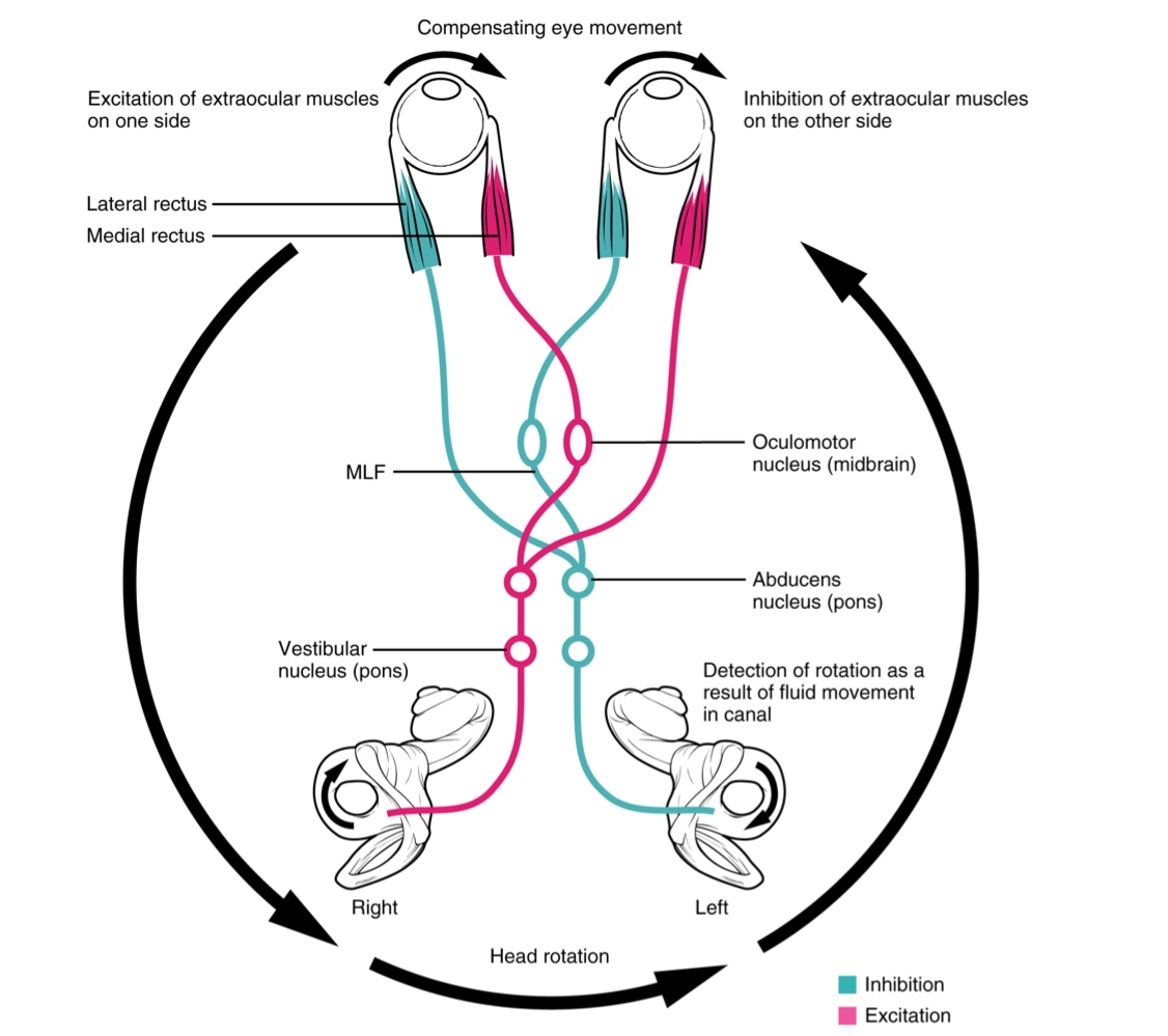
Vestibulo-Ocular Reflex (VOR):
Function: Stabilizes the visual field by keeping stimuli centered on the fovea during head rotation.
Mechanism: Semicircular canals in the inner ear detect head rotation (e.g., to the right) and signal the abducens and oculomotor nuclei to rotate the eyes in the opposite direction (e.g., to the left).
Deficits: Impairment in the VOR may indicate vestibular damage (such as Ménière’s disease) or brainstem damage affecting the MLF or related nuclei.
Nerves of the Face and Oral Cavity
Clinical Inspection: The inspection of the oral cavity and pharynx often starts with the patient saying "ah," allowing the examiner to view the fauces (the opening of the oral cavity into the pharynx).
This tests for signs of infection (e.g., tonsillitis) and the function of associated cranial nerves.
Taste and Sensation:
Gustatory Testing: The facial (VII) and glossopharyngeal (IX) nerves convey taste. Testing involves applying salty, sour, bitter, or sweet stimuli to either side of the tongue.
General Sensation and Gag Reflex: The glossopharyngeal nerve relays general sensations from the pharyngeal walls.
The gag reflex is elicited by stimulating the lateral wall of the fauces. The motor response—contraction of the pharyngeal muscles—is mediated by the vagus nerve (X).
Vagus Nerve (XX) and Speech:
Although primarily autonomic, the vagus nerve stimulates skeletal muscles in the pharynx and larynx for swallowing and speech.
Testing: Patients are asked to repeat alternating consonant sounds like "lah-kah-pah" while the examiner observes the movements of the soft palate and arches between the palate and tongue.
Salivation:
Initiated by the salivary nuclei of the medulla through the facial (VII) and glossopharyngeal (IX) nerves.
Facial Nerve: Preganglionic fibers synapse in the pterygopalatine ganglion, which projects to the submandibular and sublingual glands.
Glossopharyngeal Nerve: Synapses in the otic ganglion, which projects to the parotid gland.
Reflex Arc: Salivation in response to food is a visceral reflex, though it can be coordinated by the hypothalamus in response to the smell or sight of food.
Hypoglossal Nerve (XII):
Function: The motor nerve for most tongue muscles, excluding the palatoglossus (which is controlled by the vagus nerve).
Extrinsic muscles: Connected to other structures.
Intrinsic muscles: Contained entirely within lingual tissues.
Testing: The patient is asked to "stick out your tongue."
Deficit: If the nerve on one side is damaged, the tongue will point toward the side with the damage (protrudes incorrectly).
Loss of function of the tongue can interfere with speech and swallowing.
Clinical Significance: Because the location of the hypoglossal nerve and nucleus is near the cardiovascular, inspiratory and expiratory areas for respiration, and the vagus nuclei that regulate digestive function, a tongue that protrudes incorrectly can suggest damage in adjacent structures that have nothing to do with
Motor Nerves of the Neck
Accessory Nerve (XI):
Also referred to as the spinal accessory nerve, it innervates the sternocleidomastoid and trapezius muscles.
Muscle Functions:
Sternocleidomastoid: Contraction of both muscles flexes the head forward; individual contraction causes rotation of the head to the opposite side.
Trapezius: Acts as an antagonist to flexion, causing extension and hyperextension of the neck. It also contributes to elevating the scapula and clavicle.
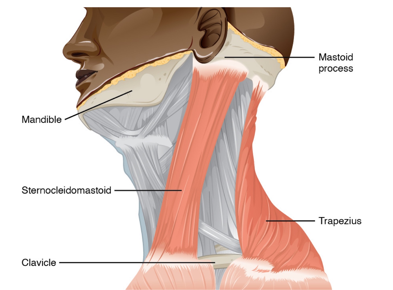
Neurological Connections: These muscles also receive input from cervical spinal nerves along with the accessory nerve.
For the sternocleidomastoid, spinal nerves provide primarily sensory projections.
For the trapezius, spinal nerves provide motor input from the spinal cord.
Clinical Testing and Assessment:
Shoulder Shrug: The trapezius is tested by asking the patient to shrug both shoulders while the examiner watches for asymmetry.
Resistance Testing: Strength is assessed by having the patient flex and extend the neck or shrug their shoulders against resistance.
Lateral Flexion: Lateral flexion of the neck toward the shoulder tests both muscles simultaneously. Differences in strength between sides suggest damage on the weaker side.
Significance: Because these muscles change the position of the head, deficits in the accessory nerve can
Ch. 16.4 The Sensory & Motor Exam
Connections between the body and the CNS occur through the spinal cord. While cranial nerves connect the head and neck directly to the brain, the spinal cord receives sensory input and sends motor commands out to the body through the spinal nerves.
Spinal Cord Configuration:
Unlike the brain, which develops into a complex series of nuclei and fiber tracts, the spinal cord retains a relatively simple tube-like structure.
Structure and Functional Localization:
Gray Matter: Surrounds the small central canal in the center of the cord.
Dorsal (Posterior) Horns: Dedicated primarily to sensory functions.
Ventral (Anterior) and Lateral Horns: Associated with motor functions.
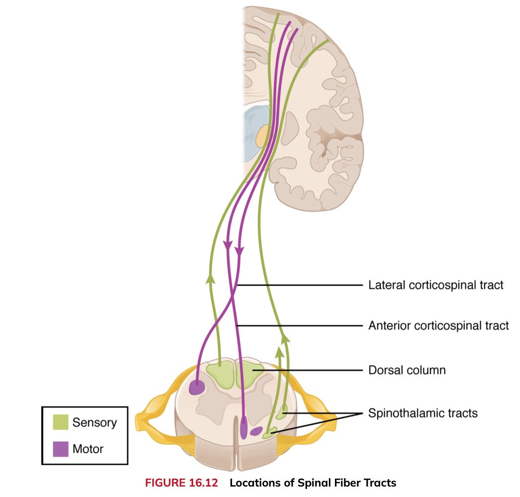
White Matter: Found on the surface and organized into three distinct columns.
Dorsal Column: Responsible for relaying sensory information upwards to the brain.
Anterior Column: Almost exclusively relays descending motor commands to the ventral horn motor neurons.
Lateral Column: Conveys both sensory and motor information between the spinal cord and the brain.
Sensory Modalities and Location
General Senses Distribution: General senses are distributed throughout the body within various organs.
Somatic Senses: Incorporated primarily into the skin, muscles, or tendons. These make up the conscious perception of how the body interacts with the environment.
Visceral Senses: Originate from nervous tissue in internal organs (e.g., heart or stomach). These usually remain below the limit of conscious perception as they assist in homeostatic regulation via the autonomic nervous system.
The Sensory Exam: Focuses on testing consciously perceived somatic senses.
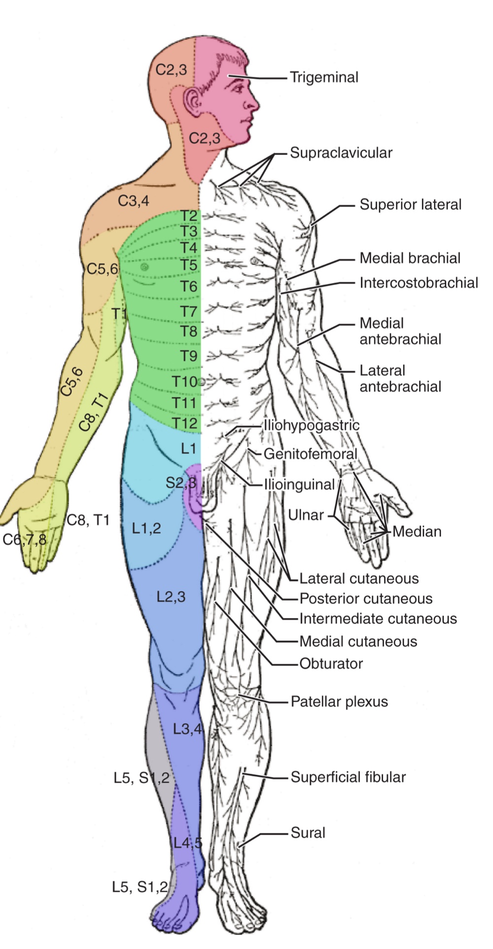
Dermatomes: Regions of the skin that connect to the spinal nerves in a topographically organized manner.
Testing Methodology: Sensory fields are tested using a light touch stimulus, such as the soft end of a cotton-tipped applicator, applied to the skin.
Directional Assessment: Testing is performed from distal to proximal locations in the appendages and lateral to medial locations in the trunk.
Patient Feedback: Patients are asked if the touch is perceptible and if there are differences in sensation across different regions.
Example: Fibers of the 8th cervical nerve (C8) innervate the fingers and the medial surface of the forearm.
CNS Correlation: These sensory paths connect to the cortical region of the postcentral gyrus where somatosensation is perceived.
Testing Specific Somatosensory Modalities:
Pain: Tested using the broken (pointed) end of a cotton-tipped applicator.
Vibration: Tested via an oscillating tuning fork placed against prominent bone features (e.g., the distal head of the ulna).
Temperature (Cold): Perceived using the neutral metal of a still tuning fork.
Tactile Movement: Assessed by drawing a stimulus across the skin for approximately 2−3 cm. The patient must identify the direction of movement.
Proprioception: Tested by moving the patient's fingers or toes and asking them to describe the sense of position and motion. If distal locations are not perceived, the test moves to increasingly proximal joints.
Major Ascending Tracts:
Dorsal Column Pathway: Conveys fine touch, vibration, and proprioceptive information.
It ascends ipsilaterally in the spinal cord and decussates in the medulla.
Spinothalamic Pathway: Primarily conveys pain and temperature.
It decussates at the level of entry in the spinal cord and ascends contralaterally.
Clinical Significance: Failure to discriminate between submodalities (e.g., mistaking pain for light touch) may point to specific errors in ascending projections,
such as those caused by a hemisection of the spinal cord.
Sensory Discrimination:
Two-point Discrimination: Assesses the density of sensory endings/receptive fields.
Calipers or forceps are used to touch the skin in two locations simultaneously to see if the patient can recognize them as distinct points.
Failure is often an indication of a dorsal column pathway deficit.
Double Simultaneous Stimulation: Two stimuli are applied to the same position on both sides of the body simultaneously.
If one side is not perceived, it indicates damage to the contralateral posterior parietal lobe (the region where conscious perception is based).
Stereognosis and Graphesthesia: Mental status subtests that also localize function to the parietal cortex.
The Romberg Test:
Procedure: The patient is asked to stand straight with feet together, achieve balance, and then close their eyes.
Mechanism: Without visual feedback, the body must rely on proprioceptive stimuli from joints and muscles, as well as inner ear data, to maintain an upright orientation.
Significance: Deficits indicate problems with dorsal column pathway proprioception or projections to the cerebellum through the spinocerebellar tract.
Muscle Strength and Voluntary Movement
The skeletomotor system is largely based on a simple, 2-cell projection from the precentral gyrus of the frontal lobe to the skeletal muscles.
Corticospinal Tract: Represents the neurons that send motor output from the primary motor cortex.
Pathway: Fibers travel through the deep white matter of the cerebrum, midbrain, and pons into the medulla.
Decussation: Most fibers decussate in the medulla and descend via the lateral (crossed) or anterior (uncrossed) columns of the spinal cord.
Synapse: Fibers synapse on motor neurons in the ventral horn, which then project to skeletal muscles to cause contraction.
Motor Neurons:
Upper Motor Neuron (UMN): The initial cell in the cerebral cortex.
Lower Motor Neuron (LMN): The cell in the spinal cord that projects to the muscle.
Voluntary movement requires both of these cells to be functioning.
Clinical Performance:
Inspection and Palpation: Muscles are checked for structural irregularities or scarring before testing function.
Muscle Tone: Assessed by moving muscles through a passive range of motion.
Hypotonicity or Flaccidity: A lack of muscle tone indicating that the LMN is failing to conduct action potentials to maintain a basal level of acetylcholine at the neuromuscular junction.
Muscle Strength: Tested by contracting muscles against resistance.
Lateral differences in strength suggest a deficit in one corticospinal tract relative to the other. An overall loss of strength without laterality may indicate a global motor system problem.
Pronator Drift: A subtest for UMN lesions (e.g., from stroke, MS, or cerebral palsy). The patient extends both arms palms up with eyes closed; an unconscious relaxation toward a pronated position indicates motor system failure.
Reflexes
Reflexes combine spinal sensory and motor components, demonstrating that the LMN is functioning properly.
Classification of Reflexes:
Deep Tendon Reflex (Stretch Reflex): Elicited by a strong tap to a tendon (e.g., knee-jerk).
Upper Extremity Tests: Biceps, brachioradialis, triceps, and flexors for the digits.
Lower Extremity Tests: Knee-jerk (quadriceps) and ankle reflex (gastrocnemius and soleus).
Mechanism: A tap stretches the muscle, activating muscle spindles that send signals through the dorsal root. These synapse directly onto ventral horn motor neurons, causing a compensatory contraction for stability.
Superficial Reflex: Elicited through gentle stimulation of the skin, causing contraction of the associated muscles.
Abdominal Reflexes: Used to target functionality in the lower thoracic spinal segments.
The Plantar Reflex and Babinski Sign:
Testing Procedure: Brushing a stimulus (usually the examiner’s fingertip) along the plantar surface of the foot.
Positive Babinski Sign: Characterized by foot dorsiflexion and the extension/splaying of the toes. This is the expected and normal response in newborn infants.
Negative Babinski Sign: In adults, descending input from the corticospinal tract modifies the response to cause curling of the toes (moderate plantar flexion). A positive sign in an adult is a clinical indicator of damage
Comparison of Upper and Lower Motor Neuron Damage
Many of the tests of motor function can indicate differences that address whether damage to the motor system is in the upper or lower motor neurons.
Upper Motor Neuron (UMN) Lesions
Signs:
Muscle weakness.
Strong deep tendon reflexes.
Decreased control of movement or slowness.
Pronator drift.
Positive Babinski sign.
Spasticity: An excess contraction in resistance to stretch. It can result in hyperflexia, which is when joints are overly flexed.
Clasp-knife response: Occurs when the patient initially resists movement but then releases, and the joint will quickly flex like a pocket knife closing.
Lower Motor Neuron (LMN) Lesions
Signs:
Paralysis or paresis (partial loss of voluntary muscle control).
Flaccid paralysis: Refers to a complete or partial loss of muscle tone. This is in contrast to the loss of control in UMN lesions, where tone is retained and spasticity is exhibited.
Fibrillation and fasciculation
compromised or lost reflexes resulting from the denervation of the muscle fibers
Ch. 16.5 The Coordination & Gait Exams
The role of the cerebellum is a subject of continuous study, though it is clearly linked to motor function and procedural memory (motor memory), such as the coordination required to ride a bicycle.
Cerebellar Learning: Neural connections within the cerebellum facilitate motor learning, a process often modeled by classical conditioning (as illustrated by Ivan Pavlov's research).
Physical Scale: The cerebellum accounts for approximately 10% of the total mass of the brain and contains varied functions critical to the motor system.
Location and Connections of the Cerebellum
The cerebellum is located in apposition to the dorsal surface of the brain stem, specifically centered on the pons.
The Pons: The name is derived from the Latin word for "bridge," referring to the thick bundle of myelinated axons on its ventral surface.
These fibers project from the gray matter of the pons into the contralateral cerebellar cortex.
Cerebellar Peduncles
Three major white matter bundles connect the cerebellum to different regions of the brain stem:
Inferior Cerebellar Peduncle (ICP): Conveys sensory input to the cerebellum, partially from the spinocerebellar tract and fibers from the inferior olive.
Middle Cerebellar Peduncle (MCP): Part of the cortico-ponto-cerebellar pathway connecting the cerebral cortex with the cerebellum (primarily targeting lateral regions).
It carries a copy of motor commands from the precentral gyrus and input from the visual cortex.
Superior Cerebellar Peduncle (SCP): The major output of the cerebellum. It projects to the red nucleus in the midbrain and the thalamus, which returns processing to the motor cortex.
Cerebellar Circuitry and Coordination
The cerebellum functions as a circuit that compares motor commands (received via the MCP) with sensory feedback (received via the ICP, including proprioception and vestibular sensations).
This comparison allows for the coordination of complex movements, such as walking. If a discrepancy is detected (e.g. slipping), the cerebellum generates corrective output through the SCP.
The red nucleus sends these corrective motor commands to the spinal cord through the rubrospinal tract.
Functional Regions and Divisions
Midline Regions: Involved in maintaining balance and coordinating gait by targeting the axial musculature.
Vermis (Spinocerebellum): Receives primary input from the dorsal columns and spinocerebellar pathways.
Flocculonodular Lobe (Vestibulocerebellum): Receives vestibular projections for equilibrium.
Lateral Hemispheres (Cerebrocerebellum): Primarily concerned with planning motor functions by targeting the appendicular musculature. It receives significant input
Cerebellar Coordination Exam Overview
Testing focuses on the coordination of appendicular musculature (limbs) and axial musculature (posture and gait).
Functionality relies on a combination of cerebral motor control and sensory feedback from somatic, visual, and vestibular systems.
Subtests for Appendicular Musculature and the Lateral Cerebellum
Tremor Check: Assessing for tremors that appear while a patient maintains a position with extended arms.
Rebound Response: Checking the body's ability to automatically return limbs to a fixed position after a sudden displacement. The cerebellum adjusts descending motor commands based on proprioceptive feedback to regain position.
Check Reflex: Testing the ability to halt a muscle contraction immediately upon the removal of resistance. This prevents overexertion, similar to the adjustment required when lifting an object that is lighter than expected.
Alternating Movements and Antagonistic Muscle Groups
Finger-to-Nose and Toe-to-Finger Tests: These require switching between opposing muscle groups to touch a target. This involves coordinated movement across multiple joints, including the elbow,knee,shoulder,hip,wrist,andankle
Rapid Alternating Movements:
Upper Extremities: Sequential finger-to-thumb touching or rapidly flipping a hand to pat a surface with the palm and then the back.
Lower Extremities: Sliding the heel down the shin from the knee to the ankle.
Speech Coordination: Repeating consonants like "lah-kah-pah" to test rapid movements of the tongue, lips, and palate.
Cerebellar Integration and Execution
Coordination requires constant communication between the motor cortex and the cerebellum through the pons.
The cerebrocerebellum utilizes visual and sensory feedback processed through the thalamus to plan and adjust motor commands.
Posture and Gait
Gait is considered a subtest of the coordination exam or a separate component that addresses walking and balance.
These activities test the functions of the spinocerebellum and the vestibulocerebellum.
Station Subtest:
Assesses the patient's ability to maintain balance and posture while standing and during movements like hopping on 1 foot.
It focuses on foot placement and baseline stability.
Comparison to Romberg Test: Unlike the Romberg test, where the patient's eyes are closed to test proprioception, the station subtest is performed with the eyes open.
Walking Assessments:
Normal Gait: Evaluates distance walking while observing foot placement and the movement of the arms relative to the body.
Tandem Gait: Requires the patient to walk in a straight line by placing the heel of one foot against the toe of the other.
Advanced Balance Tests: Includes walking specifically on the heels or on the toes to test further coordination.
Ataxia
A movement disorder of the cerebellum characterized by a loss of coordination in voluntary movements.
Sensory Ataxia: Refers to balance problems arising from deficits in proprioception and equilibrium rather than direct motor failure.
Common Causes:
Exogenous Substances: Exposure to alcohol, drugs like ketamine, or mercury can lead to reversible ataxia.
Focal Lesions: Damage from strokes affecting cerebellar arteries, tumors, or physical trauma to the back of the head and neck.
Genetic and Hereditary Conditions: Chronic conditions that lead to the degeneration of the cerebellum or spinal cord, malformations of the brain, or metabolic disorders like Wilson’s disease, characterized by the abnormal accumulation of copper.