Innate Immune System
Immunology Lecture 2: The Innate Immune System
Objectives of Lecture 2
Complement System Pathways
Understand the alternative and lectin pathways and their contributions to innate immunity.
Identify key components and differences between the pathways.
Professional Phagocytes
Characteristics and functions of macrophages and neutrophils.
Mechanisms of how these cells reach infection sites and their roles in innate immunity.
Importance of dendritic cells (DCs) in both innate and adaptive immunity.
Sentinel Cells Recognition
Mechanisms by which sentinel cells detect invaders.
Role of Interferon and NK Cells
Understand the functions of interferon and natural killer (NK) cells in viral infections.
Recognize the cooperative nature of innate immunity.
Complement System
Overview
Definition: A collection of about 20 proteins in serum that combat invaders.
Activation Pathways:
Classical Pathway: Triggered by antibodies.
Alternative Pathway: Evolved before the classical pathway.
Lectin Pathway: May be the most significant.
Activation Process
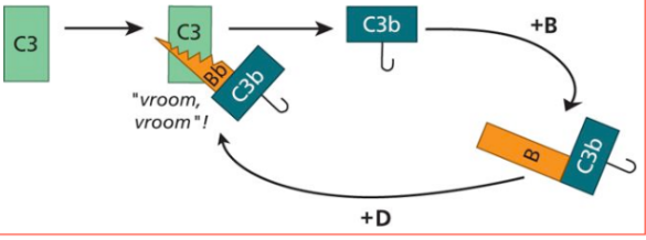
C3 Protein: Spontaneously cleaved into C3a and C3b.
C3b Binding: Binds to hydroxyl and amino groups on foreign cells.
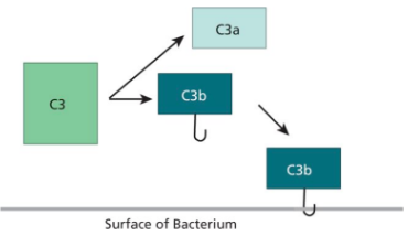
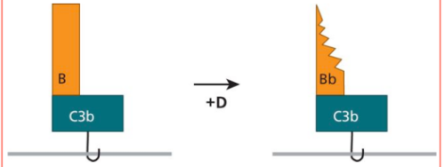
Addition: Complement protein B is attached to C3B, and protein D clips off B to make C3bBb (C3 Convertase)
Amplification: C3 convertase (C3bBb) formation leads to more C3 cleavage.
Creates more C3b, which joins with more B to become C3 convertases
Membrane Attack Complex (MAC): C3 Convertase can cleave complement protein C5 into C5a and C5b
C5b binds to the bacterial cell, and recruits C6,C7,C8,C9.
This forms pore called a membrane attack complex (MAC) that can assemble in exposed membranes on the outer surface of an organism
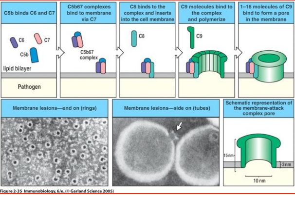
The organism undergoes lysis and dies.
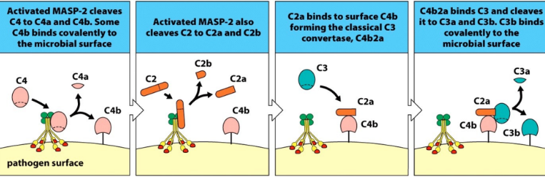
Lectin Pathway
Mannose is a sugar found on bacteria, fungi, viruses, and parasites. It is not found on local human cells, making this pathway more specialized
Mannose-Binding Lectin (MBL): Binds to mannose on pathogens, activating MASPs to generate C3 convertase.
MASPs = Mannose-binding lectin associate serine proteases
Generate C3 convertase (C2aC4b)
C3 Convertase cleaves C3, creating C3b.
What follows is the exact same process as the alternative pathway.
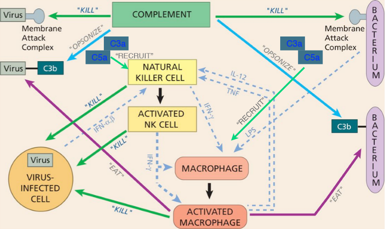
Functions of Complement
Opsonization: Modified C3b (iC3b) aids in marking pathogens for phagocytosis.
Chemoattractants: C3a and C5a recruit phagocytes to infection sites.
Professional Phagocytes
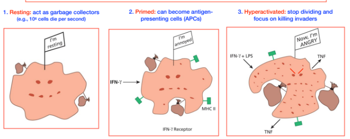
Macrophages
Location: Reside in tissues (skin, lungs, intestines).
States:
Resting: Garbage collectors.
Most of life span is spent as garbage collectors.
Primed: Can present antigens.
Contain major histocompatibility complex II (MHC II) - signals helper T-cells.
Interferon gamma (IFN - gamma) from Natural Killer cells (NK) or dendritic cells stimulates the macrophage
Hyperactivated: Focus on killing invaders.
Neutrophils
Location: Found in the bloodstream, short-lived as they are toxic and can damage tissues
Functions:
Kill invaders and die by apoptosis.
They do not present antigens
Produce cytokines to recruit other immune cells.
Can form neutrophil extracellular traps (NETs) to trap pathogens.
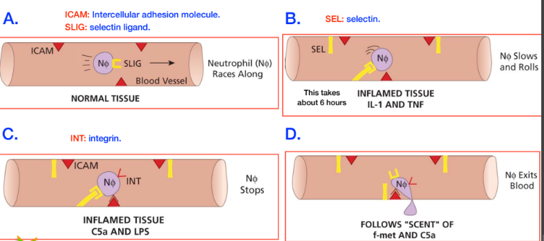
Recruitment Mechanism
Neutrophils exit the bloodstream through a regulated process involving selectins and integrins, ensuring controlled tissue damage.
Steps:
Neutrophils are lined with SLIG (selectin ligand). When inflamed tissue, IL-1 (interleukin) and TNF (tumor necrosis factor) are produced, blood vessels produce SEL (selectin)
SLIG binds to selectin, slowing down the neutrophils in the blood stream. Process takes about 6 hours
Once bound to the cell, the neutrophil expresses INT.
Chemoattractants C5a and LPS (lipopolysaccharide) are signalled, which means the neutrophil will express INT
INT binds to ICAM (intercellular adhesion molecule) and holds the neutrophil in place
The neutrophil leaves the blood stream, following the “scent” of f-met and C5a
formyl methionine (f-met) is expressed on bacteria.
3 types of molecules must be expressed before the neutrophil leaves the blood stream: interleukins (IL-1), TNF, LPS, and Complement proteins (C5a). This prevents neutrophils from leaving without infection and destroying normal tissues.
Sentinel Cells
Recognition Mechanisms
Pattern Recognition Receptors (PRRs): Detect pathogen-associated molecular patterns (PAMPs) and damage-associated molecular patterns (DAMPs).
DAMPS are released from dying cels
Toll-like Receptors (TLRs): Key PRRs that recognize specific PAMPs, such as LPS from Gram-negative bacteria.
As stated, TLRs are a type of PRR
TLRs can be on the cell of macrophages of dendritic cells (DCs)
DCs have TLRS and recognize PAMPS
Importance of PRRs
Recognize general, shared characteristics of many invaders of the same class
TLR4 recognizes LPS on all gram-negative bacteria
LPS is required for gram negative bacteria, it is a conserved trait
Interferon and NK Cells
Interferon Response
Type 1 Interferons (α and β): Produced by plasmacytoid dendritic cells (pDCs) and macrophages, upon viral detection, inducing antiviral protein production in neighboring cells.
IFN-alpha or beta Bind to the receptors of neighboring cells, and induce the production of antiviral proteins
Uninfected warned cells continuing being themselves, but infected warned cells commited suicide via apoptosis (controlled cell death)
NK Cells
Characteristics: Short-lived lymphocytes that respond to infected cells.
On call from the blood like neutrophils, and use a similar mechanism to leave the blood stream.
In tissues, they divide to boost their numbers.
Functions:
Receive cytokine signals from other immune cells (like neutrophils) or infected cells (cells signalling IF-α or IF- β), or bacteria (secreting LPS). They then Secrete cytokines (e.g., IFN-γ) to stimulate macrophages.
Induce apoptosis in infected cells via Fas/Fas ligand or injecting granzyme using perforin proteins.
How do NK cells identify target cells?
NK cells have no T-Cell receptors (TCRs), but two other receptors on target cells signal NK to act on that cell:
MHC I - inhibitory signal, tells the NK cell not to kill. This signal is found on all human cells
Surface proteins/carbohydrates - expressed when a cell is stressed (when infected) signals the NK cell to kill
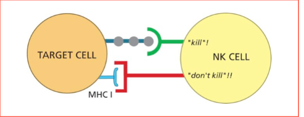
Some viruses prevent MHC I from being expressed, twarts the CTLs (killer T cells), but it does not thwart an NK cell, as the NK cell still sees the protein/carb signal.
Cooperative Efforts in Innate Immunity
Collaboration: Macrophages alert neutrophils to exit the bloodstream.
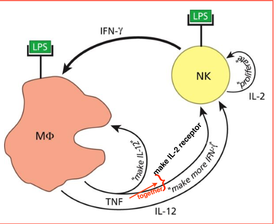
Cytokine Signaling: NK cells produce IFN-γ after being stimulated by LPS from gram negative bacteria, which activates macrophages, creating a feedback loop for enhanced immune response.
IFN-γ hyperactivates the macrophage, which then makes TNF (tumor necrosis factor), which signals itself to make cytokine IL-12
Continuing the Cycle: TNF and IL-12 stimulate NK cells to produce more IFN-γ (stimulating more macrophages), and to produce the IL-2 receptor
IL-2 binds to NK cells, making them proliferate to create more cells.
Complement protein signalling - iC3b on target organisms stimulate macrophages to create complement proteins C3, B, D, which are used to start the alternative pathway.
Resource Management: The innate response is proportional to the infection size, ensuring efficient resource use.
Viruses can hide inside cells, making them more of a challenge for the innate immune response
Summary of Innate Immunity
Neutrophils and Dendritic Cells: Key players in the innate immune response, with distinct roles in pathogen recognition and response.
Overall Function: The innate immune system acts as the first line of defense, utilizing various cells and proteins to combat infections effectively