Comprehensive Review Notes: Human Anatomy, Cell Biology, Histology, and the Integumentary System
Body Organization, Terminology, and Homeostasis
Anatomy vs. Physiology: Anatomy studies body structures and their physical relationships; Physiology studies how those structures function. The Principle of Complementarity states that function always reflects structure.
Levels of Biological Organization: Chemical level (atoms and molecules) Cellular level Tissue level Organ level Organ system level Organismal level.

Characteristics of Life: Energy production/consumption (ATP primary energy source, heterotrophic intake), Growth/repair, Adaptation/responsiveness, and Reproduction (essential for species survival).
Survival Needs: Nutrients, oxygen, water, normal body temperature, and appropriate atmospheric pressure.
Homeostasis: The active maintenance of stable internal conditions around established physiological set points.
Control Loop Components: Stimulus Receptor/Sensor (detects change) Afferent pathway Control Center (integrates signal) Efferent pathway Effector (executes response).

*   **Negative Feedback**: Reverses or reduces the direction of the original stimulus to return the variable to its homeostatic set point (e.g., thermoregulation via sweating/vasodilation, insulin regulation of blood glucose).
*   **Positive Feedback**: Amplifies or accelerates the original stimulus, pushing the variable further from its initial state to control infrequent, non-continuous events (e.g., oxytocin during labor contractions, platelet plug formation during blood clotting).
Anatomical Position: Standing upright, feet together, eyes facing forward, arms at sides, palms facing forward.
Directional Terms: Anterior (ventral) / Posterior (dorsal), Superior (cranial) / Inferior (caudal), Medial / Lateral, Proximal / Distal (limb references relative to attachment point), Superficial / Deep.
Body Planes & Cavities:
Planes: Transverse (horizontal), Frontal (coronal), Sagittal / Midsagittal.
Dorsal Cavity: Cranial cavity (contains brain) and Spinal cavity (contains spinal cord).
Ventral Cavity: Thoracic cavity (Pleural cavities for lungs, Pericardial cavity for heart, Superior mediastinum) and Abdominopelvic cavity (Abdominal cavity, Pelvic cavity). Anatomically divided into 4 quadrants or 9 regions.
Cellular Structure, Transport, and Communication
Primary Biomolecules:
Nucleic Acids: Nucleotide monomers linked by phosphodiester bonds (DNA, mRNA, tRNA, rRNA).
Proteins: Amino acid monomers linked by peptide bonds; execute cellular tasks and structural functions.
Lipids: Phospholipids (membrane structure), Cholesterol (membrane stabilization and steroid precursor), Acylglycerols (energy storage), and Waxes (protective barriers).
Carbohydrates: Monosaccharide monomers linked by glycosidic linkages; function in energy storage and cellular recognition tags.
Plasma Membrane Structure:
Fluid Mosaic Model: A thin () phospholipid bilayer embedded with cholesterol (), integral proteins, peripheral proteins, and surface carbohydrates (glycocalyx).
Fluidity Factors: Higher temperatures increase fluidity; saturated fatty acids decrease fluidity; unsaturated fatty acids increase fluidity. Cholesterol acts as a temperature buffer.
Membrane Transport:
Permeability: Permeable to small, nonpolar solutes (oxygen, carbon dioxide, steroid hormones); Impermeable to large, polar solutes and charged ions (carbohydrates, proteins, ions).
Passive Transport (down concentration gradient, no ATP): Simple diffusion, Osmosis (water movement causing isotonic, hypertonic, or hypotonic cell volume changes), and Facilitated diffusion (utilizing channel proteins or carrier proteins). Channel types include voltage-gated, ligand-gated, and mechanosensitive channels.
Active Transport (against concentration gradient, requires ATP): Primary active transport (e.g., Sodium-potassium pump pumping out and in per ATP), Secondary active transport (cotransport), and Vesicular transport.
Modes of Cell Signaling:
Autocrine: Soluble messenger binds to receptors on the same cell that secreted it.
Paracrine: Soluble messenger acts on nearby target cells.
Endocrine: Hormones travel through the bloodstream to act on distant target cells.
Cell Junctions:
Adherens Junctions: Mechanical attachment using cadherins and actin filaments.
Desmosomes: Provide structural elasticity and cell-to-cell adhesion using cadherins and intermediate filaments.
Hemidesmosomes: Anchor the basal surface of epithelial cells to the extracellular matrix (ECM) using integrins and keratin.
Tight Junctions: Formed by occludin molecules to seal intercellular spaces and prevent passage of molecules between cells.
Gap Junctions: Composed of connexin monomers forming connexons that create hydrophilic channels for direct intercellular communication.
Cell Life Cycle:
Interphase: Cell growth and protein synthesis (), DNA replication ( phase), and preparation for division ().
Mitotic Phase (): Mitosis (Prophase, Metaphase, Anaphase, Telophase) resulting in nuclear division, followed by Cytokinesis (cytoplasmic division) to generate two identical daughter cells.

Histology and Tissue Types
Epithelial Tissue:
Characteristics: Tightly packed cells held by junctions, distinct apical and basal polarity, avascular but innervated, and highly regenerative.
Classification: Simple (1 layer), Stratified ( layers), or Pseudostratified; Squamous (flat), Cuboidal (cube-like), Columnar (tall), or Transitional (variable/distensible shape).

*   Glandular Classification: Endocrine (ductless, secretes hormones into capillaries) vs. Exocrine (secretes products into ducts or onto surfaces).
*   Exocrine Secretion Mechanisms: Merocrine (exocytosis; e.g., salivary glands), Apocrine (pinching off apical cytoplasm; e.g., mammary glands), Holocrine (whole cell rupture; e.g., sebaceous glands).
Connective Tissue:
Characteristics: Cells embedded within an extracellular matrix (ground substance + fibers); derived from embryonic mesenchyme.
Cell Types: Fibroblasts/fibrocytes, Osteoblasts/osteocytes, Chondroblasts/chondrocytes, Adipocytes, and Blood cells.
Fiber Types: Collagen (thick, resists tension, most abundant), Elastin (long, branched, allows stretch and recoil), Reticulin (short, thin, forms flexible meshwork).
Classes: Loose CT (Areolar, Adipose, Reticular), Dense CT (Regular, Irregular, Elastic), Cartilage (Hyaline, Elastic, Fibrocartilage), Bone (calcified collagen matrix), and Blood (cells in plasma matrix).
Muscle Tissue:
Skeletal Muscle: Voluntary, striated, multinucleated, elongated myotubes attached to skeleton.
Cardiac Muscle: Involuntary, striated, branched cells joined by intercalated discs with central single nuclei in heart walls.
Smooth Muscle: Involuntary, non-striated, spindle-shaped cells arranged in sheets within walls of hollow organs.
Nervous Tissue:
Neurons: Excitable, conductive cells that receive and transmit electrical signals.
Glial Cells: Non-conductive support cells providing structural protection and nutrients.
Body Membranes:
Cutaneous Membrane: Skin; dry epithelial/connective sheet exposed to air.
Mucous Membranes: Wet sheets lining body cavities open to the exterior (digestive, respiratory, urogenital tracts).
Serous Membranes: Wet double-layered sheets lining closed ventral body cavities (Pleurae, Pericardium, Peritoneum).
Tissue Repair & Development:
Repair Sequence: Inflammation (clotting/scab formation) Organization/Regeneration (granulation tissue/capillary ingrowth) Fibrosis (scar formation).
Regenerative Capacity: Excellent (epithelia, bone, blood-forming, areolar, dense irregular CT); Moderate (smooth muscle, dense regular CT); Weak (skeletal muscle, cartilage); None (cardiac muscle, central nervous system tissue).
Germ Layers: Ectoderm, Mesoderm, Endoderm.
The Integumentary System
Integumentary Functions: Protection (physical, chemical, biological barrier), Thermoregulation (sweating, vasodilation/vasoconstriction), Sensation, Vitamin D synthesis (UV conversion of 7-dehydrocholesterol to cholecalciferol/Vitamin , converted to active calcitriol), Excretion, and Blood reservoir.
Skin Structure:
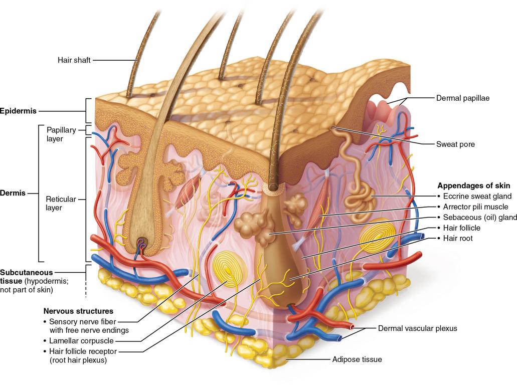
* **Epidermis**: Keratinized stratified squamous epithelium.
* Cell Types: Keratinocytes (main type, structural keratin), Melanocytes (melanin pigment synthesis), Dendritic / Langerhans cells (immune antigen presentation in stratum spinosum), Tactile / Merkel cells (mechanoreceptors for fine touch in stratum basale).
* Layers (deep to superficial): Stratum basale (single row of dividing cuboidal cells), Stratum spinosum ( rows of keratinocytes), Stratum granulosum ( rows accumulating keratohyalin), Stratum lucidum (dead clear layer present only in thick skin), Stratum corneum ( layers of dead, flattened keratinized cells).
* **Dermis**: Vascular connective tissue layer supporting the epidermis.
* Papillary Layer: Loose connective tissue with fine collagen/elastic fibers, dermal papillae, and capillary loops.
* Reticular Layer: Dense irregular connective tissue with thick collagen bundles forming cleavage lines (tension lines).
* Dermal Receptors: Meissner's corpuscles (light touch/vibration), Lamellated corpuscles (deep pressure/vibration), Ruffini corpuscles (pressure/distortion).
* **Hypodermis**: Subcutaneous adipose tissue layer below the dermis (anchors skin, not part of integument).
Appendages of the Skin:
Hair: Grows from hair matrix in the hair bulb. Shaft structure contains Medulla (soft keratin), Cortex (hard keratin), and Cuticle (hard keratin). Growth phases: Anagen (active growth), Catagen (end of active growth), Telogen (resting phase). Contraction of the arrector pili muscle pulls hair upright causing goosebumps.
Nails: Plates of hard keratin produced by the nail matrix beneath the eponychium (cuticle); average growth rate is for fingernails.
Sebaceous Glands: Holocrine glands that secrete lipid-rich sebum into hair follicles for lubrication and antimicrobial protection.
Sudoriferous Glands: Apocrine sweat glands (axillary and pubic regions, secrete into hair follicles, bacterial breakdown produces odor) and Merocrine / Eccrine sweat glands (widespread, secrete directly onto skin for thermoregulation).
Skin Clinical Conditions & Aging:
Color Sign Diagnostics: Cyanosis (blue/low oxygen), Erythema (redness/inflammation), Pallor (pale/anemia), Jaundice (yellow/liver dysfunction), Ecchymosis (bruising).
Skin Cancers: Basal cell carcinoma (most common, least malignant, stratum basale invasion), Squamous cell carcinoma (keratinocytes of stratum spinosum, potential metastasis), Melanoma (melanocytes, highly metastatic; screened using ABCD rule: Asymmetry, Border irregularity, Color variation, Diameter > 6\,\text{mm}).
Burns: Evaluated using the Rule of Nines ( regions at each, plus perineum at ).
Classification: 1st-degree (epidermal damage only, redness/edema), 2nd-degree (partial thickness, epidermal and dermal damage, blisters), 3rd-degree (full thickness, nerve endings destroyed, painless burn site, requires skin grafting).
Critical thresholds: > 25\% body with 2nd-degree, > 10\% body with 3rd-degree, or 3rd-degree on face, hands, or feet.

* Aging Changes: Epidermal thinning, reduced dermal collagen/elasticity (wrinkling), decreased glandular activity, thinning hair, and slowed nail growth.