BIO 110 | LESSON 1 - LESSON 5

Lesson 1: Microscope

Microscope

  • Magnifying device used to look at cells

PEOPLE TO REMEMBER

Anton van Leeuwenhoek

  • Dutch Scientist

  • Creator of the Microscope

  • Protozoans → Live Cells

  • Father of Microscopy

Robert Hooke

  • First coined the word “cells”

  • 1665: First to view the cell wall on a cork-like structure

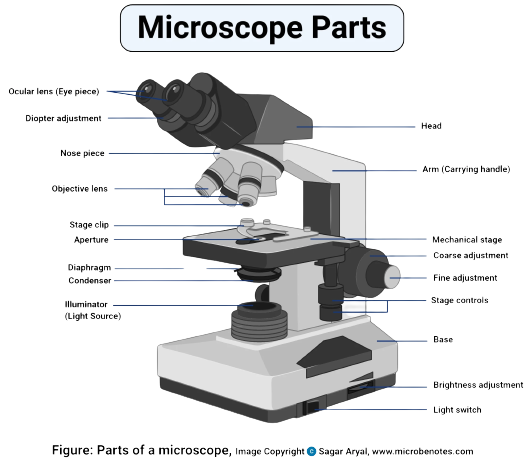
PARTS OF THE MICROSCOPE

Mechanical Parts

  • Support/Strength

  • To help move around the microscope

  • Base, Arm, Draw Tube

  • Base

  • Arm

  • Revolving Piece

  • Nose Piece

  • Stage Controls

  • Mechanical Stage

  • Head

Illuminating Parts

  • Capturing the light from the source

2 TYPES

  • Mirror

  • Condenser (electronic)

  • Illuminator

  • Light Switch

  • Brightness Adjustment

  • Diaphram

    • Controls the light entering

  • Condenser

    • Captures Light

Magnifying Parts

  • Magnifying

4 TYPES OF LENSES

  • High Power Option (HPO)

  • Low Power Option (LPO)

  • Oil Immersion Option (OIO)

  • Scanning Option (SO)

  • Objective lens

  • Eye Piece

  • Coarse Adjustment (Low Power)

  • Fine Adjustment (High Power)

NOTES:

Gram Negative Bacteria

  • Pink in color

  • More resistant to most available antibiotics

  • Harder to cure

  • Has extra outer membrane and Has more rigid and compact layering

Gram Positive Bacteria

  • Purple

  • Has thicker peptidoglycan

Rods / Kokai = Shape of Organism

PARTS OF THE MICROSCOPE (SPECIFIC)

Eye Piece/Occular Lense

  • used to look through the microscope found at the top of the microscope.

  • Its standard magnification is 10x with an optional eyepiece having magnifications from 5x to 30x.

Objective Lenses

  • 40x to 100x

4 TYPES OF LENSES

  • High Power Option (HPO) (Blue)

  • Low Power Option (LPO) (Yellow)

  • Oil Immersion Option (OIO)(Light Yellow)

  • Scanning Option (SO) (Red)

Body Tube

  • carries the eyepiece just above the objective lens.

  • It is flexible and can be rotated for maximum visualization, for variance in distance.

  • For monocular microscopes, they are non-flexible.

Adjustment Knobs

  • Used to focus the image

  • To move up and down

Fine Adjustment (High Power)

Coarse Adjustment (Low Power)

Base

  • Holds up the microscope

Arm

  • Support to the head

  • Used when carrying the microscope

Revolving Nosepiece/Turret

  • Holds objective lenses

  • Used to revolve the objective lenses

illuminator

  • Captures light from an external source

  • Low voltage (100v)

Iris Diaphragm

  • a circular disk that controls the amount of light that reaches the specimen

Condenser

  • to collect and focus light from the illuminator into the specimen

Aperture

  • a hole on the microscope stage, through which the transmitted light from the source reaches the stage

Mechanical Stage

  • Platform for the slide to rest

Stage control

  • To move the slides placed on the stage

Stage Clips

  • Holds the slide in place


Lesson 2: Characteristics of Life

Cell

  • Basic unit of life

  • Cellula: Tiny Boxes

  • Is in all living things

  • Basic structural and functional unit

  • Smallest unit of life

  • Replicates independently

  • Can vary in different shapes, sizes, and functions

DIFFERENT CHARACTERISTICS

Reproduction

  • Produce new cells of the same kind through cell division

Development

  • Is not merely characterized by a literal increase in girth

  • Refers to the irreversible increase in the number of cells

  • A multitude of cell divisions will change an organisms form frfom simple to complex

Movement

  • Interacting with the environment to have a higher chance of surviviing

  • Must be able to change its physical position

Responsiveness

  • React accordingly to their stimuli.

Ex: when you get cold, you try to warm yourself up.

Metabolism

  • Conversion of energy from food

Homeostasis

  • Can maintain a stable internal environment in the face of changing external conditions

Feedback Loop

  • Reaction to any disturbance in the cell's bodily functions

  • Restores the latter to its original state

Organization

Organelles

  • Makes up the cell

  • Smaller units

  • Each performs a specific function

  • Cannot live or stand along (dependent)

  • Cells → Tissues → Organs →etc.

Genes

  • Having their own genetic identity

Evolution

  • Cells with better adaptations with respect to their environment will have higher chance of surviving

  • Can ake in single-celled bacteria over short periods

LIFES LEVELS OF ORGANIZATION

  • Cells → Tissues → Organ → Organ System → Organism

ORIGINS OF LIFE

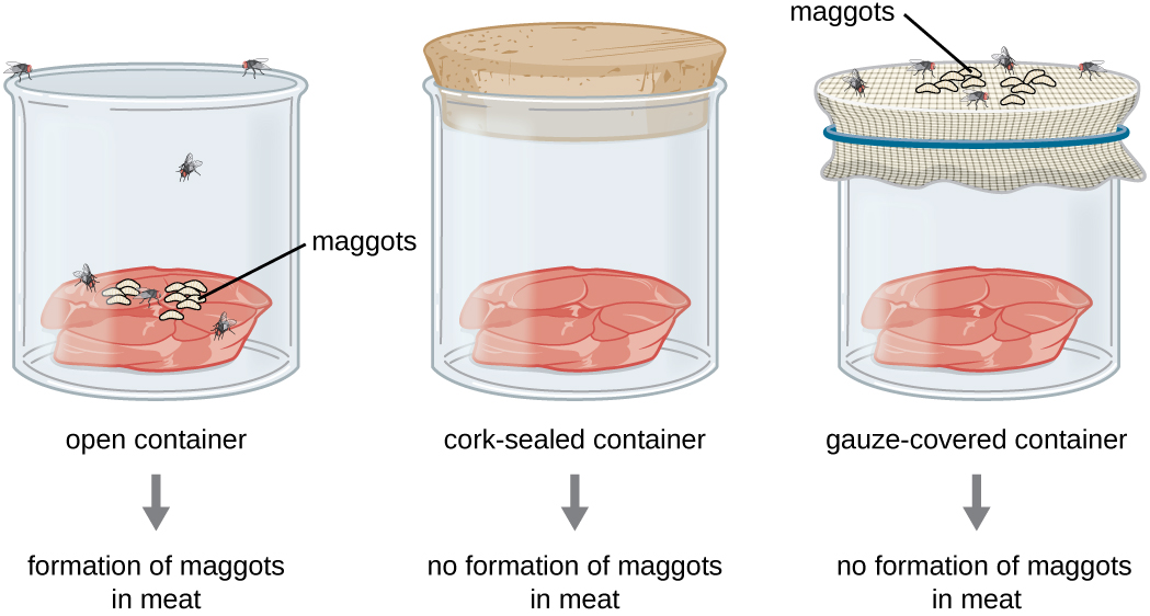
Francesco Redi’s Experiment

(Biogenesis)

3 Meat Set Up

  • Meat 1: Unsealed

  • Meat 2: Sealed w/ Cork

  • Meat 3: Sealed w/ Gauze

Results

  • Meat 1: Flies and Maggots were found directly on the meat

  • Meat 2: There were no Flies nor Maggots found since it cannot be smelled by the flies.

  • Meat 3: Attracted Flies and Maggots due to its smell but is not on the meat itself due to the barrier of gauze

Conclusion

  • Life arises from living things

Notes:

  • Maggots: Fly Eggs

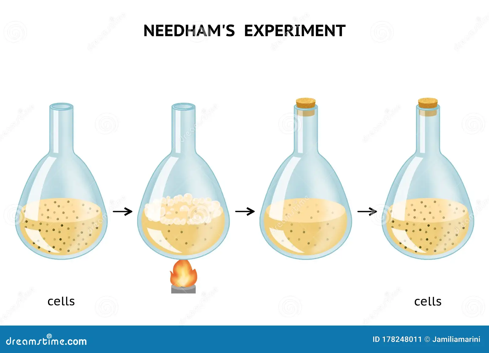
John Needham’s Experiment

(Abiogenesis)

About

  • It is similar to pasteurizing

  • Continuously boiling

Process

  • In a flask, the broth was brought to a boil for a certain period of time.

  • It was covered right after boiling to avoid contamination

Results

  • Microorganisms have formed

Conclusion

  • Life is caused by spontaneous generation or by non-living organisms

Notes:

  • The liquid was not heated enough

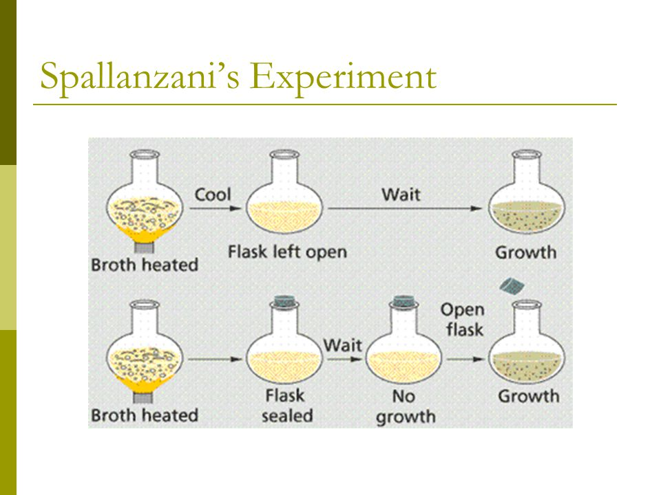
Lazzaro Spallanzani’s Experiment

(Biogenesis)

2 Sets of Broth

  • Set A: Broth with NO SEAL

  • Set B: Broth WITH SEAL

Process

  • Both broths were heated at the same time.

Results

  • Set A: With the flask open, there was growth seen

  • Set B: With the flask sealed, growth was absent not until the seal was removed where growth was visible

Conclusion

  • Life occurs when something enters the flask

  • Life Occurs from Living Things

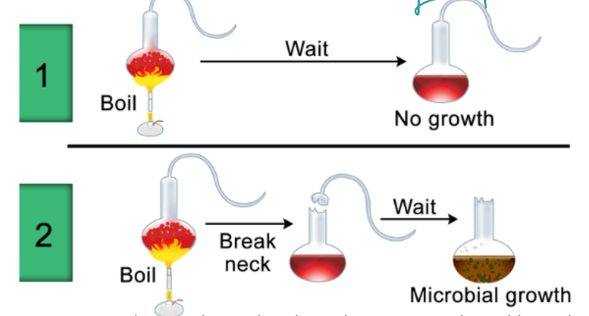
Louis Pasteur’s Experiment

(Biogenesis)

Curved Bottled Neck

  • A curved bottleneck was used to conduct this experiment

Process

  • The bottle was heated

Results

  • Because of the shape of the bottle, the microorganisms were trapped on the neck.

  • This is because of the moisture that was formed in the spout.

  • This led to breaking/taking off the long spout, wherein the microorganisms were able to flow freely in the container

Divine Creation

About

  • The universe was created by a supreme being

Spontaneous Origin

(Abiogenesis)

About

  • Life evolved from an inanimate matter

  • First assembled from underwater volcanic vents, surface of clay sediments, deep in earth’s crust, under frozen oceans

Simple Molecules + Simple Molecules = Complex Molecules

Primordial Soup

  • Complex biological compounds

  • Lightning triggers

Conclusion

  • Life is formed from Non-Living things

Svante Arrhenius’ Panspermia

(Abiogenesis)

About

  • Made out of meteor/cosmic dust

BIOGENESIS V.S. ABIOGENESIS

Biogenesis

Abiogenesis

  • Life exists from life

  • Did not start from life

  • Started from non-living things

Existence of Life

King Charles II

  • Instructed Robert Hooke to study about the Natural World

Robert Hooke

  • Viewed the cork of an oak tree

  • Cellulate: Cells

  • First to view dead cells

  • “Micrographia” is his best selling Novel about microscopy

Anton Van Leeuwenhoek

  • “Father of Microscopy”

  • Viewed live cells

    • Animalcules (little animals)

  • Protozoa

It took 200 years for the general public to accept that living things are composed of cells

Lesson 3: Cell Theory

Cell

  • Basic unit of life

  • Cellula: Tiny Boxes

  • Is in all living things

  • Basic structural and functional unit

  • Smallest unit of life

  • Replicates independently

  • Can vary in different shapes, sizes, and functions

Cell Theory

  • Earliest foundation of the study of life

  • Basic unifying foundation

POSTULATES OF CELLS

  • Cells are the Smallest Unit of Life. All organisms are made up of cells (Matthias Schleiden)

  • Cell is the basic unit of organization of all organisms (Theodore Schwan)

  • Cells Arise from Previous Cells (Rudolf Virchow)

PEOPLE TO REMEMBER

Matthias Jakob Schleiden

  • “Professor of Botany” in University of Jena, Germany

  • 1318: Studying plants under microscope

  • “All plants tissues are composed of cells”

Robert Brown

  • Scottish Botanist

  • Discovered the Nucleus

Brownian Motion

  • Random movement of macroscopic particles

Theodore Schwan

  • Specialized in Zoology

  • Presence of cells in animal tissues

Rudolf Ludwig Karl Virchow

  • Studied how cells play as role models in body diseases

Omnis Cellula e Cellula

  • Cells arise from pre-existing cells

Robert Remak

  • Jewish Scientist

  • Tried to prove the idea of cell division by hardening the cell membrane

MODERN CELL THEORY

Energy flows within cells

Genetic makeup of the cells of organisms that belong to the same species is the same

Cells carry and pass on heredity units to the offspring during cell division

The activity of an organism depends on the activity of independent cells

Cell Shape

  • Depends on its functions

  • Defined by the geometrical information of the space occupied by the cell and is determined by its external boundaries

CELL SHAPES:

  • Round

  • Elongated

  • Spherical

  • Spindle Shaped

  • Branched

SICKLE CELL ANEMIA

  • Blood cloth clogs

Cell Size

  • Small Surface Area = More Efficient and Effective

  • Moves faster around the cell when it is smaller

  • Cell Size = Surface Area : Volume

  • Object larger, volume rises faster

  • Higher Surface Area : Volume = Higher Efficiency

Lesson 4: Basic Cell Types

BASIC CELL TYPES

Descriptions

Eukaryotic

Prokaryotic

Name Origin

Comes from the Prefix

  • “Eu”: true

  • “karyo”: Nucleus

Comes from the Prefix

  • “Pro”: Before

  • “karyo”: Nucleus

Nucleus?

Double Membrane Bound Nucleus

  • Delimits the nucleus from cytoplasm

  • Cells DNA

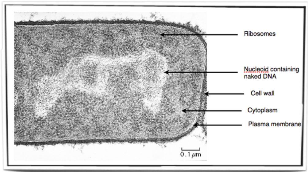
Nucleiod

  • Central open part where the cells DNA is found

Structure

Large and Complex Membrane-Bound Organelles

  • For respiration and photosynthesis

Small and Simple

Cell Membrane

Similar composition

Hydrophilic (Water Loving) Hydrophobic (Water Allergic)

Cell Name

Eukaryotes

BIGGER than prokaryotes

Organisms

Prokaryotes

SMALLER than Eukaryotes

Unicellular

Different Cell

  • Animal and plant protozoa

  • Fungi organisms

  • Bacteria

  • Archaea

Type of Cell

Multi-Cellular or Unicellular

Uni-Cellular or Single-Celled

Number of Chromosomes

Diploid

(Two copies of each chromosome)

46 Chromosomes

Haploid

(One copy of each chromosome)

23 Chromosomes

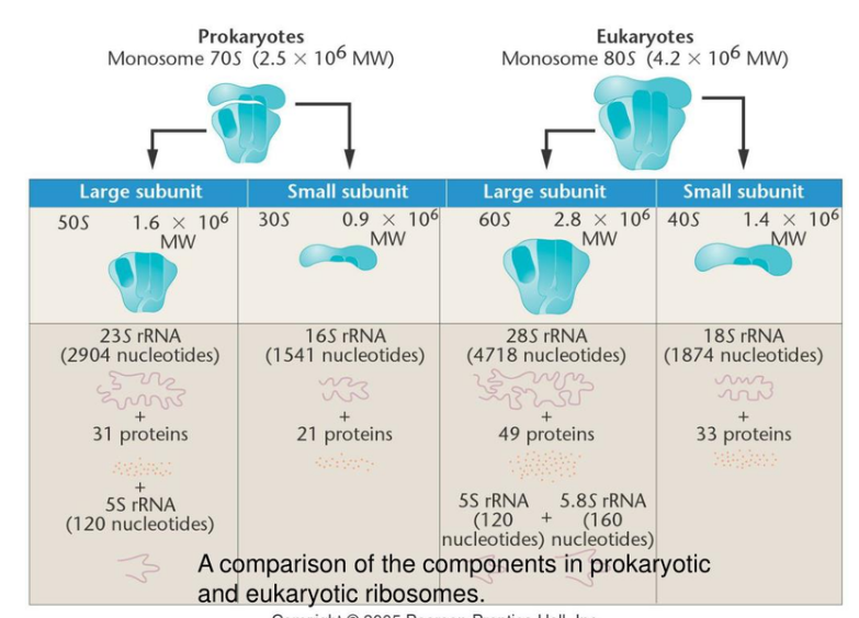
Nucleotides

120 Nucleotides

160 Nucleotides

Reproduction

Sexual and Asexual

Asexual

Organnelles

More Organnelles

Less Organnelles

DNA

Identical genetic code encoded in DNA

Process of Transcription and Translation

Same Process and only similar in those parts. Produces Protein at the end.

ATP

Same events in metabolic pathways and energy storage

Photosynthesis

Same mechanisms as plants (cynobacteria)

Endosymbiotic

Eukaryotic evolved from prokaryotic

Prokaryotic → Eukaryotic

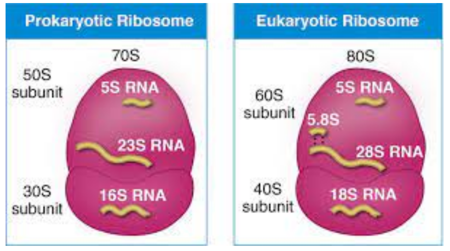
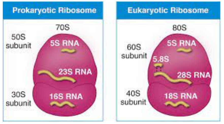
Ribosomes

  • Production and protein of a cell

Svedberg (S)

  • Rate of settling down of particles in a specific medium

  • Higher S Value = Heavier/Larger the Molecule

RIBOSOMES

Descriptions

Eukaryotic

Prokaryotic

Size

Bigger

Smaller

“S” Value

80s

70s

Speed

Faster

Slower

Images

RNA

  • Ribonucleic acid

  • Creataes protein

  • Counter part of DNA

rRNA

  • Ribosomes

Nucleotides

  • In amino acids

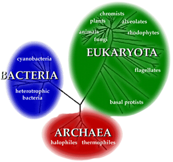
Three Domain System

  • Eubacteria: True Bacteria

  • Archaea: NOT True Bacteria

    • Possesses Similarities with eukaryotes

    • Introns and utilizes enzymes in metabolic pathways that eukaryotes have

Eukaryotic Cells

Prokaryotic Cells

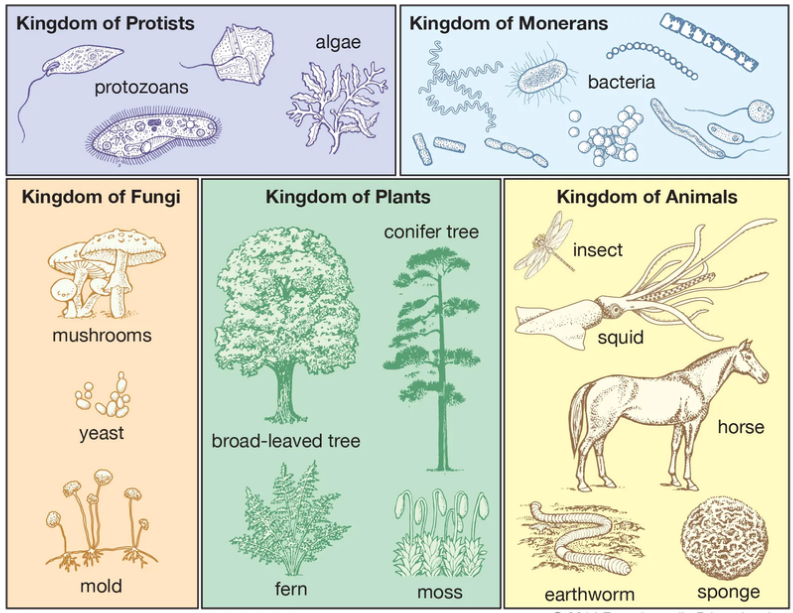
5 Kingdoms

Kingdom of Protists

Kingdom of Fungi

Kingdom of Plants

Kingdom of Animals

Kingdom Monera

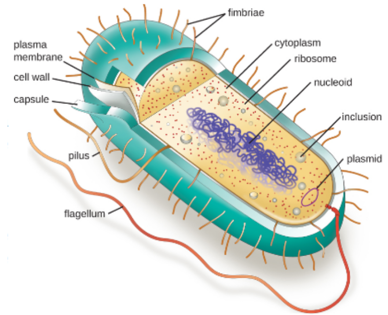
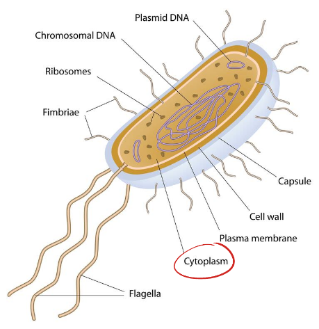
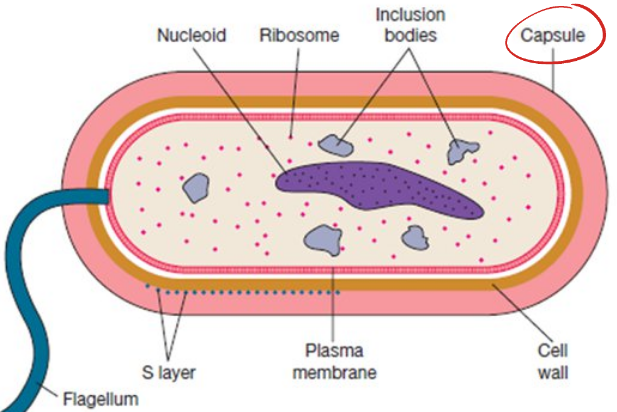
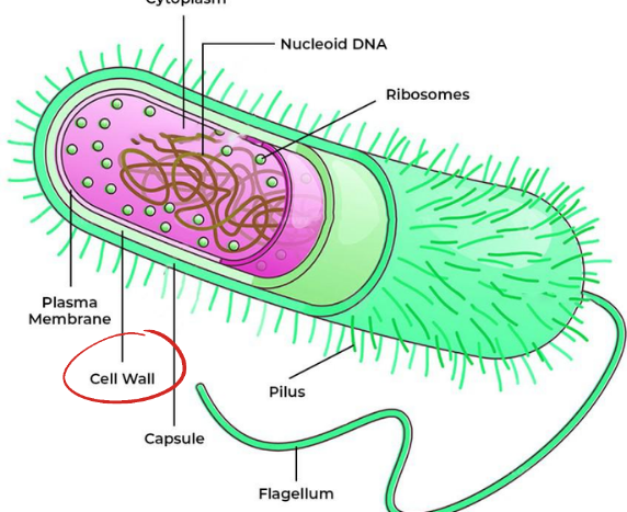
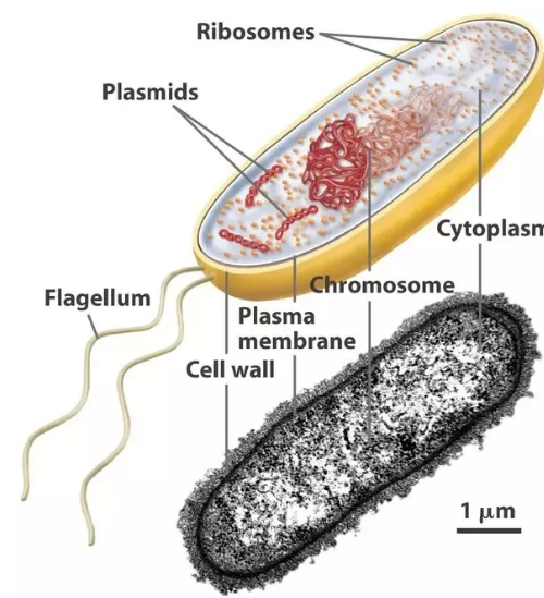
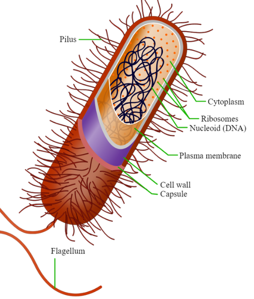
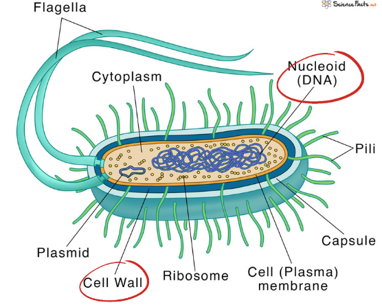
Structure of Prokaryotic Cells

STRUCTURE OF PROKARYOTIC CELLS

CYTOPLASM

  • Jell-like substance

  • Composed of mainly water

  • Contains enzymes, salts, cell components, and various organic molecules

  • Most chemical reactions occur here

Example:

  • Being in a swimming pool

OUTER COVERING

Capsule

  • Outermost covering

  • Additional outer covering

  • Protects whenever cell is in a undesirable location

  • Assists in retaining moisture

  • Adhere to surfaces and nutrients

Cell Wall

  • Protective layer

  • Surrounds some cells

  • Gives them shape and rigidity

  • Located outside the cell membrane

  • Prevents Osmotic Lysis

    • Bursting due to increase of volume

  • Other structures are present in prokaryotic species, but not in others

Peptidoglycan

  • Complex molecule

  • made up of Amino Acids and Sugar Polymers

Plant Cell

  • The only Eukaryotic Organism with a cell wall

Plasma Cell Membrane

  • Selectively permeable membrane

  • Regulates the entranceand exit of materials

Movement of Prokaryotic Cells

MOVEMENT OF PROKARYOTIC CELLS

Pili

ABOUT:

  • Hair-like structures on the surface of the cell

2 TYPES OF CELLS

Fimbriae

  • Shorter

  • Helps bacteria attach to surfaces

Sex Pili

  • Aid in exchange of genetic information

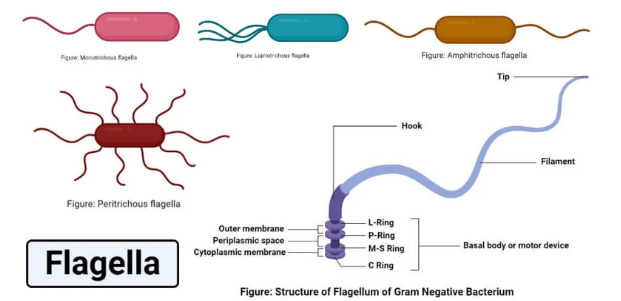
Flagella

  • Long whip-like protrusions

  • Attached to the cell that aid in cell locomotion

Prokaryotic Genetic Materials

GENETIC MATERIALS OF PROKARYOTIC CELLS

Nucleiod Region

  • Area of the cytoplasm

  • Contains RNA Molecule

RNA Molecule

  • Ribonucleic acid

  • Creataes protein

  • Counter part of DNA

Plasmids

  • Circular

  • Extra-chromosomal DNA

  • Found in cytoplasm

  • Provides genetic advantages

    • Ex: Antibiotic Resistance

NOTES:

DIFFERENCE OF GRAM POSITIVE AND GRAM NEGATIVE

Gram Positive Bacteria

Gram Negative Bacteria

  • Purple

  • Has thicker

  • Pink in color

  • More resistant to most available antibiotics

  • Harder to cure

  • Has extra outer membrane

  • Has more rigid and compact layering

SOMATIC CELLS (DIPLOID): BODY CELLS
GAMETE (HAPLOID): SEX CELLS

Lesson 5: Structures of Eukaryotic Cells

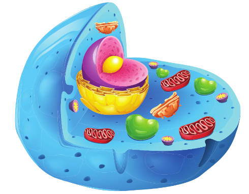
Cell Organelles
  • Little organs

  • Specialized sub-cellular structures

  • Perform one or more jobs inside of the cell

Structures of Eukaryotic Cells

Cell Membrane

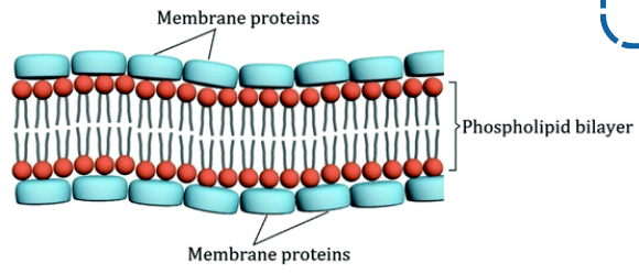
  • Thin and outermost layer

  • Separates the cell from its external environment

  • Semipermeable Membrane

  • Regulates substance to Absorb, Secrete, Excrete

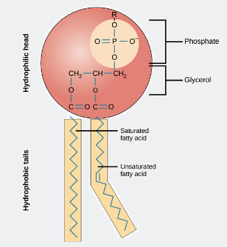
  • Phosphate Head : Hydrophilic

  • Lipid Tail : Hydrophobic

2 MODELS

Classic Model

Hugh Davson & James Danielli

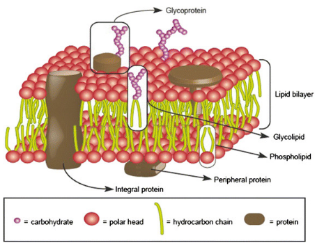
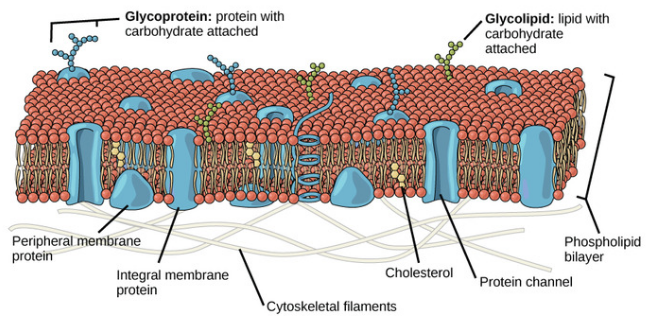
Fluid Mosaic Model

Jonathan Singer & Garth Nich

  • Sandwiched by membrane

  • Does not move

COMPONENTS OF CELL MEMBRANE

Rigidity

  • Cholesterol

  • Sterol

Cellular Recognition

  • Gycloprotein

  • Glycolipid

Glyco- : Carbon

Cell-Cell Recognition

  • Distinguishes cells

  • Channel Proteins

  • Transport Proteins

  • Recognition, Adhesion, Receptor, Electron Transfer Protein, Glycocalyx

Cholesterol

  • Makes cell membranes flexible

  • Less fluid

  • Less permeable to water-soluble substances

    • Ions & Simple Sugars

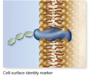
Carbohydrates

  • Attached to the membrane

  • Identification tags

  • Enables cells to distinguish each other

MEMBRANE PROTEINS

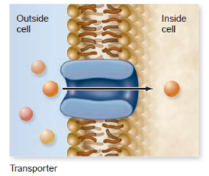
Transport Proteins

  • Extends from the phospholipid layer

  • Help materials cross the membrane

Channel Protein

  • Import or export of materials

  • Expel waters

Cell Recognition

  • Enables cells to distinguish their own cells

Pathogens

  • Harmful bacteria

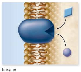
Enzymatic Proteins

  • Metabolic reactions

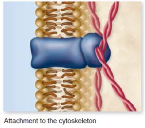
Cytoskeleton Protein

  • Muscle and Skeleton to the cell

  • Shape and motility

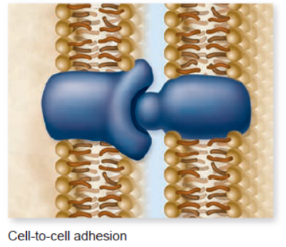
Junction Protein

  • Cell-to-cell adhesion

  • Communication with other cells

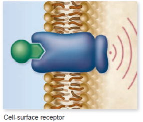
Receptor Protein

  • Exchange of signals

  • Signals from the nucleus

Ligand

  • Different molecules stick

  • Electron pair donors

Nucleus

  • Plural: Nuclei

  • Control center of the cell

  • Storage of genetic information

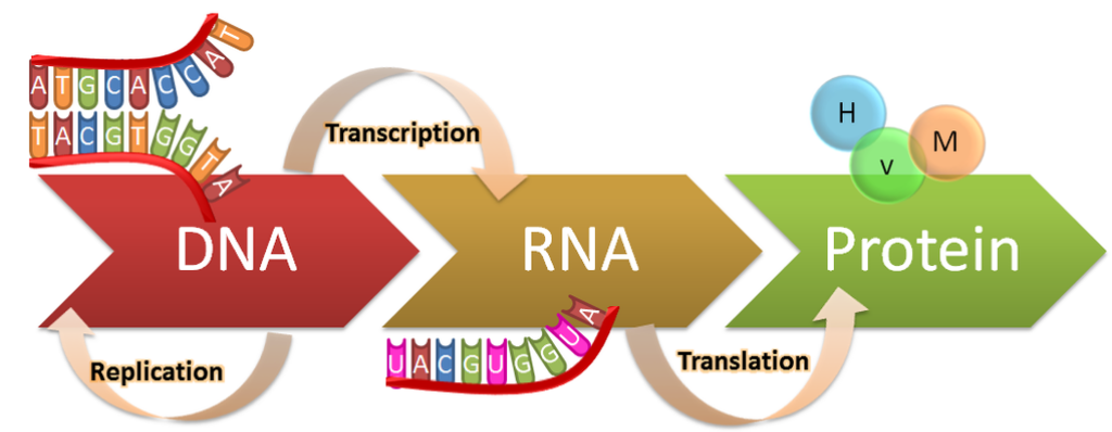
  • Sites where nucleic acids (DNA and RNA) synthesize (Central Dogma)

Histone

  • Specialized DNA to make chromatin

Nucleolus

  • Structure

  • Where ribosomes are made

ANATOMY OF NUCLEUS

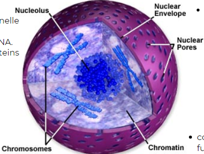
Nuclear Envelope

  • Boundary of the nucleus

  • Permeable for macromolecules (protein)

Perinuclear Shape

  • Shape in between inner and outer nuclear membrane

Nuclear Pores

  • Where substances move in and out of the nucleus

  • Passageway of microorganisms

Chromatin

  • Combination of DNA and Protein

  • Further condense to form chromosomes during cell division

Chromosomes

  • Two chromatids that are connected via centromere

Nucleolus

  • Spherical membrane-free organelle

  • Contains fibrils and granules

Fibrils

  • Contains DNA coding for RNA

Granules

  • Contains rRNA with proteins from cytoplasm

CYTOPLASM

  • Largest interior part of the cell

  • Protection for outer body

  • Contains Cytosol

Cytosol

  • Semi-fluid substance

  • Where organelles are suspended

  • Contains electrolytes, metabolites, RNA, Protein

ROLES OF CYTOPLASM

  • Where molecules first pass through

  • Where organelles move within around


CYTOPLASMIC ORGANELLES

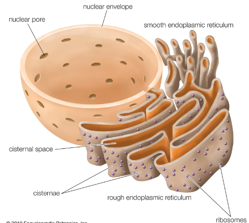
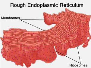
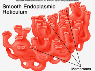
ENDOPLASMIC RETICULUM

  • Membrane Bound Organelle

  • Forms a network of interconnected sacs called cisterna

  • Intercellular highway for the transport of molecules within a cell

  • Its amount depends on the function of the cell

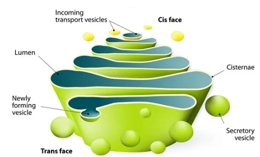
Lumen

  • Cisternal space

Rough Endoplasmic Reticulum

  • Presence of ribosomes on its membrane surface

  • Produces proteins

More RER = Large Amount of Protein

Smooth Endoplasmic Reticulum

  • Absence of ribosomes

  • Produces lipids

  • Gland Cells: synthesizing of steroids

  • Liver Cells: breakdown of toxic substances

  • Muscles: Regulation of calcium levels

GOLGI APARATUS

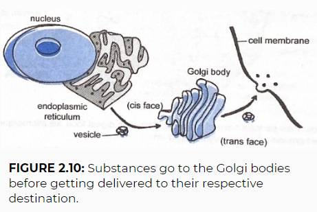
  • Works closely with the Endoplasmic Reticulum

  • Processing, packaging, and sorting of secretory materials within a cells

Exocytosis

  • Exiting of materials

FACES OF GOLGI APPARATUS

Cis Face

  • Facing the Endoplasmic Reticulum (ER)

Trans Face

  • Facing the Cytoplasm or Call Membrane

HOW IT WORKS

Step 1

Substance from ER enters the LUMEN

Step 2

Substance pinches off to become a vesicle

Step 3

Vesicle moves towards the cis face

Step 4

Golgi Bodies may modify substances by putting “tags” for recognition

Step 5

Vesicles containing the modified substances will exit at trans face

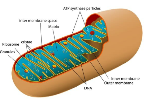
MITOCHONDRION

  • Power plant of the cell

  • Double membrane organelle

  • Reproduction of Adenosine Triphospate (ATP)

  • Has its own DNA and Ribosomes

PARTS OF MITOCHONDRION

Outer Membrane

  • covering

Inner Membrane

  • Creates cristae

Matrix

  • Contains different enzymes important in producing ATP

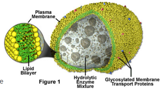
LYSOSOME

  • Garbage collectors of the cell

  • small , spherical, membrane-bound organelle

  • Originated from the ER

  • Contains Hydrololytic Enzymes

  • Uses water to break down substances

  • The digested food and recycled components are released back to the cytosol

  • Indigestible foods will become residual bodies

FUNCTIONS OF HYDROLYTIC ENZYMES

  • Digest Food

  • Recylcle old components of the cells

  • Kill invading mico-organisms

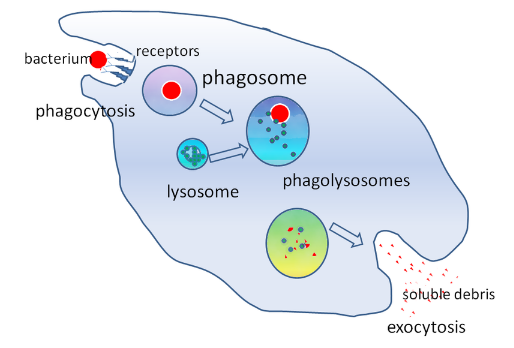
PHAGOSOMES

  • Vesicle in the cytoplasm of the cell

  • Contains phagosomus particle

  • Enclosed within a part of the cell membrane

Phagosomus Particle

  • To fight bacterias

Step 1

  • Lysosomes fuse with Phagosomes

Step 2

  • Releases its hydrolytic enzymes to kill micro-organisms inside

Step 3

  • Debris released in the process can stimulate the immune system to remember the micro-organisms

CELLULAR MACROMOLECULES

RIBOSOMES

  • Small, dense structures

  • Helps in the assembly of proteins in the cell

  • Are not membrane bound

  • Distribution of ribosomes in cells depends on how the proteins will be used

FORMS

Found freely in cytoplasm

Attached in rough ER

Attached in Polyribosome

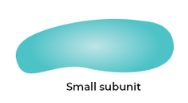
2 SUB UNITS

Small Subunit

Big Subunit

CENTRAL DOGMA OF LIFE

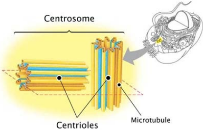
CENTROSOME & CENTRIOLES

Microtubule

  • 1 Tube

Centrioles

  • Set of microtubules

  • Assist in cell division

  • Surrounded with pericentriolar materials

  • Only visible in actively dividing animal cells

Pericentriolar Materials

  • Enables the growth of microtubules

  • Formation of spindel fiber

Centrosome

  • Combination of Centrosomes

  • Part of the cytoplasm

  • Produces microtubules

  • Animals only (main organizing center

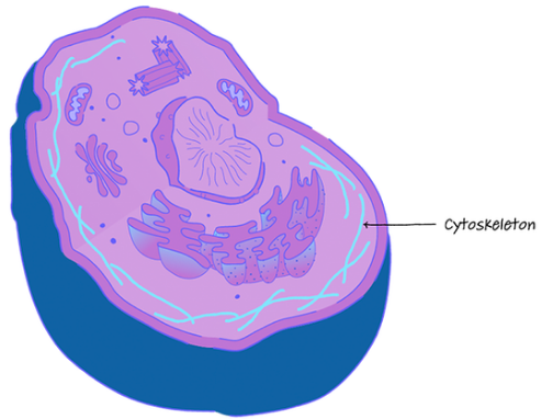
CYTOSKELETON

  • Provides motility and strength to the cell

  • Network of filaments and tubules that extent throughout the cell

TYPES OF FIBERS

Microtubules

  • Long, slender, protein tubes

  • Composed of linear polymers of tubulin

Ex: spindel apparatus

Microfilaments

  • Supports the cell in maintaining their shape and structure

Ex: spindel fibers

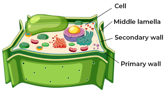
CELL WALL

  • Located outside the cell membrane

  • Made up of cellulose

Cellulose

  • Composed of a long chain of carbohydrates (polysaccharides)

Roles

  • Maintain the shape

  • Protection

  • Helps regulate cells uptake of water

DEVELOPMENT OF CELL WALL

Step 1

Newly formed plants have thin and flexible primary cell walls

Step 2

As the growth stops, secondary cells either:

  • Thicken the primary walls

  • Depositing new layers which contain more cellulose and ligning

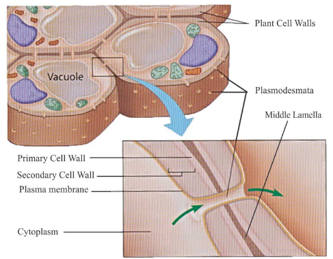
Plasmodesmata

  • Channels the cell wall

  • Allows exchange of substance between adjacent cells

Extra info

Most plant cells have primary and secondary cell wall.

Substances are transported to another plasmodesmata

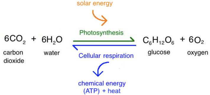
PHOTOSYNTHESIS

PROCESS OF PHOTOSYNTHESIS

CHLOROPLASTS

  • Site of photosynthesis in plants

  • Double-membrane organelle

  • Converts light energy into chemical energy

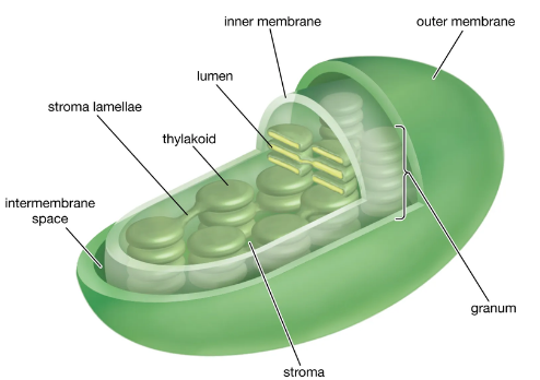
ANATOMY OF CHLOROPLASTS

Stroma

  • A colorless fluid surrounding grana

Grana

  • Densely layered stacks of thylakoids (10 to 20)

Thylakoids

  • Sites of conversion of light energy to chemical energy

Chlorophyll

  • Responsible for plants color

Lumen

  • Site of several reactions

VACUOLE

  • membrane-bound organelle

  • Can be found in both animal and plant cells

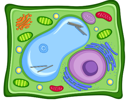
DIFFERENCES OF ANIMAL & PLANT CELL

Animal Cells

  • Temporary storage

Plant Call

  • Large central vacuole

  • Maintains turgor pressure to prevent wilting

Turgor Pressure

  • Pressure exerted to the cell wall by water moving into the cell

Modifications of the Cell

Cell Modifications

  • specialized features

  • Re-acquired by teh cell after cell division

  • Helps the cells in different and beneficial ways

KINDS OF CELL MODIFICATIONS

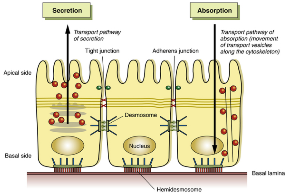
Apical Modifications

(TOP)

Basal Modifications

(Bottom)

Lateral Modifications

(Sides)

APICAL MODIFICATIONS (TOP)

CELL APPENDAGES

  • Locomotion of the cells

  • Locomotive action is due to the microtubules

Axoneme

  • Central core surrounded by an extension of the cell membrane

  • Connected to the Basal Body

FLAGELLUM

  • plural : Flagella

  • Moves in whiplike manner

Structure

  • Same axial structure like cilia

  • Longer in size

Major Parts: Basal Body, Hook Filament

Example: tail of sepermatozoa

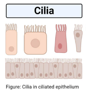
CILIA

  • singular: cilium

  • Require power and recovery strokes

Structure

  • Short hair like structure/projections

  • Each cilium is connected to the basal body and extends from the free surface

  • Core is composed of microtubules in a specific manner

Example:

  • lining of trachea

  • Fallopian tube

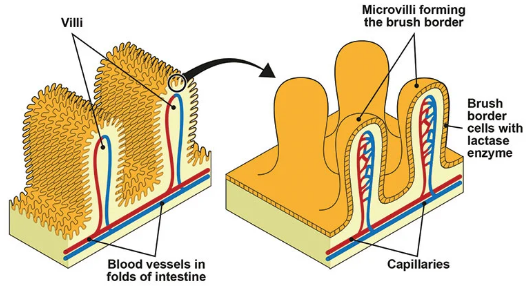
VILI

  • Finger-like projections

  • Arise from the epithelial layer in some organs

  • Help increase surface area

  • Faster and more efficient absorption

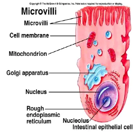
MICRO VILI

  • brush/striated boarder

  • Fingerlike extensions for increasing the surface area of the membrane

  • absorption nutrients

Example: Intestinal Cell

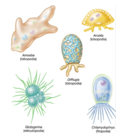
PSEUDOPODS

  • “False Feet”

  • Temporary

  • Irregular lobes

  • Formed by amoebas and other eukaryotic cells

  • Bulge outward to move the cell or engulf prey.

TYPE OF PSEUDOPODS

Filopodia

  • lender actin-based structures

  • serve sensory and locomotory functions

  • mostly found in multicellular organisms.

Axopodia

  • Long and thin protrusions from the cells

  • Rigid and flexible

Reticulopodia/Rhizopodia

  • Thread like

  • Branch and fuse to form a network that is extremely dynamic

Lobopodia

  • Finger-liketubular pseudepodia

  • Consists of ecto and endoplasm

  • Most common type

BASAL MODIFICATIONS

DESMOSOMES/HEMIDESMOSOMES

  • Anchoring junction on the basal surface of the cell

  • Rivet-like links between the cytoskeleton and extracellular matrix components such as the basal lamina that underlie epithelia.

  • Primarily composed of keratin, integrins and cadherins.

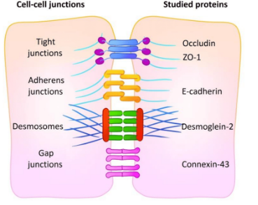
Anchoring Junction

  • Anchors junction on the lateral surface of the cell

Tight Junction

  • Acts as barriers

  • Regulates hte movement of water and soultes between epithelial layers

Gap Junction

  • Communicating junctions

  • Closable channels

  • Connects the cytoplasm of adjoining animal cells