CARDIAC CYCLE
The Cardiac Cycle
Phases of Contraction and Relaxation: The cardiac cycle alternates between systole (contraction and emptying) and diastole (relaxation and filling).
Systolic Pressure: Refers to the peak pressure in blood vessels during systole.
Diastolic Pressure: The lowest pressure during ventricular diastole.
ECG Correlation:
Atrial Systole: Starts after the P wave.
Ventricular Systole: Begins near the end of the R wave and ends just after the T wave.
The cycle will be explained by correlating
ECG readings,
intracardiac pressures,
blood volume changes,
cardiac valve activity,
and heart sounds.
Slide 6-13: Phases of the Cardiac Cycle
The following sequence details each phase of the cardiac cycle:
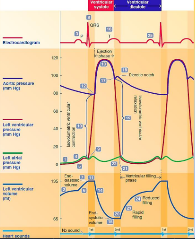
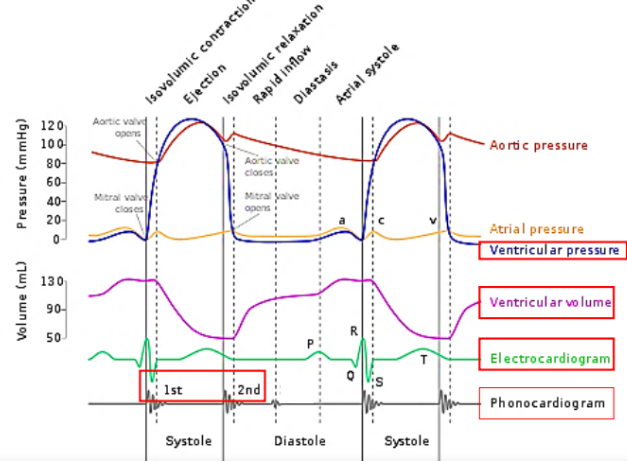
Mid Ventricular Diastole:
Corresponds to the TP interval on the ECG, where both atria and ventricles are relaxed.
Blood passively flows from the venous system into the atria and through open AV valves into the ventricles.
Ventricular volume gradually increases due to passive filling.
Late Ventricular Diastole:
The SA node reaches threshold, causing atrial depolarization (seen as the P wave on the ECG).
Atrial contraction follows, increasing atrial pressure and pushing additional blood into the ventricles.
The AV valve remains open as atrial pressure exceeds ventricular pressure, allowing complete ventricular filling.
End of Ventricular Diastole:
Atrial contraction concludes, and ventricular filling is complete.
The volume in the ventricle at this stage is termed end-diastolic volume (EDV), averaging approximately 135 mL.
Ventricular Excitation and Onset of Ventricular Systole:
The QRS complex signifies ventricular depolarization, initiating ventricular contraction.
Ventricular pressure rises sharply exceeding atrial pressure as the ventricles begin to contract, closing the AV valve and producing the first heart sound (S1).
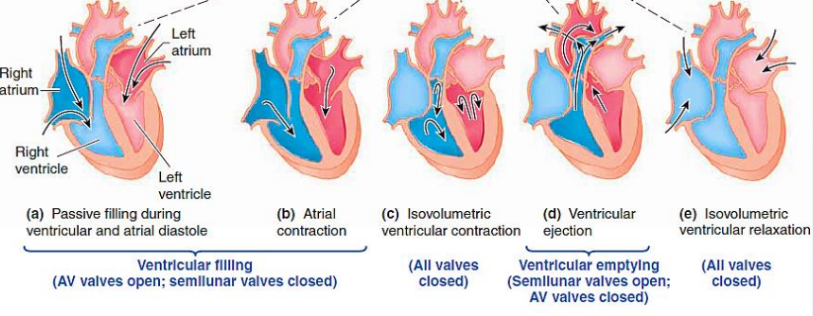
Isovolumetric Ventricular Contraction:
During this brief phase, both the AV and aortic valves are closed, creating a closed chamber.
Ventricular pressure rises without a change in blood volume.
Ventricular Ejection:
As ventricular pressure surpasses aortic pressure, the aortic valve opens, initiating blood ejection into the aorta.
Stroke Volume (SV): The amount of blood ejected per beat, approximately 70 mL.
This phase marks the active ejection component of ventricular systole.
End of Ventricular Systole:
The ventricles do not completely empty; the remaining blood volume is termed end-systolic volume (ESV), around 65 mL.
Ventricular Repolarization and Onset of Ventricular Diastole:
The T wave on the ECG indicates ventricular repolarization.
As ventricular pressure falls below aortic pressure, the aortic and pulmonary valves close, producing the second heart sound (S2).
Isovolumetric Ventricular Relaxation:
Both the aortic valve and AV valve remain closed as ventricular pressure is still higher than atrial pressure, resulting in no change in blood volume.
Ventricular Filling:
Once ventricular pressure drops below atrial pressure, the AV valve opens, allowing blood to flow from the atria into the ventricles.
The Ventricular Diastole Phase: Encompasses both isovolumetric relaxation and the filling period, reinitiating the cardiac cycle as the SA node fires again.
Slide 14: Duration of the Cardiac Cycle
Resting Cardiac Cycle Duration: One cycle lasts 0.8 seconds when at rest.
Ventricular Systole: 0.3 seconds.
Ventricular Diastole: 0.5 seconds.