Appendicular Skeletal Muscles
A. Muscles Acting on the Upper Arm & Shoulder
Deltoid Muscle: Superficial superolateral triangular muscle that caps the shoulder
Action: flexes, extends, abduct, medially & laterally rotates arm
Coracobrachialis Muscle: Most medial & deep arm muscle that attaches to the humeral shaft (brachial region) & coracoid process, hence its name
Action: flexes & medially rotates arm
Latissimus Dorsi Muscle: Superficial, posterior, & broad; extends from the waist to the axilla; attached to the thoracolumbar fascia poseriorly; latissimus means broadest, dorsi means back
Action: extends, adducts & medially rotates arm; prime mover of the shoulder joint, along with the pectoralis major muscle
Pectoralis Major Muscle: Superficial, thick, & fleshy chest muscle of the mammary region
Action: flexes, adducts, & medially rotates arm; another prime mover of the shoulder joint, along with the latissimus dorsi muscle
Pectoralis Minor Muscle: Deep chest muscle with 3 heads that connects to a few superior ribs to the coracoid process
Action: works with the serratus anterior muscle to move scapula bone anterior & laterally
Serratus Anterior Muscle: Superficial & superolateral fan-shaped muscle that has a serrated appearance; originates from all or nearly all the ribs
Action: works with the pectoralis minor muscle to move scapula anterior & laterally
Rotator Cuff Muscles:
Action: 4 intermediate muscles that collectively stabilize the glenohumeral joint
Supraspinatus Muscle: located posteriorly, superior to the scapular spine
Action: aids deltoid muscle in abduction of the arm
Infraspinatus Muscle: located posteriorly, inferior to the scapular spine
Action: rotates arm laterally
Teres Minor Muscle: located posteriorly, inferior to the infraspinatus muscle, but superior to the teres major muscle
Action: rotates arm laterally
Subscapularis Muscle: located superficial to the subscapular fossa; the only muscle out of the 4 that is located anteriorly
Action: rotates arm medially
Teres Major Muscle: Intermediate & posterior muscle, located inferior to the teres minor muscle
Action: extends & medially rotates arm
B. Muscles Acting on the Forearm & Hand
Upper Arm Contribution (to forearm & elbow movements)
Brachialis Muscle: Deep to the biceps brachii muscle on the anterior side & deep to the triceps brachii muscle on the posterior side
Action: prime mover of elbow flexion
Biceps Brachii Muscle (1 muscle with 2 heads): (plural: bicipites) Superficial & appears as a large anterior bulge on the upper arm; it has 2 heads - a lateral long head & a medial short head
Action: rapid or forceful supination (powerful supinator) of forearm as it works with the supinator muscle); also synergistic (enhances) with the brachialis muscle in elbow flexion
Attached to the biceps brachii aponeurosis that extends to the anterior forearm.
Triceps Brachii Muscle (1 muscle with 3 heads): (plural: tricipites) Superficial & posterior muscle with 3 heads - a long head (located medially), a lateral head (which is next to the long head), & a medial head (distally located in the humerus; a section of it can be seen better on the anterior side deep to the biceps brachii & brachialis muscles, since it is covered on the posterior side by both the long & lateral heads)
Action: prime mover of elbow extension along with the anconeus muscle.
Lateral Forearm
Brachioradialis Muscle: Large fleshy mass on the most lateral side of the forearm; it originates from the distal end of the humerus & inserts to the distal end of the radius; think of it as a lateral border that separates the flexor side from the extensor side
Action: flexes the elbow
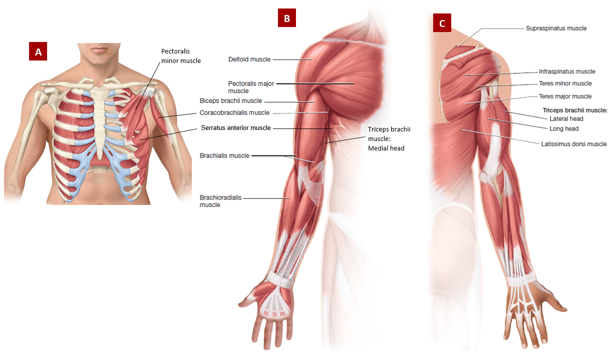
Figure 3.20 Muscles acting on the upper arm & shoulder: (A) Deep anterior view, (B) Right superficial anterior view, (C) Right superficial & intermediate posterior view
Seen in Both Anterior & Posterior Forearm
Supinator Muscle: This muscle is so deep that parts of it can be seen on both the deep levels of the flexor & extensor side; it is wrapped around the radius bone & located near the anterior & posterior side of the elbow
Action: supinates forearm; works with the biceps brachii muscle for supination
Anterior Forearm (Flexor Side)
Superficial Muscle Layer (4 muscles)
Note: The superficial muscles listed below are in lateral to medial order
Mnemonic: P-F-P-F or Pass, Fail, Pass, Fail
Pronator Teres Muscle: Small & proximal; located next to the large fleshy brachioradialis muscle; it is located near the antecubital region
Action: assists the pronator quadratus muscle with pronation
Flexor Carpi Radialis Muscle: Its tendon attaches to the 2nd & 3rd metacarpals
Action: flexes the wrists anteriorly; aids in radial flexion of the wrist
Palmaris Longus Muscle: Weakly developed, sometimes absent; has a significantly long tendon attached to a fibrous triangular broad sheet called palmar aponeurosis
Action: anchors skin & fascia of palmar region
Palmar Aponeurosis: superficial to the palm covering the tendons & muscles of the hand; if the palmaris longus muscle is absent, it attaches directly to the flexor retinaculum
Flexor Carpi Ulnaris Muscle: Medial; located next to the extensor carpi ulnaris muscle, wherein both muscles border the medial forearm; its tendon attaches to the pisiform & hamate bones
Action: flexes the wrists anteriorly; aids in ulnar flexion of the wrist
Intermediate Layer (1 muscle)
Flexor Digitorum Superficialis Muscle: Has 4 tendons attached from the 2nd to 5th fingers
Action: flexes the wrists, knuckles, & fingers, except for the thumb
Deep Layer (3 muscles)
Flexor Digitorum Profundus Muscle: Medial; has 4 tendons attached from 2nd to 5th fingers
Action: flexes the wrists, knuckles, fingers, except for the thumb
Flexor Pollicis Longus Muscle: Lateral; has a long tendon attached to the thumb; pollicis refers to the thumb
Action: flexes the thumb
Pronator Quadratus Muscle: Small & a square-shaped muscle located near the wrist
Action: prime mover of forearm pronation
Flexor Retinaculum: Wristband-like sheet where most tendons of the anterior forearm muscles passes under it; the only exception is the tendon of the palmaris longus muscle which pass over it
Action: stabilizes the tendons & carpal region on the flexor side
Palmar Hand Muscles: Anterior (4 muscle groups)
Thenar Muscle Group: Fleshy mass of muscles located at the base of the thumb
The flexor pollicis brevis & abductor pollicis brevis muscles are found in this group.
Action: group of muscles concerned with thumb's precise movements
Hypothenar Muscle Group: Fleshy mass of muscles located at the base of the little finger
Action: group of muscles concerned with the little finger's precise movements
Lumbrical Muscles: Superficial muscles located at the palmar side of the metacarpals
Action: group of superficial muscles that extends the knuckles
Palmar Interosseous Muscles: (plural: interossei) Deep muscles located at the palmar side of the metacarpals
Action: group of deep muscles that adduct fingers; important for grip strength
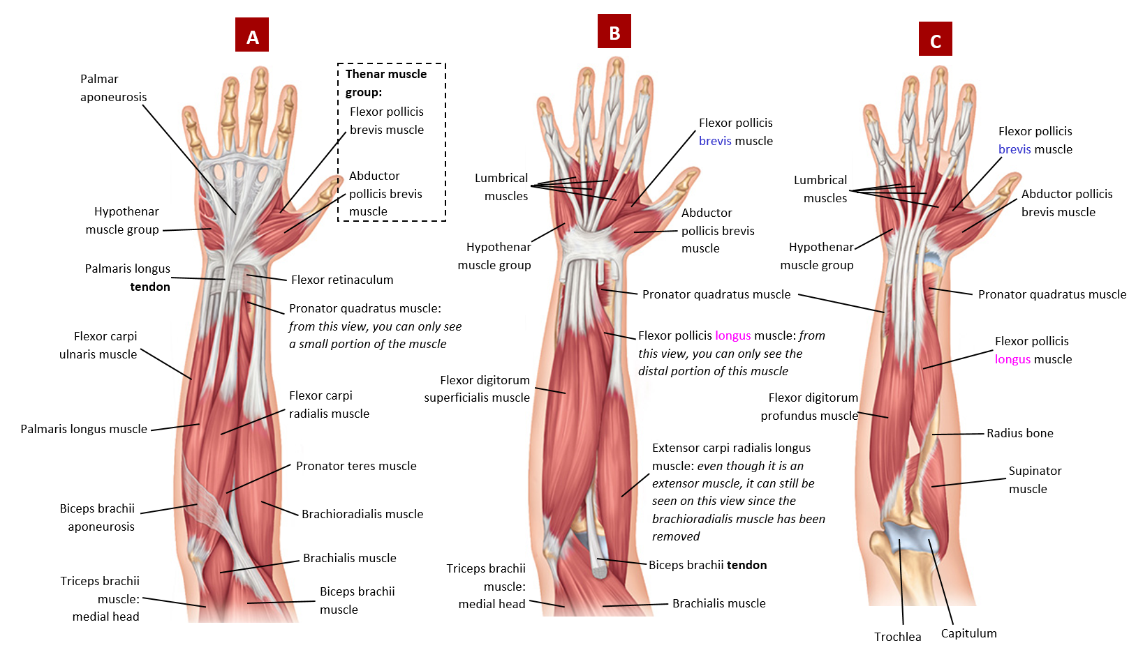
Figure 3.21 Right flexor forearm muscles on the anterior side with some upper arm muscles & structures: (A) Superficial, (B) Intermediate, (C) Deep
Posterior Forearm (Extensors)
Both Superficial & Deep (1 muscle)
Anconeus Muscle: Small triangular muscle near the elbow
Action: extends the elbow; works with the triceps brachii muscle in elbow extension
Superficial (5 muscles)
Note: The superficial muscles listed below are in lateral to medial order
Extensor Carpi Radialis Longus Muscle: Located next to & slightly overlaps with the brachioradialis muscle, pay extra attention to the separation between these two muscles; carpi refers to the carpal bones (wrist) where its tendon distally attaches to
Action: extends the wrist; aids in radial flexion of wrist
Extensor Carpi Radialis Brevis Muscle: Located very close to the extensor capri radialis longus muscle; brevis means short; pay attention to the separation between the two muscles
Action: extends the wrist; aids in radial flexion of wrist
Extensor Digitorum Muscle: Wide muscle that has 4 tendons attached to the 2nd to 5th fingers
Action: extends the wrist, knuckles, fingers, except for the thumb
Extensor Digiti Minimi Muscle: A thin muscle that is in very close proximity of the extensor digitorum muscle; do not confuse with the extensor carpi ulnaris muscle - pay attention to where their tendons insert to
Action: extends the wrist & all joints of the little finger only
Extensor Carpi Ulnaris Muscle: It is located next to the flexor carpi ulnaris muscle, wherein these muscles are both the most medial forearm muscles; pay attention to seeing the ulna bone visible between these two muscles
Action: extends wrist when fist is clenched; aids in ulnar flexion of wrist
Deep (4 muscles)
Note: The deep muscles listed below are in proximal to distal order (elbow to thumb)
Pollicis Muscles Mnemonic: Longus-Brevis-Longus OR Abductor-Extensor-Extensor
Abductor Pollicis Longus Muscle: Deep; on superficial view, you can see a small part of this muscle & its tendon attaching to the thumb
Action: extends & abducts the thumb
Extensor Pollicis Brevis Muscle: Deep; on superficial view, you can see a small part of this muscle &its tendon attaching to the thumb
Action: extends the thumb & its knuckle
Extensor Pollicis Longus Muscle: Deep; but on superficial view, you can see a small part of this muscle or just its tendon sticking out & attaching to the thumb
Action: extends, adducts, & laterally rotates the thumb
Extensor Indicis Muscle: Deep with a tendon attaching to the index finger
Action: extends the wrist & index (pointing) finger
Extensor Retinaculum: Wristband-like sheet where tendons of the posterior forearm muscles pass under it
Action: stabilizes the tendons & carpal region on the extensor side
Dorsal Hand Muscles: Posterior (1 muscle group)
Dorsal Interosseous Muscles: Located on the metacarpals of the hand but on the dorsal side; aka the back of the hand muscles
Action: group of muscles that abduct the fingers; important for grip strength
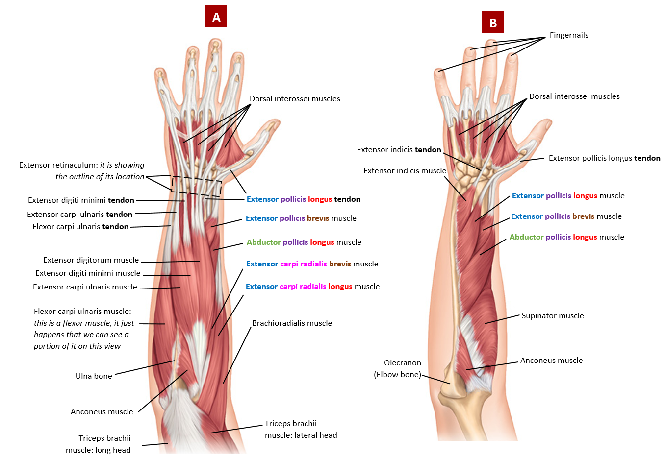
Figure 3.21 Left extensor forearm muscles on the posterior side with some upper arm muscles & bone structures: (A) Superficial, (B) Deep
Appendicular Skeletal Muscles: Lower Extremities
C. Anterior Muscles of the Hip
Iliopsoas Muscles Group (2 muscles):
Group Action: Prime mover for flexing the thigh.
Iliacus Muscle: Superficial to the iliac fossa
Psoas Major Muscle: Thick round muscle arising from the thoracic & lumbar regions
Psoas Minor Muscle: Superficial to the psoas major muscle; only present in some individuals
Action: assists with hip flexion
D. Anterior Compartment Muscles of the Thigh (Knee Extensors)
Quadriceps Femoris Muscle Group (4 muscles & 4 heads): All 4 muscles converge to a single patellar tendon (aka quadriceps femoris tendon); note: do not confuse with the patellar ligament
Group Action: prime movers of knee extension; the most powerful muscle group in the body
Rectus Femoris Muscle: Superficial
Action: also flexes thigh at hip
Vastus Lateralis Muscle: (plural: vasti) Superficial
Action: also stabilizes the patella bone
Vastus Medialis Muscle: Superficial
Action: also stabilizes the patella bone
Vastus Intermedius Muscle: Deep to the rectus femoris muscle; Note: do not name it as the vastus intermedialis muscle, it will be in marked as incorrect
Action: just extends the knee
E. Medial Compartment Muscles of the Thigh (Thigh ADductors)
Adductor Muscle Group (5 muscles): Take note that 4 out of the 5 muscles in this group are attached to the pubis' inferior ramus; 1 out of the 5 is attached to the superior ramus; remember, the pubis is one of the bones of the hemipelvis, which is located inferiorly & anteriorly
Group Action: act primarily as the adductor of the thigh
Mnemonic: PG-AddMLB
Pectineus Muscle: Most superior & a superficial short muscle within this group; attaches to the pubis' superior ramus
Gracilis Muscle: Most medial (inner thigh), superficial; attaches to the pubis' inferior ramus
Adductor Magnus Muscle: Deep muscle of the 3 muscles named adductor in this group on its anterior side, but deep to the hamstrings muscle group from the posterior side; biggest muscle of the group; attaches to the pubis' inferior ramus
Adductor Longus Muscle: Superficial of the 3 muscles named adductor in this group; attaches to the pubis' inferior ramus
Adductor Brevis Muscle: Intermediate muscle of the 3 muscles named adductor in this group; attaches to the pubis' inferior ramus
F. Muscles Originating on the Lateral Side of the Thigh (Thigh ABductors)
Abductor Muscle Group (2 muscles):
Group Action: act primarily as the abductor of the thigh
Tensor Fasciae Latae Muscle (TFL): (plural: tensor fascia lata) Superficial muscle attached to the iliac crest superiorly & to the iliotibial band inferiorly
Action: also aids in knee extension
Sartorius muscle: Superficial strap-like muscle crossing the quadriceps femoris muscle group, from the lateral side (anterior superior iliac spine) of the hip, to the medial side of the knee (proximal tibial end); longest muscle of the body
Action: also a weak knee flexor
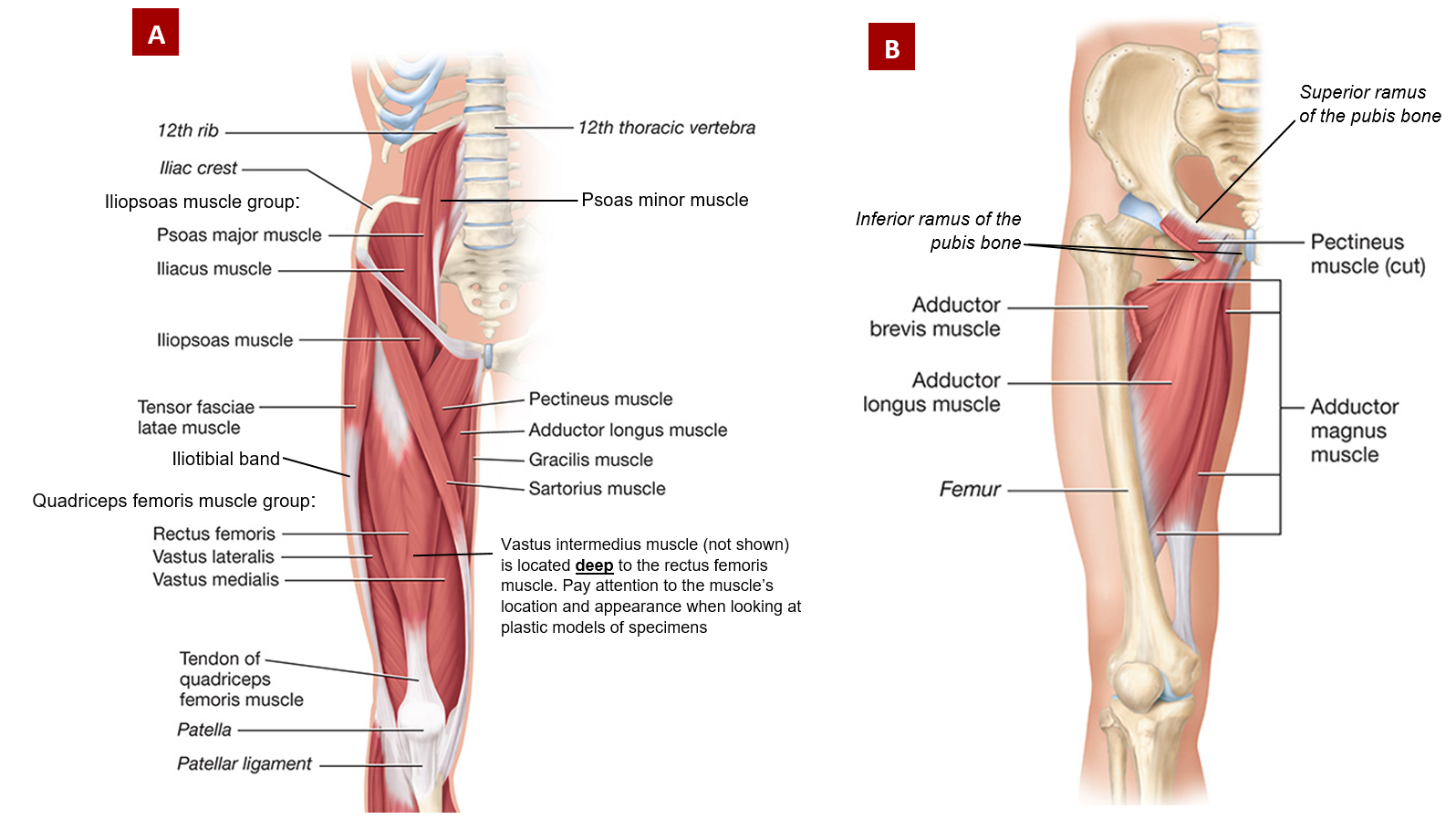
Figure 3.23 Right anterior muscles, muscle groups, & bone structures of the hip & thigh: (A) Superficial, (B) Deep
G. Posterior Muscles of the Hip
Gluteus Muscle Group (3 muscles): (plural: glutei)
Group Action: aids in stabilizing the upper body & pelvis
Gluteus Maximus Muscle: Superficial large muscle
Action: major extensor of thigh
Gluteus Medius Muscle: Intermediate lateral muscle
Action: medially rotates thigh
Gluteus Minimus Muscle: Deepest smallest of the three
Action: medially rotates thigh
Lateral Rotators Muscle Group (6 muscles): Found deep to the glutei maximus & medius muscles only; same deep level but inferior to the gluteus minimus muscle
Group Action: opposes medial rotation by the glutei medius & minumus muscles only, hence they're named the lateral rotators
Mnemonic: P-GOGO-Q
Piriformis Muscle: Most superior of all the muscles in this group; pear-shaped muscle
Gemellus Superior Muscle: (plural: gemelli) Superior to both the obturator muscles; gemellus means twins
Obturator Externus Muscle: Fan shaped; originates on the lateral/external side of the obturator foramen & inserts by the greater trochanter
Gemellus Inferior Muscle: Inferior to both the obturator muscles
Obturator Internus Muscle: Fan shaped; originates on the medial/internal side of the obturator foramen & inserts by the greater trochanter
Quadratus Femoris Muscle: Most inferior muscle in this group; rectangular-shape
H. Posterior Compartment Muscles of the Thigh (Knee Flexors)
Hamstrings Muscle Group (3 muscles):
Group Action: flexes the knee
Biceps Femoris Muscle: Lateral muscle with 2 heads; long head is superficial to the short head; femoris refers to the femoral region
Semitendinosus Muscle: Medial; superficial to the semimembranosus muscle; named after its unusually long tendon; pay attention to its correct spelling, semitendonosus is incorrect
Semimembranosus Muscle: Medial; deep to the semitendinosus muscle; pay attention to its correct spelling, semimembrinosus is incorrect
Popliteus Muscle: Acts on the knee
Action: unlocks knee to allow flexion
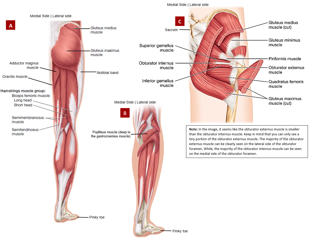
Figure 3.25 Right posterior muscles, muscle groups, & structures of the hip & thigh: (A) Superficial, (B) Deep knee, (C) Deep hip
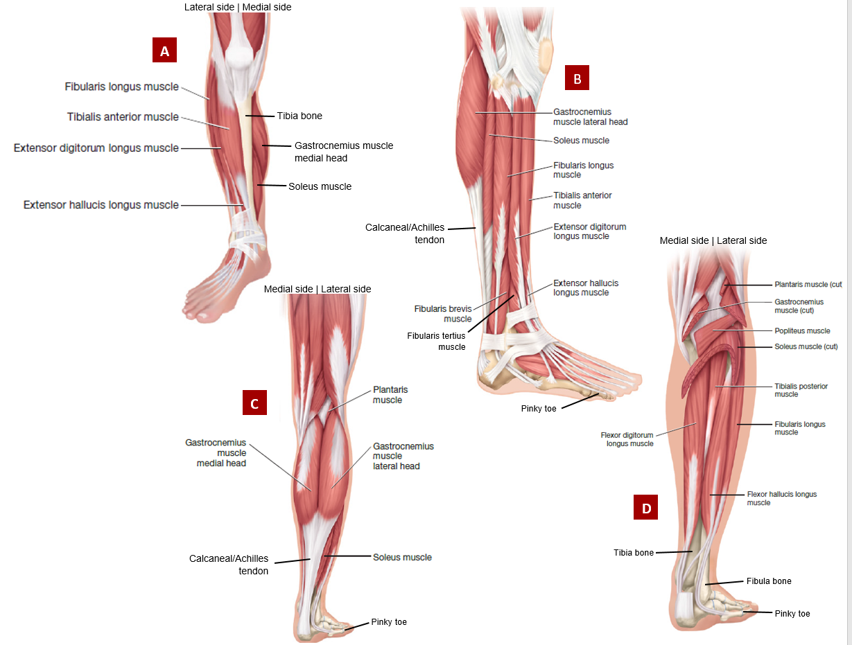
I. Anterior Compartment Muscles of the Lower Leg (Extensors)
Dorsiflexor Muscle Group (4 muscles):
Superficial (Medial to Lateral)
Mnemonic: FEET
Note: Both of these muscles attach to the tibia bone
Tibialis Anterior Muscle: Medial; next to the tibia bone; its tendon attaches to the big toe
Action: dorsiflexes (points toes towards the sky) & inverts foot (soles of feet facing medially)
Extensor Digitorum Longus Muscle: Lateral; tendons attach to the toes except the big toe
Action: extension of toes & dorsiflexes foot
Deep (Medial to Lateral)
Note: Both of these muscles attach to the fibula bone
Extensor hallucis longus muscle: Medial; its tendon attaches to the big toe; hallucis refers to the big toe
Action: extension of big toe & dorsiflexes foot
Fibularis Tertius: Lateral to the extensor hallucis longus muscle; its tendon attaches to the little toe
Action: dorsiflexes & everts foot during walking
J. Posterior Compartment Muscles of the Lower Leg (Flexors)
1 Superficial & 1 Deep
Triceps Surae Muscle Group (2 muscles & 3 heads): Both muscles in this group inserts to the calcaneus bone via the strongest tendon in the body called the Achilles or Calcaneal tendon; surae refers to the calf region
Gastrocnemius Muscle: Superficial muscle with 2 heads - medial & lateral heads
Action: plantar flexes (points toes toward to the ground) foot at ankle joint
Soleus Muscle: Deep to the gastrocnemius muscle
Action: plantar flexes foot at ankle joint
Superficial (1 muscle)
Plantaris Muscle: Superficial; originates from the lateral supracondylar line; relatively unimportant & absent for some people; its tendon is often used for tendon grafts needed in other parts of the body
Action: weak assist to the triceps surae muscle group for plantar flexion
Deep (3 muscles)
Medial to Lateral
Flexor digitorum longus muscle: Most medial of the 3 deep muscles
Action: flexes 2nd toe (long toe) to the 5th toe (pinky toe)
Tibialis posterior muscle: Middle of the 3 deep muscle
Action: inverts foot
Flexor hallucis longus muscle: Most lateral of the 3 deep muscles
Action: flexes big toe
K. Lateral Compartment Muscles of the Lower Leg (Flexors Too)
Fibularis Longus Muscle: Superficial to the fibularis brevis muscle
Action: plantar flexes & everts the foot (sole facing laterally)
Fibularis Brevis Muscle: Deep to the fibularis longus muscle
Action: plantar flexes & everts the foot