M2L5: Cell signalling
Overview of signalling
Isogenic cells may respond differently to microenvironmental cues
Cells discern signals from their environment and different cell types may respond to a given signalling molecule differently
Eg. TGF-β stimulates fibroblast differentiation into myofibroblasts but promotes EMT in epithelial cells
Cross talk between pathways activated in response to different signals determines the overall cell response and fate
Stimuli for sensors can be generic or cell type specific
Metabolites (amino acids, lipids, nucleotides)
Small molecules (NO, CO2, xenobiotics)
PRRs (dsRNA, ssRNA, DNA, LPS)
Mechanical (ECM, cell to cell pressure)
Hormones
Light
Smell
Sensors - receptors (plasma membrane, organelle), TFs (cytosolic, organelle, inner PM)
Transducer (signalling cascade, subcellular relocalisation)
Effectors (activity of a protein eg. IP3R and GLUT4, TFs)
Signal transduction usually involves PTMs that act as a switch
TFs are the effectors of most signalling pathways as they can regulate a diverse set of genes and orchestrate a wider response
Signalling pathways do not have to be linear, they can converge, diverge or cross talk
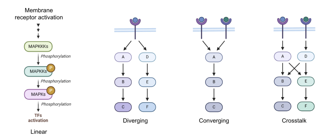
Signal amplification, eg phosphorylation cascade
Signal transduction requires protein-protein interaction or interaction with messenger molecules for transduction specificity
Adaptor proteins act as scaffold to help bring together multiple proteins - assists in relaying the signal downstream for better efficiency and specificity
Grb2: binds to p-Y on RTF through SH2 and the FEG Sos through SH3 domain
Shc: binds to p-Y on RTK through phospho-tyrosine binding (PTB) domain and to Grb2 via SH2
IRS: Binds IR through PTB and POP2/3 though PH domain
Integrins: structural, scaffolding and catalytic function
Cells use diverse molecules (ligands) to communicate - ligand-receptor interactions elicit intracellular signalling
Target receptor of ligand can be:
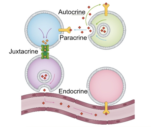
Self (autocrine)
Self without secretion (intracrine)
Nearby cells (paracrine)
Physically contected cells (juxtacrine)
Distant cells through circulation (endocrine)
Solubility and stability of signalling molecules reflect type of signalling used
Contact dependent signalling:
Cell surface signalling (tight junction and immune cells activation)
Cell adhesion molecule (ECM)
Metabolites as signalling molecules
Small molecule metabolites (NO, Trp, ATP, succinate, alpha-KA, OA, PA, lactate) can act in intracrine and paracrine signalling molecule and and even endocrine (lactate)
Evolution of small molecule metabolites as signalling molecules as opposed to proteins in some circumstances may be to save energy (they do not need to be specifically synthesised from scratch for signalling as they are already available) and to communicate about the status/needs of the cell more easily based on the pathways that trigger certain metabolites to be produced
Cell cell communication enable coordinate regulation for tissue homeostasis and organ function
In development:
Signalling molecules secreted from a single source (ventral side) can create morphogen gradient that enables cellular patterning during development
Opposing gradients of signalling molecules can endow crypt cells with positional identity to adopt a specific cell lineage
Cell-cell contacts also contribute to positional identity
Differential receptor expression further reinforce positional boundaries
Range of receptors (each specific for restricted set of ligands)
Cell surface transmembrane receptors - extracellular, transmembrane, intracellular domains
Intracellular receptors - for ligands originated extracellularly (passive diffusion or active uptake) or intracellularly
Nuclear receptor - steroid or thyroid hormone receptors, retinoic acid and orphan receptors
IP3 receptor (intracellular calcium channels located in the ER, release Ca2+ into the cytosol which activates various cellular processes)
First/second messengers
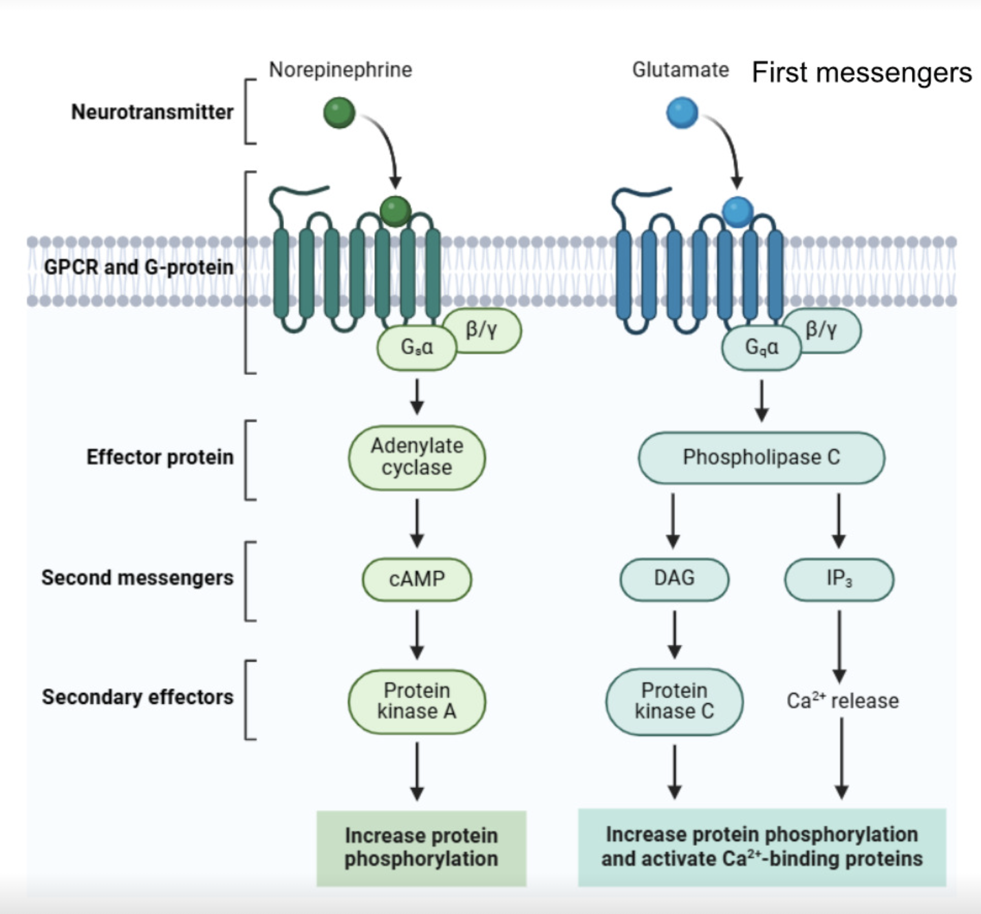
First messengers - extracellular signalling molecule, eg. hormones, neurotransmitters
Effector protein - membrane-associated enzyme or channel activated by the receptor
Second messenger - small intracellular molecules that amplify and distribute the signal within the cell
Small, easily diffusible, rapid integration of upstream signals and amplification/spreading of downstream signals
Secondary effector - intracellular protein activated by the second messenger which performs the functional cellular response
Examples (second messenger —> secondary effector):
Cyclic nucleotides (cAMP —> PKA, cGMP —> PKG)
Ions (Ca2+, Na+)
Phospholipid derived (IP3, DAG —> PKC)
Gases (NO)
G-protein coupled receptors (GPCRs)
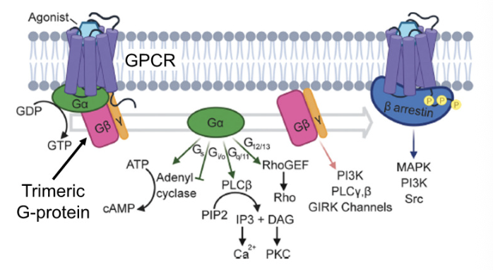
Largest family of transmembrane receptors which are coupled to a trimeric G protein (guanine nucleotide binding protein) consisting of α, β, and γ subunits
Binding of extracellular ligand induces conformational changes in receptors —> Gα GDP-to-GTP exchange for activation
Vertebrate GPCR - glutamate (G) family, rhodopsin (R) family,
adhesion (A) family, frizzled/taste2 (F) family, and secretin (S)
family
Depending on which type of G protein is coupled to the receptor, different downstream pathways amay be activated
Gs, Gi/o —> activates/inhibits cAMP pathway
Gq/11 —> IP3/DAG
G12/13 —> Rho GEF
GPCR signalling is terminated via receptor phosphorylation by GPCR kinases (GRKs) which recruits β-arrestin to bind, blocking interaction with downstream signalling molecules and causing receptor desensitisation/internalisation
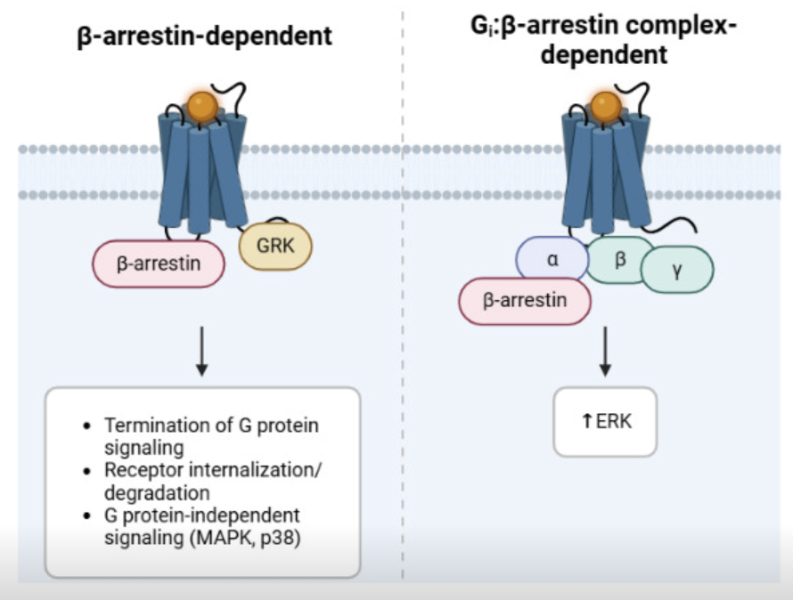
β-arrestin also initiates secondary GPCR signalling as it acts as a scaffold for other signalling molecules, regulating different gene expression programs
If the first round of signalling is not terminated properly then the receptor may remain active for longer, causing prolonged or abnormal β-arrestin recruitment, reading to secondary signalling from endosomes or pathological signalling
Wnt/β-catenin pathway
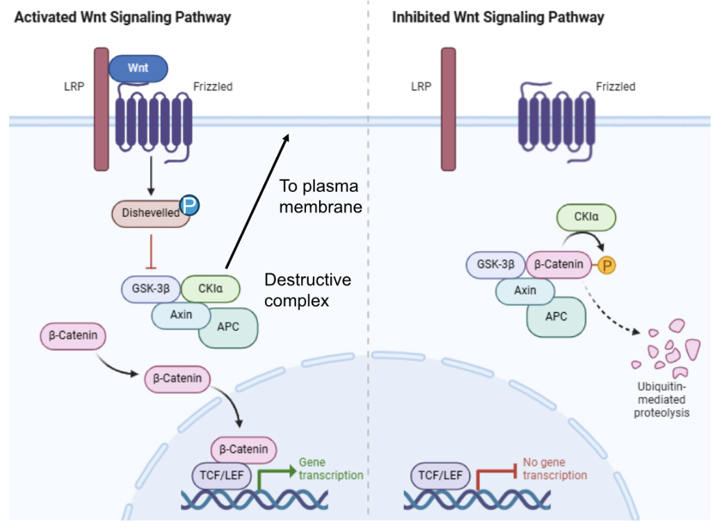
Interface between Frizzled GPCR and LRP5/6 coreceptor proteins provides a platform for Wnt signalling molecules to bind
Canonical Wnt pathway regulates cell fate specification and migration in embryonic development and tissue homeostasis
Dishevelled (Dsh) - highly conserved protein containing amino terminal DIX domain, central PDZ domain, C terminal DEP domain
DIX and PDZ domains inhibit GSK3 activity
GSK3 is a component of a destructive complex which phosphorylates and targets β-catenin for ubiquitin-mediated proteolytic degradation in the absence of Wnt signalling
Constant turnover of β-catenin rather than synthesising it only when needed is to support its rapid activation
In the presence of Wnt signals, β-catenin degradation is inhibited which allows it to enter the nucleus and act as a coactivator of gene expression with TCF/LEF family of transciption factors
Non-canonical Wnt pathways do not use LRP5/6 or β-catenin and involve G protein-mediated regulation of tissue polarity/planar cell polarity (PCP) signalling and Ca2+ signalling
Receptor tyrosine kinases (RTKs)
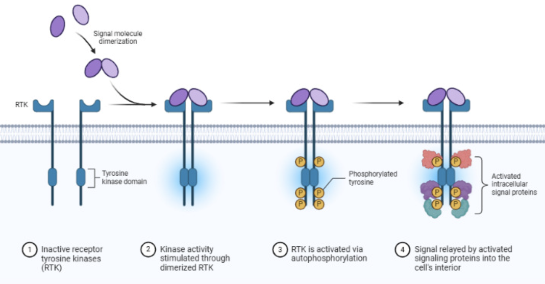
Examples - VEGFR, EGFR, PDGFR, IR and EGFR
Single pass α-helix transmembrane proteins
Ligand binding induces receptor dimerisation and reciprocal phosphorylation on tyrosine residues which act as docking sites for adaptor proteins
Dimerised receptors recruit and activate multiple intracellular signalling molecules for signal amplification
Homodimerisation - identical RTKs dimerise to activate standard signalling eg. canonical MAPK, PI3K pathways
Heterodimerisation - two different RTKs (either from the same family or different families of RTKs) dimerise which leads to signalling diversity
eg. EGFR/HER2, HGF/C-MET and AXL/EGFR
This may also be a mechanism of drug resistance by causing redundancy when honing in on one receptor in a targeted therapy
As it is difficult to overcome ligand-receptor binding, termination usually involves receptor internalisation via the clathrin coated pathway
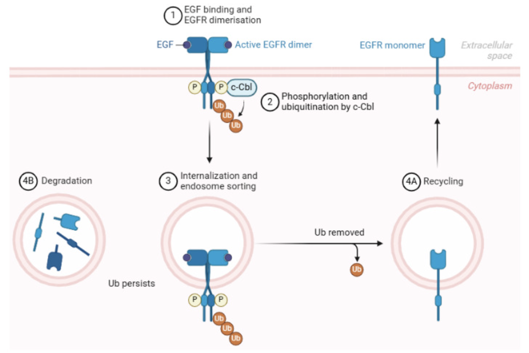
Sorting to lysosomes for degradation or recycling to the membrane, helps limits the signal intensity and duration
Following internalisation the receptor is still bound to the ligand and can still signal from the endosome until it is degraded
This may be exploited to give rise to therapeutic resistance
Ras signalling
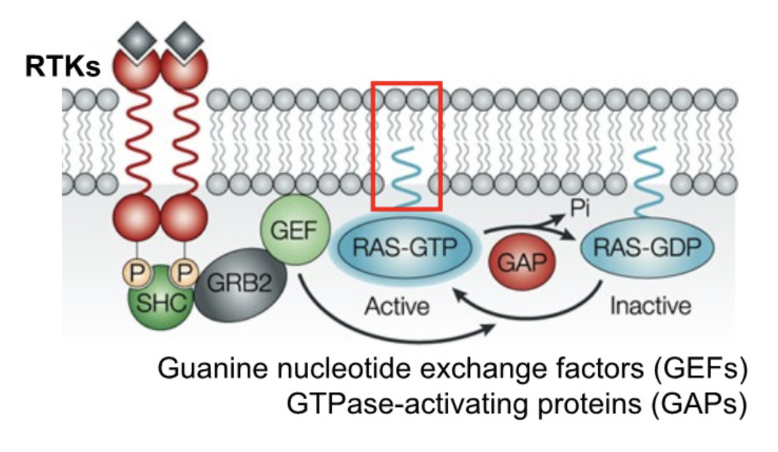
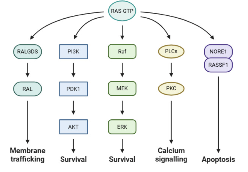
Small GTPase (monomeric G protein) - HRAS, KRAS, NRAS etc
Molecular switch that cycles between active (GTP-bound) and inactive (GDP-bound) state
Other families - Rho, Rab, Arf, Ran
Farnesylation and palmitoylation anchors Ras to PM - critical for function
Activating mutations in cancer cancer types
Considered undruggable until recent efforts placed in targeting MAPK and PI3K/AKT pathways
Sotorasib targeting G12C (covalent inhibitors), but this is found only in a small subset of cancer (especially lung)
Possible because cysteine substitution at G12 position produces a side chain with a reactive sulfhydryl group, enabling covalent bonding with an electrophilic group in the inhibitor sotorasib
Targeting G12D, G12V, G13D, and Q61H remain a challenge
MAPK signalling
Regulates growth, differentiation and stress response, including immune response
Signalling can be acute (proliferation or transient transcriptional activation) or sustained (differentiation, senescence, apoptosis)
Signal flow: Receptor (e.g. RTK or GPCR) → Ras (small GTPase) → Raf (MAPKKK) → MEK (MAPKK) → ERK (MAPK) → transcription factors / target proteins
Main MAPKs - extracellular signal regulated kinase (ERK), p38 stress-activated protein kinase (SAPK), c-Jun N-terminal kinase (JNK)
Negatively regulated by dual-specificity phosphatases (DUSPs) - some influence subcellular MAPK localisation by binding and retaining them in specific cellular compartments
Dual specificity - can recognise Ser/Thr and also Tyr
ERK targeted by vemurafenib, dabrafenib, trametinib in cancer therapy
MAPK small molecule inhibitors often fail due to redundancy
PI3K/AKT - principal pathway regulating proliferation, survival, and metabolism
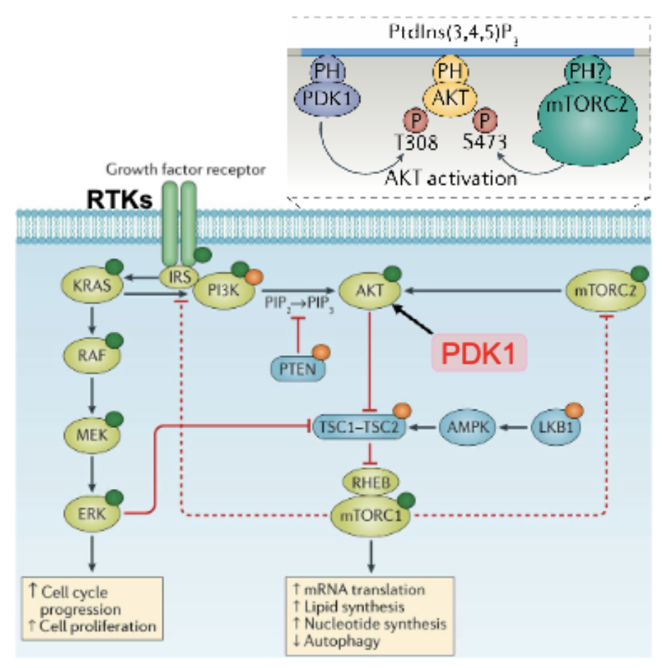
Activation of RTKs and GPCRs recruit PI3K which phosphorylates PIP2 into PIP3 at the plasma membrane
PIP3 acts as a docking site for Akt using its PH domain
Akt is activated by PDK1 via PIP3 and mTORC2 (PM localised)
Active Akt phosphorylates cytosolic and nuclear substrates for growth and survival
Tumour suppressor PTEM dephosphorylates PIP3 to PIP2
Contact dependent RTKs
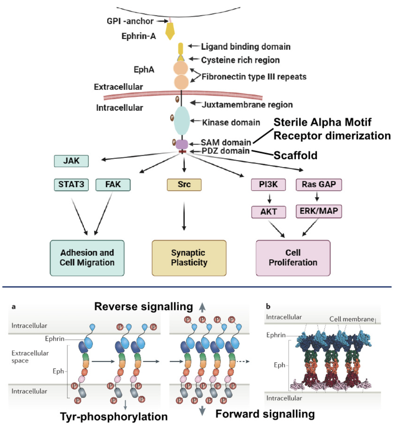
Activated by ligands on adjacent cells or ECM
Important for cell adhesion. migration, tissue patterning/homeostasis, angiogenesis, synaptic plasticity and immunity
Eph receptors are the largest family of RTKs, binding to ephrins on adjacent cells, classified according to ligand affinity as EphA or EphB
Eprin-A - anchored to the cell, membrane by glycosylphosphatidylinositol (GPI) linkage and lacks cytoplasmic domain
Ephrin-B - attached to cell membrane by single TM domain containing a short PDZ-binding motif, Dig1 and zo-1
PDZ anchors receptor to cytoskeleton
Ephrin-Eph oligomerisation and signalling is bidirectional
Cell-matrix and matrix-cell communication
Integrins - heterodimeric TM proteins named according to their alpha/beta subunit compositions and mediated interaction with ECM and cell surface adhesion proteins, eg collagen (outside-in)
Involves Rho, Rac, and cytoskeletal rearrangement
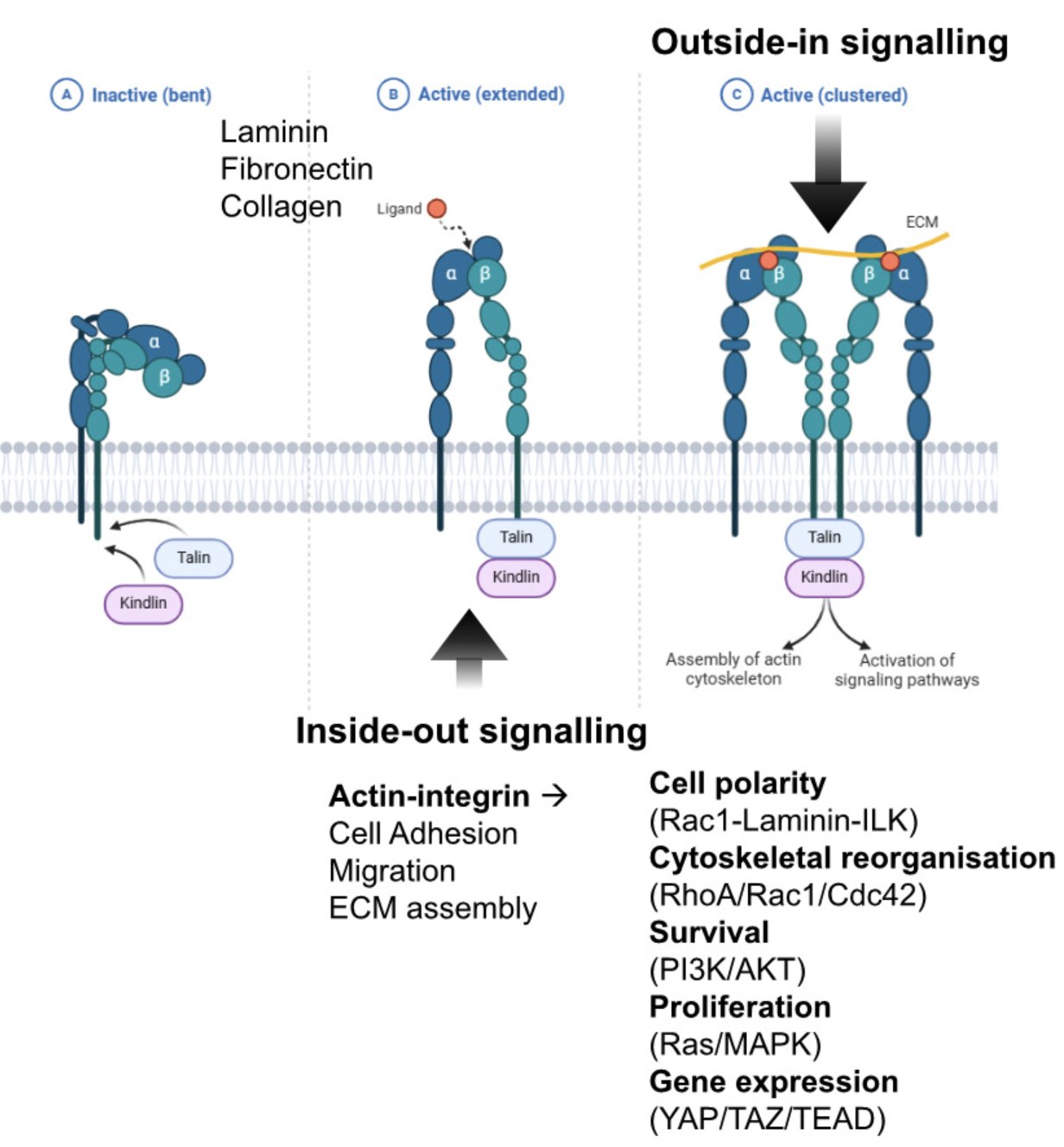
Anoikis is induced by loss of contact to the ECM
Activation of the integrin pathway when there is cell-ECM contact prevents this program of cell death
Inside out signals can induce integrins to adopt a high affinity extended conformation for their appropriate ligands
Normally integrin can be sequestered in the kinked form - not exposing its ligand binding domain outside the cell, can not sense what is outside
If the cell wants to sense what is outside, it can activate this signalling to open up the kinked conformation to promote ligand binding
Involved in mechanotransduction from ECM adhesion to cytoskeleton to nuclear envelope, all to regulate survival
Integrin β4 indirectly interacts with actin cytoskeleton through long cytoplasmic tail
Internalisation of integrin bound ligand help transmit signal intracellularly
Integrin can cooperate/amplify RTK signalling (eg EGFR) - integration of survival, growth, proliferation signals
JAK/STAT pathway
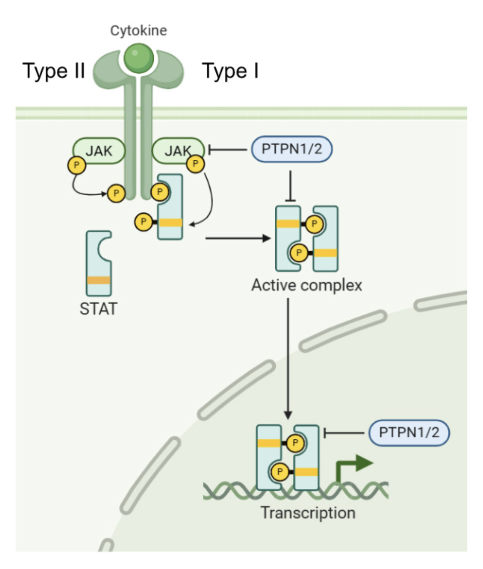
Cytokine receptors whose kinase activity is dependent on tightly associated tyrosine kinase Janus kinase (JAK) on the cytoplasm
Cytokines (eg. TPO, IL, IFN, EPI, prolactin, growth hormones) bind to the receptor, cauing receptor dimerisation and JAK cross-phosphorylation
Phosphorylation provides docking sites for signal transducers and activators of transcription (STATs) which gets phosphorylated by JAK
Phosphorylated STATs dimerise and translocate into the nucleus to activate gene expression
Non-receptor protein tyrosine phosphatases (PTPNs) attenuate the signal
7 STATs can be recruited to different combinations of Type I/II cytokine receptors to induce a ligand specific response
TGF-β/BMP/SMAD pathway
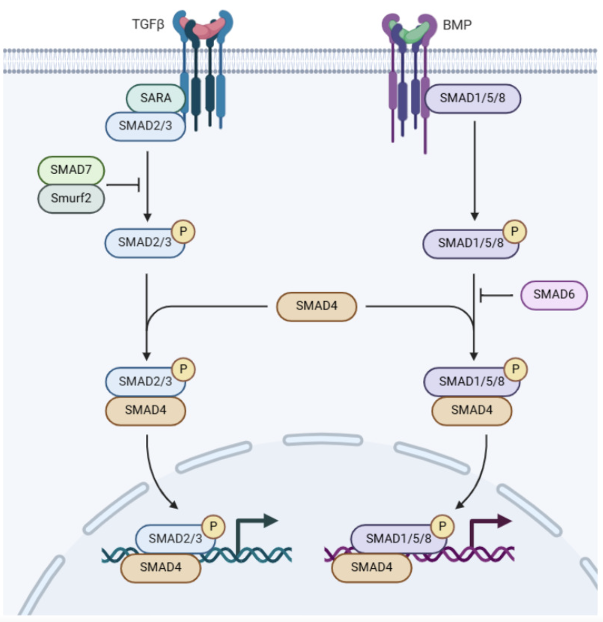
Dual specificity Ser/Thr and Tyr kinase receptor - tetramer of two type I and type II
Ligands include three TGF-β, BMPs and two activins
Ligand binding to type II receptors enable them to phosphorylate and activate type I
Activated type I phosphorylates C terminal of receptor-activated SMAD2/3 (R-SMADs) which complex with co-SMAD (SMAD4) to enter the nucleus and activate gene expression
Crucial role in development in development and tissue homeostasis and can promote fibrosis and metastasis in disease
Inhibitory SMADs (I-SMADs) eg SMAD6/7 compete with R-SMADs for binding and activation by the receptor
Receptor, transducer and activator of transcription
Some proteins can act as receptor, transducer of signal and effector
Notch signalling
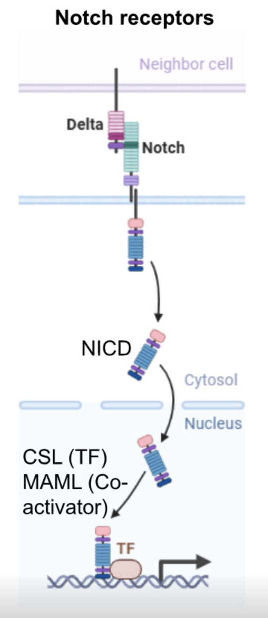
Notch receptor interacts delta (ligand on other cell), causing proteolytic cleavage of Notch receptor and release of its intracellular domain
Intracellular domain has co-factors of transcription factors Nuclear relocalisation of intracellular domain drives gene expression
Accumulation of unfolded/misfolded protein in the ER (UPR)
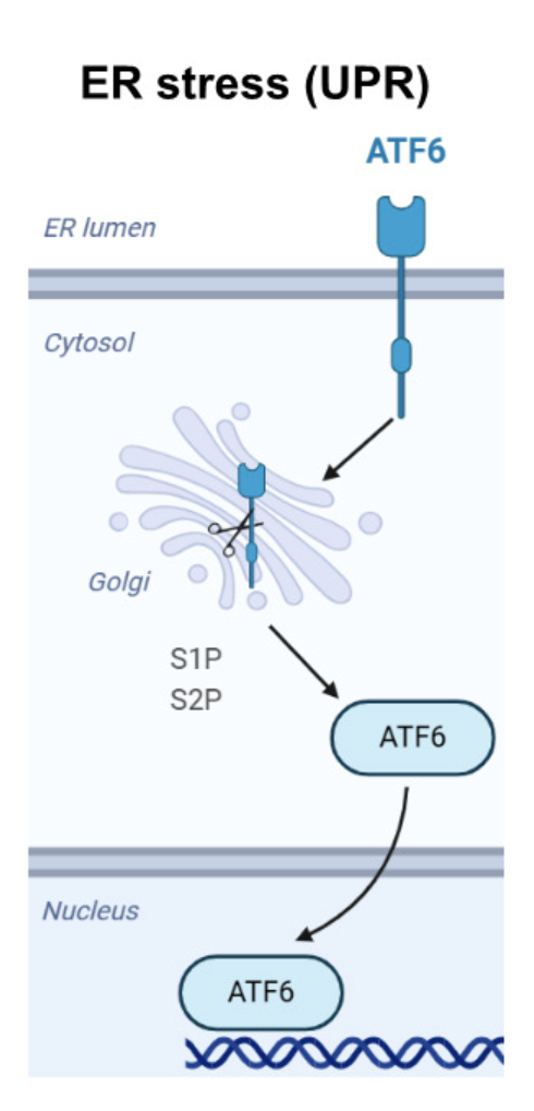
Dissociation of chaperone protein BiP from ATF6
ATF6 transported to Golgi
Proteolytic cleavage by Site-1 and Site-2 proteases (S1P and S2P)
Upon cleavage it is released as an active transcription factor p50ATF6 in the cytoplasm
Translocated to nucleus and activates ER-associated degradation (ERAD)/BiP
Sterol regulatory element binding proteins (SREBEPs)
Signalling during starvation
mTORC is a nexus downstream glucose and amino acid sensing
mTORC1 promotes growth
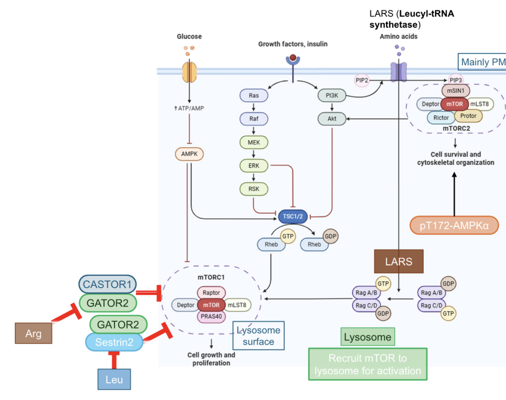
Promotes protein synthesis through S6K1 activation and 4E-BP1 inhibition, enabling proliferation
This regulation happens normally at the surface of lysosomes - amino acid sensing can happen here due to role of lysosomes in degrading/recycling proteins
Coupling mTORC1 with the lysosome, allows it to sense whether there are sufficient amino acid building blocks for protein synthesis
Translation is also coupled to growth/survival pathways so that growth is not only initiated by nutrient sufficiency but also external signals for growth
Glucose is sensed by ATP:ADP/AMP ratio which indicates the metabolic status
Inhibits autophagy through ULK1, ATG13 and TFE3/B
Pro proliferative signalling activates tuberous sclerosis complex 2 (TSC2, tuberin), a GTPase activating protein (GAP) which inhibits mTORC1
GATOR2 complex is a positive regulator of mTORC1
mTORC2 promotes survival and regulates movement by regulating AKT
Multitude of stresses integrate at the ISR (integrated stress pathway)
Stresses - ER stress, infection, lack of nutrients, Cyt c release…
ISR downregulates global translation due to phosphorylation of eIF2α (essential for translation initiation as it brings Met-tRNA to the ribosome) by stress-activated kinases (PERK, GCN2, PKR, HRI)
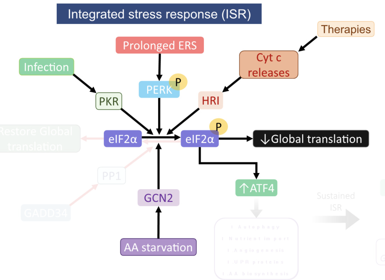
Phosphorylated eIF2α reduces global translation to conserve energy and activates ATF4 transcription factor which mitigates stress by activating CARE genes which promote amino acid synthesis, antioxidant response, chaperone expression, autophagy, cell survival, or autophagy for extensive damage