Protein Structure
protein structure is the 3-D shape polypeptides take on
primary structure: squence of polypeptides
secondary structure: alpha helixs and beta pletead sheets
tertiary structure: is fully folded 3-D shape of a polypeptide
myoglobin: final is this strucutre
quaternary structure: final shape of protein, can be made up of subunits
subunits: fully folded tertiary structure of single polypeptide
hemoglobin have subuints
carbon has tetrahedral geometry: with angle of 109.5 degrees
aminos acids have chirality
chirality: not identical to its mirror image
stereoisomers: molecules with same chemical bonds but different arrangement of them in space
in nature amino acids are found in L form
some D-amino acids are found in bacterial walls
glycine is achiral because its R group is another H
discerning between L & D amino acids
clockwise spelling when H is pointing toward you is in L form, spells CORN
carboxyl, R group, amino group
if counter clockwise is D amino acid
3-D drawing
wedges project out towards you
dashes point backwards
line is in line with plane
3-D polypeptide
r groups are in opposite directions, trans configuration
favored as R groups are not hindering one another
can fold in cis configuration (same) but would hinder each other
peptide bonds with proline form in cis configuration 3-10%
each resiude retains tetrahedral shape
main carbon atoms lie within same plane
key features of polypeptides
bond that joins alpha Carbon to carbon of carbonyl is psi bond
trident symbol
bond that joins alpha carbon to amide is phi bond
symbol of circle with capital i through it
trans config when both phi and psi bond have 180 degree rotation
alternating r groups
cis config when both phi and psi bonds have 0 degrees rotation
r groups on same side
conformational flexibility for protein comes from 360 degree rotation around phi and psi bonds
peptide bond is planar as protein folds and rigid
due to partial double bond character amide
N can donate electrons to C which pushes them to O resulting in
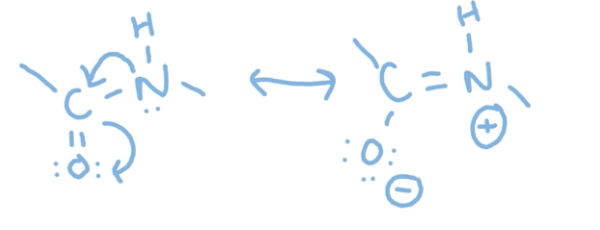
amides have resoance
creates the double bond which are rigid so no rotation
polypeptide can be viewed as series of rigid planes that can rotate at alpha carbon
Secondary protein structure
forms as a result of polypeptides maximizing the number of H-bonds it can make between its carbonyls and amides from its backbone
common type is alpha helix and beta pleated sheet
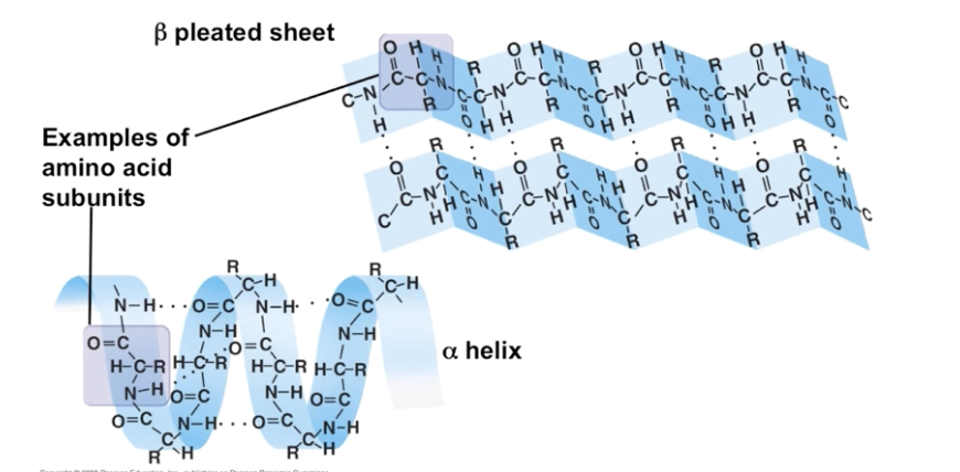
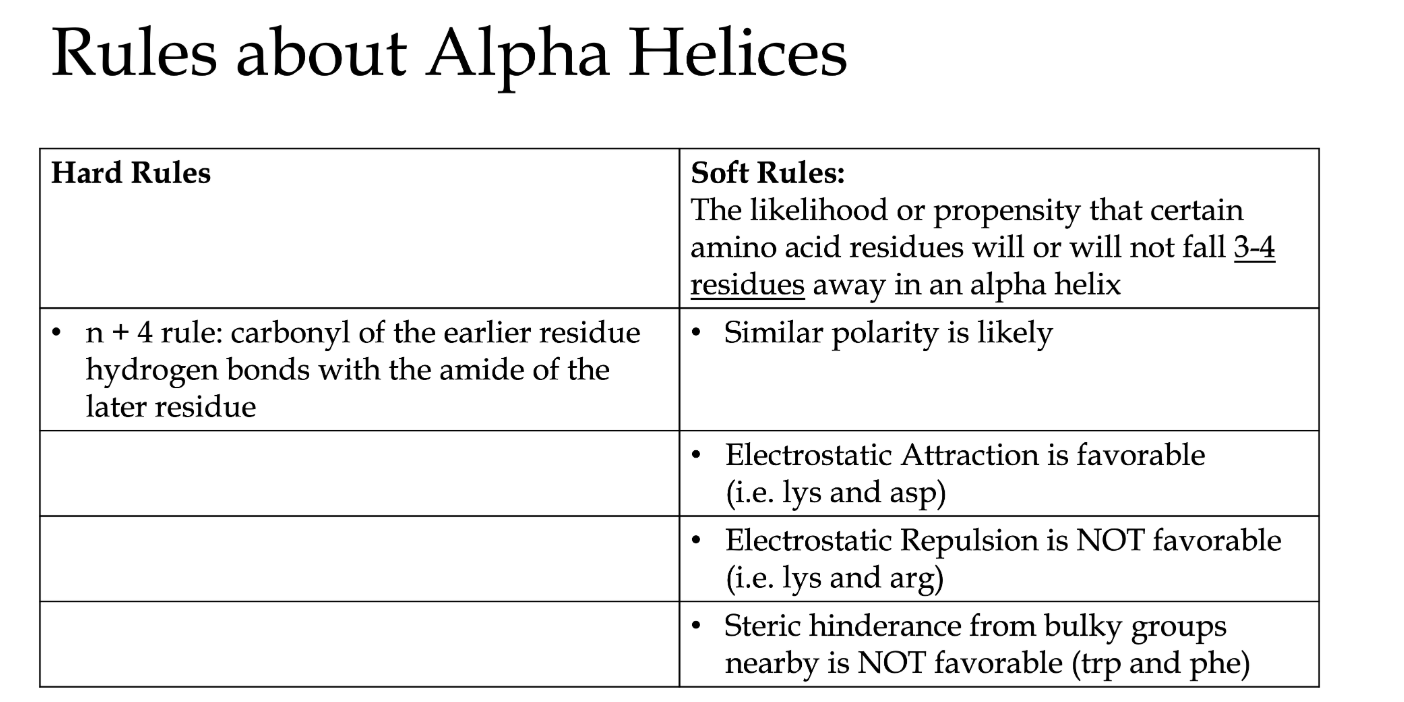
alpha helices: coiled portion of polypeptides, result of carbonyl of eariler residue H bonds with amide of the later residue that is 4 away
called n+4 rule
makes a complete turn every 3.6 residues
R groups are positioned 100 degree angle from one another
360 degree rotation has 3-4 residues
have a right hand turn like screws
on a right handed spiral staircase you can keep your right hand on the bannister and go down them the whole time
myoglobin made of 7 alpha helices
carbonyl’s point down and amides point up
perfect H-bond
hydrophobic portion of protein (R group) point inward
hydrophillic point outward and interact with aqueous solvent
not a hard rule but every 3-4 residues share polarity
Pro and Gly least likely to be a part of alpha helix
don’t have prolines
can’t Hydrogen bone so presence creates a destabilizing kink
amino acid residues with oppsite charges found 3-4 resiudes away, are stabilizing due to favorable electrostatic attraction
amino acid residues with same charges found 3-4 residues away can be destabilizing due to unfavorable electrostatic repulsion
amino acid residues with bulky R group found 3-4 residues away can be destabilizing due to steric hinderance
beta pleated sheets
form when two or more polypeptides H bond with each other; two forms anti-parallel and parallel
anti-parallel: happen when polypeptide folds in on itself, explains the direction of protein
H bonds occur between carbonyl and aimdes
two strands run in opposing direction (like DNA)
parallel: need a lot more polypeptide between the two starands to make the sheets line up in parallel fashion
pleats happen from alpha carbons fully extending and alternating up and down
r groups do this to
porin made of beta pleated sheets
found in bacteria cell membrane for transport of stuff
antiparallel shape
result of extended polypeptide, R groups point in opposite directions
beta turns (tight turns): connect 2 antiparallel strands together(makes 180 degree turn)
Hydrogen bond between 3 residues away
proline and glycine often found
imino nitrogen of Pro can take on the cis from which makes for a tight turn
can’t connect two parallel strands
helps fully understand its job
steric hinderence: when atoms/molecules try to take up same space it causes electron clouds to overlap, causing repulsive force and influences which bond angles are more likely to occur
not favorable
tertiary structure: refers to 3-D arrangement of all atoms in folded polypeptide; may be the final protein of it may be subunit of complete final protein
folds upon itself, may include alpha helix and beta pleated sheets
secondary and tertiary structures began to take form as the ribosome is folding the translating
sequence is responsible the protein folding
native conformation is the folded and functional form of the protein
denature state, is the unfolded and there is no activity or function
can happen due to
temperature increase
pH increase/decrease
salt concentration changes
solvent changes
doesn’t break covalent bonds in polypeptide (order is preserved) interferes with IM interactions
anfinsen’s dogma: 3D structure determined by seqence of amino acids
even if protein was denatured they can still refold
the unfolded protein in entropically favorable, as it has more disorder
protein folding is an energtically favorable process due to gibbs free energy
delta G = delta H - TdeltaS
negative gibbs free energy means reaction is spontaneous/exergonic
positive gibbs free means reaction is non-spontaneous/endogonic
negative enthalpy means exothermic
positive enthalpy means endothermic
negative entropy means more ordered
positive entropy means more disordered
things that make protein folding entropically favorable
hydrophobic effect: exergonic; biggest factor that makes protein folding happen
unfolded is entropically favorable for polypeptide but unfavorable for water around because of non polar R groups
Clathrate: water molecules encaging a nonpolar solute very order bad for entropy, but folding means breaking free of this as the non polar R group fold inside and polar are outside better for water
overcomes the entropy
formation of new IMF and disulfide bonds: collectively contribute
makes the enthalpy negative
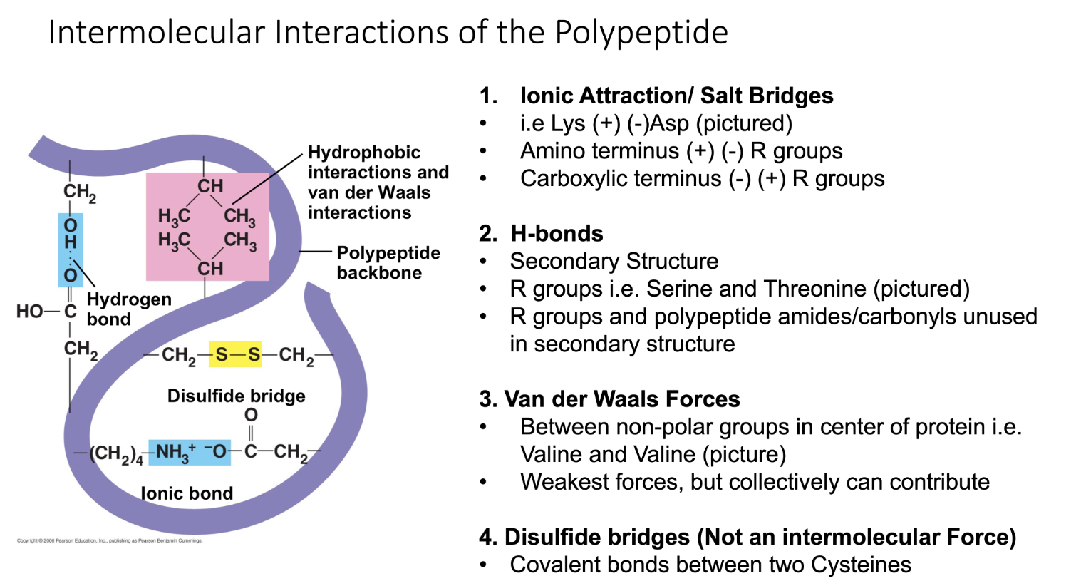
IMF
ionic attraction: strongest
occur between R groups of positive and negative charged
H-bonds: less strong then inonic
secondary structures, R groups, R groups and polypeptide amides/carbonyls unused in secondary structures
Van der Waals: weakest force of all, but can collectively contribute; NP molecules take on temporary dipoles
between NP groups in center of protein
can be ion-Induced Dipole, dipole induced dipole, dispersion
Di-sulfide bonds between cysteines
not IMF interaction
occurs between two cysteines
quaternary structure: final protein made up of 2 or more subunits
each subunit is its own polypeptide that folds into its own tertiary structure
subunits may be identical or may be different
hydrophobic effect and IMF keep the subunits togethers