out of class readings/vids
action potentials/neurons!
cns is brain and spinal cord, connects to pns.
The nervous system is composed of two basic cell types: glial cells (also known as glia) and neurons. Glial cells, which outnumber neurons ten to one, are traditionally thought to play a supportive role to neurons, both physically and metabolically. Glial cells provide scaffolding on which the nervous system is built, help neurons line up closely with each other to allow neuronal communication, provide insulation to neurons, transport nutrients and waste products, and mediate immune responses. Neurons, on the other hand, serve as interconnected information processors that are essential for all of the tasks of the nervous system. This section briefly describes the structure and function of neurons.
info input = dendrite, info output = axon
info convayed left to right through synapses (pre and postsynaptic neuron - which never physically touch), focus on chemical synapses but there are other types. presynaptic neuron has NT vesicles that release NT into the cleft to cause a change in post synaptic cell
neg inside cell compared to outside
A neuron’s outer surface is made up of a semipermeable membrane. This membrane allows smaller molecules and molecules without an electrical charge to pass through it, while stopping larger or highly charged molecules.
In the resting state, sodium (Na+) is at higher concentrations outside the cell, so it will tend to move into the cell. cant pass
Potassium (K+), on the other hand, is more concentrated inside the cell, and will tend to move out of the cell. can pass through membrane
unstable resting potential, membrane keeps it unstable (most important factor of unstability is the semipermeable membrane) maintain negative outside compared to positive inside due to concentration gradients and electrical gradients which creates dif voltage in/out of cell. 3 na out /2 k in pump also contributes to resting potential but not as much.
The nucleus of the neuron is located in the soma, or cell body. The soma has branching extensions known as dendrites. The neuron is a small information processor, and dendrites serve as input sites where signals are received from other neurons. These signals are transmitted electrically across the soma and down a major extension from the soma known as the axon, which ends at multiple terminal buttons. The terminal buttons contain synaptic vesicles that house neurotransmitters, the chemical messengers of the nervous system.
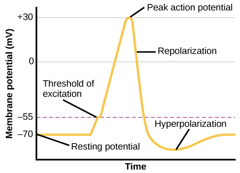
action potential: the electrical signal that typically moves from the cell body down the axon to the axon terminals. all or nothing event.
start with negative membrane potential at rest, as na opens it gets more positive (depolarization). at peak the na channels close and k channels open (repolarization) and k rushes outside of cell, returning it to resting. k overshoot a bit (hyperpolarization). seen most prominently in axons, build in dendrites and soma
Because it is all or none, the action potential is recreated, or propagated, at its full strength at every point along the axon. Much like the lit fuse of a firecracker, it does not fade away as it travels down the axon. It is this all-or-none property that explains the fact that your brain perceives an injury to a distant body part like your toe as equally painful as one to your nose.
neurotransmitter
involved in
Potential Effect on Behavior
Acetylcholine
Muscle action, memory
Increased arousal, enhanced cognition
Beta-endorphin
Pain, pleasure
Decreased anxiety, decreased tension
Dopamine
Mood, sleep, learning
Increased pleasure, suppressed appetite
Gamma-aminobutyric acid (GABA)
Brain function, sleep
Decreased anxiety, decreased tension
Glutamate
Memory, learning
Increased learning, enhanced memory
Norepinephrine
Heart, intestines, alertness
Increased arousal, suppressed appetite
Serotonin
Mood, sleep
Modulated mood, suppressed appetite
Agonists are chemicals that mimic a neurotransmitter at the receptor site and, thus, strengthen its effects.
An antagonist, on the other hand, blocks or impedes the normal activity of a neurotransmitter at the receptor.
Parkinson's disease, a progressive nervous system disorder, is associated with low levels of dopamine. Therefore dopamine agonists, which mimic the effects of dopamine by binding to dopamine receptors, are one treatment strategy.
In contrast to agonists and antagonists, which both operate by binding to receptor sites, reuptake inhibitors prevent unused neurotransmitters from being transported back to the neuron. This leaves more neurotransmitters in the synapse for a longer time, increasing its effects. Depression, which has been consistently linked with reduced serotonin levels, is commonly treated with selective serotonin reuptake inhibitors (SSRIs). By preventing reuptake, SSRIs strengthen the effect of serotonin, giving it more time to interact with serotonin receptors on dendrites. Common SSRIs on the market today include Prozac, Paxil, and Zoloft.
- NTs are limited, are reuptaken, broken down by enzymes, or drift away from synapse
2 CNS conductance/action potential
ions flow down electrochemical gradient - electrostatic forces states opposites attract and like repels like, and chemical states that ions flow from HI→LO
permiability is mass flowing through a membrane
conductance is charge flowing through a membrane (defined as 1/resistance) (high conductance = low resistance)
movement of mass and charge occurs simoltaneously, inc permeability leads to inc conductivity BUT IT IS NOT a linear relationship though so 2x permeability does NOT always = 2x conductivity
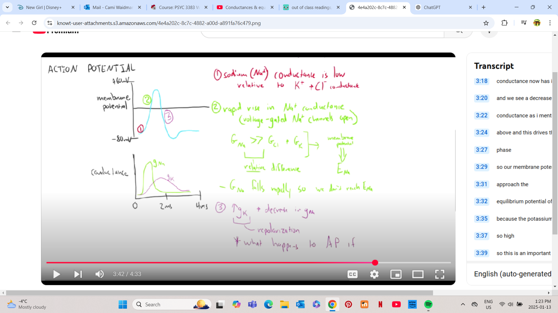
in action potentials: depolarization - na conductance is low relative to k and cl, at repolarization there is an inc in conductivity and permeability, gna (conductance) is much greater than cl and k relative conductance (absolute na conductance does not matter, only matters relative to other involved ions) and na approaches its equilibrium BUT we don’t reach equilibrium cuz the channels close, gk increases as k gets more conductive which drives repolarization and k approaches equilibrium. hyperpolarization is high relative gk as it approaches equilibrium. now we have reached the end of the action potential.
gk (relative conduciton of potassium) is found by: gk= gk (ion involved)/ gtotal (of all ions involved)
if a given ion can easily pass through the membrane, its conductance is high and it will tend towards equilibrium (which is its lowest energy state)
ON EXAM: what happens to the action potential if the potassium channels are blocked? my answer (not sure if this is right) Blocking potassium channels slows or prevents repolarization, as potassium moving out of cell—critical for returning the membrane potential toward the resting level—is impaired. This results in a prolonged action potential and reduces or eliminates hyperpolarization, as increased potassium conductance cannot occur. The membrane potential remains depolarized for longer, disrupting the cell's ability to reset and fire subsequent action potentials effectively.
2.5 CNS overview video
cns contains brain and spinal cord
spinal cord has 2 functions: 1) pass info from brain to periphery (motor going out/sensory coming in to brain), 2) drives sensory reflexes and rhythmic movements (made by neurons contained in the spinal cord NOT brain - ie doctor knee hit/ hot object withdrawing hand from something hot BEFORE your brain realizes it is hot - these are reflex circuits contained within the spinal cord!) (rhythmic motor patterns in walking, breathing, intestinal contractions, and more are driven by neurons contained in the spinal cord!)
interactive vid q’s: a sensory reflex like the pain withdrawl reflex occurs because of neuronal activation in the spinal cord, because the short pathway to the spinal cord from the PNS allows for rapid responses
spinal cord is made of 30 segments called vertebrae
each vertebra has perephiral nerves that go out to periphery that either send motor signals out to body from brain OR sensory signals to the brain
longitudinal fissure separates brain into two halves/hemispheres
left brained/right brained people is a misconception, both hemispheres have similar functions. a bit of lateralization in language production but that is shaped by environment
corpus callosum is in between both hemispheres and allows info to go from one side of the brain to the other
corpus callosum is sometimes cut to treat epilepsy, which allows us to observe the effects (split brain studies)
Brain Regions - vid 3
bottom up follows evolutionary history (oldest @ bottom, newest @ top)
brain stem = basic physiological processes required for survival (breathing, circulation, digestion, swallowing) and a transfer pathway for neuro signals to pass between spinal cord and brain
cerebellum = motor control, drives motor memories and movements
brainstem and cerebellum are most primitive/oldest
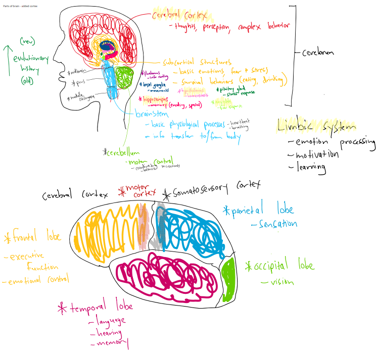
top of brain contains cortex (newest) = wrinkly outer layer responsible for thoughts, perceptions, complex behaviours. without it we would not have culture, communication, language, empathy, or understanding of ourselves and the world
between the newest and oldest lies the sub cortical structures. they drive basic emotions like fear and stress. mediate behaviours important for survival like eating and drinking
thalamus = info router
above thalamus is basal ganglia, controls movements - breaks down in parkinsons patients
hippocampus sticks out from under thalamus = info gets consolidated, spatial memory is encoded
endocrine system releases hormones into body to regulate physiological processes: hypothalamus = homeostasis (stable state, body temp, circadian rhythms, glucose levels - motivated behaviours: thirst hunger and aggression), pituitary gland sends out hormones and drives our stress response. posterior pituitary gland is considered part of the brain
amygdala = fear response
from bottom to top: medulla oblongota, pons, midbrain = basal physiological processes / HR / intestinal movements
sub cortical structures and cortex = the cerebrum, which is where integration happens (brain makes sense of incoming info)
cerebral cortex makes up 80% of our brains volume and is made of 4 lobes
frontal lobe = executive function, forming thoughts, making decision, emotional control
parietal lobe = sensory
occipital lobe = visual processing
temporal lobe = language production, hearing, memory formation
somatosensory cortex = boarder between frontal and anterior parietal lobe (where touch sensory info comes from)
motor cortex = posterior part of frontal lobe and parietal lobe (where motor plans are generated and sent out
limbic system = cortex, hypothalamus, hippocampus, amygdala → all function together to drive emotion processing, learning, motivation
Blood Vessels in the brain - vid 3.5
2 arteries bring blood from the heart to the brain: pair of vertebral arteries and pair of carotid arteries, which converge at the circle of willis, a confluence allowing for redundant routing of blood through the brain, so a single blocked vessel may not impede blood flow
3 main cerebral artieries branch off the circle of willis:
anterior cerebral artery - supplies blood to frontal lobe. symptom of stroke here is opposite side leg weakness/paralysis
posterior cerebral artery - supplies blood to temporal and occipital and thalamus and brainstem, strokes here are embolic and symptoms of stroke affect vision on opposite side of body including decreased visual field, color blindness, hallucinations. contralateral (opposite side) paralysis can also result
middle cerebral artery - supplies blood to frontal lobe and lateral surface of parietal and temporal lobes (areas covering primary motor and sensory areas of the upper body/face/hand/arm/throat/ and speech in the dominant hemisphere. this artery is most commonly involved in strokes. branches out from lateral sulcus