Module 6 Integumentary
Module Overview
Module 6: Integumentary System
Chapters: 24 and 25
Copyright: Elsevier, Inc. (2023)
Learning Objectives
Complete skin assessment procedure.
Identify diagnostic evaluations for skin conditions.
Describe treatments for surgical wounds.
Nursing care for clients with skin cancer using the nursing process.
Summarize prevention teachings for skin cancer.
Explain ABCDs of mole assessment.
Discuss nursing care for fungal, bacterial, and viral infections, infestations, and insect bites.
Nursing management for Steven Johnson Syndrome.
Identify pharmacological therapies, indications, and common adverse reactions.
Structures of the Skin
Layers of the Skin
Epidermis
Stratum Corneum: The outermost layer, consisting of dead, flattened skin cells that provide a protective barrier against environmental damage and water loss.
Stratum Lucidum: Found only in thick skin (e.g., palms and soles), this translucent layer offers an additional barrier to friction.
Stratum Granulosum: This layer contains keratinocytes that begin to die and become more flattened. It plays a vital role in waterproofing the skin.
Stratum Spinosum: Composed of keratinocytes linked by desmosomes, this layer provides strength and flexibility.
Stratum Germinativum (Basale): The deepest layer, where new skin cells are generated. This layer contains melanocytes, which contribute to skin pigmentation.
Dermis
Papillary Layer: The upper part of the dermis, this layer contains loose connective tissue, blood vessels, and sensory receptors. It contributes to the formation of fingerprints through dermal papillae.
Reticular Layer: The thicker, deeper layer composed of dense irregular connective tissue, providing strength and elasticity. It houses glands, hair follicles, and larger blood vessels.
Subcutaneous Tissue (Hypodermis)
Composed mainly of loose connective tissue and fat, this layer functions as insulation, energy storage, and a shock absorber. It anchors the skin to underlying structures like muscles and bones, facilitating movement.
Skin Appendages
Nails
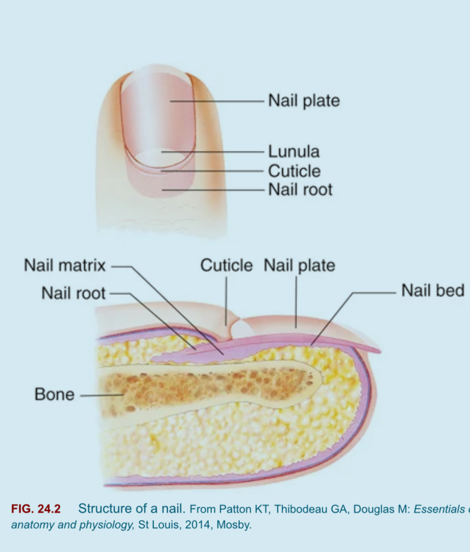 Nails are made of heavily keratinized cells and consist of several key parts:
Nail Plate: The hard, visible part of the nail, made of keratin.
Nail Bed: The skin beneath the nail plate, where the nail rests.
Nail Matrix: The tissue under the base of the nail (cuticle area) that produces new cells for nail growth.
Lunula: The crescent-shaped, white area at the base of the nail plate, representing the visible part of the nail matrix.
Cuticle: The layer of dead skin at the base of the nail that protects the nail matrix.
Types of Sweat Glands
Sweat glands are crucial for thermoregulation and skin hydration. There are two main types:
Eccrine Glands:
Function: Produce a watery sweat that helps regulate body temperature.
Location: Found all over the body, with a high density in the palms, soles, and forehead.
Example: Sweat produced during exercise.
Apocrine Glands:
Function: Produce a thicker, milky sweat that is rich in proteins.
Location: Primarily in the axillary (armpit) and groin areas; become active during puberty.
Example: Sweat that contributes to body odor when broken down by bacteria on the skin.
Skin Anatomy
Epidermal Structure
Stratum Corneum: Outer layer; dead skin cells.
Stratum Germinativum: Basal cell layer where cell generation occurs.
Melanocytes: Cells producing melanin (pigment).
Dermal Structure
Sebaceous Gland: Produces oil (sebum).
Sweat Glands: Regulate body temperature.
Hair Follicles: Encases hair roots.
Nerves and Blood Vessels: Provide sensation and circulation.
Cell Types in the Epidermis and Dermis
Epidermis Cell Types:
Keratinocytes: The predominant cell type; these cells produce keratin, a protein that waterproofs and protects the skin. They migrate from the stratum germinativum to the surface, undergoing a lifecycle of shedding and renewal.
Melanocytes: Located in the stratum germinativum, these cells produce melanin, the pigment responsible for skin color and UV protection.
Langerhans Cells: Immune cells found in the epidermis that play a role in recognizing and presenting antigens to the immune system, aiding in skin defense.
Merkel Cells: Found in the stratum basale, these cells function as mechanoreceptors and are involved in the sensation of touch.
Dermis Cell Types:
Fibroblasts: The primary cell type in the dermis, fibroblasts produce collagen and elastin, which provide strength and elasticity to the skin.
Mast Cells: Involved in inflammatory responses, these cells release histamine and other mediators during allergic reactions.
Macrophages: Part of the immune system, macrophages help to engulf and digest pathogens and debris in the dermal layer.
Basement Membrane
The basement membrane is a thin, fibrous extracellular matrix that separates the epidermis from the dermis. It consists of two layers:
Basal Lamina: The upper layer, produced by epithelial cells, contains laminins and collagen type IV, providing structural support.
Reticular Lamina: The deeper layer, made by fibroblasts in the dermis, offers anchorage to the underlying connective tissue.This membrane plays a critical role in skin integrity, serving as a foundation for cell migration and influencing cellular behavior.
Functions of the Integumentary System
Protection: Acts as a barrier against pathogens.
Insulation: Helps maintain body temperature.
Sensory Perception: Detects environmental changes.
Heat Regulation: Maintains homeostasis.
Vitamin D Synthesis: Crucial for calcium metabolism.
Skin Assessment Guide
General Assessment
Begins at first contact:
Overall condition of skin and hair noted.
Continuation through exam.
Subjective Data Collection
Key health information:
Health history
note any extended UV exposure
Medications
Previous treatments
Functional health patterns
Objective Data Collection
Physical Assessment:
Systematic inspection
Identification of primary/secondary lesions.
Gerontologic Considerations for the Integumentary System
As individuals age, several changes occur in the integumentary system, which may affect skin health and function:
Thinning of the Skin:
The epidermis becomes thinner, leading to increased susceptibility to injury and wounds.
Decreased collagen and elastin in the dermis reduces strength and resilience.
Dryness and Decreased Oil Production:
A reduction in sebaceous gland activity can result in dry skin, increasing the risk of dermatitis and skin infections.
Increased Bruising and Fragility:
Blood vessels become more fragile, causing easy bruising and more considerable recovery time from injuries.
Changes in Sensation:
Reduced numbers of nerve endings lead to diminished sensation, increasing the risk of unnoticed injuries or conditions like pressure ulcers.
Skin Cancer Risk:
The risk of developing skin cancers, such as basal cell carcinoma and squamous cell carcinoma, increases due to accumulated sun exposure and age-related changes.
Delayed Healing:
The healing process slows down due to reduced blood flow and nutrient supply, increasing the risk of infection and complications in wound healing.
Nursing Implications:
Assess skin conditions regularly and educate patients about sun protection and skin care routines to minimize damage.
Encourage hydration and the use of moisturizers to combat dryness.
Be vigilant in monitoring skin for signs of cancer, particularly in areas with a history of sun exposure.
Educate patients and caregivers about the importance of regular skin assessments to catch issues early.
Lesion Assessment
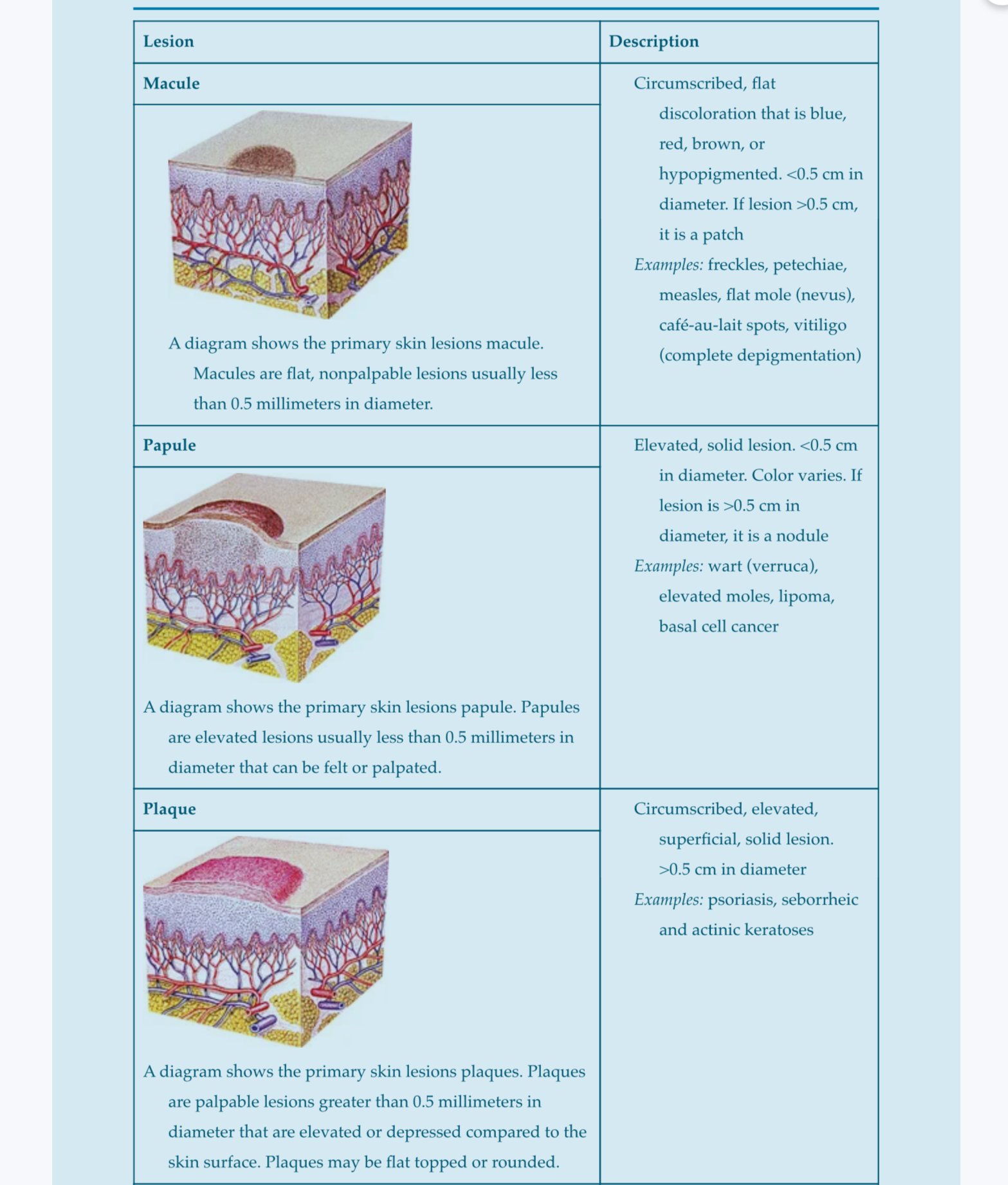
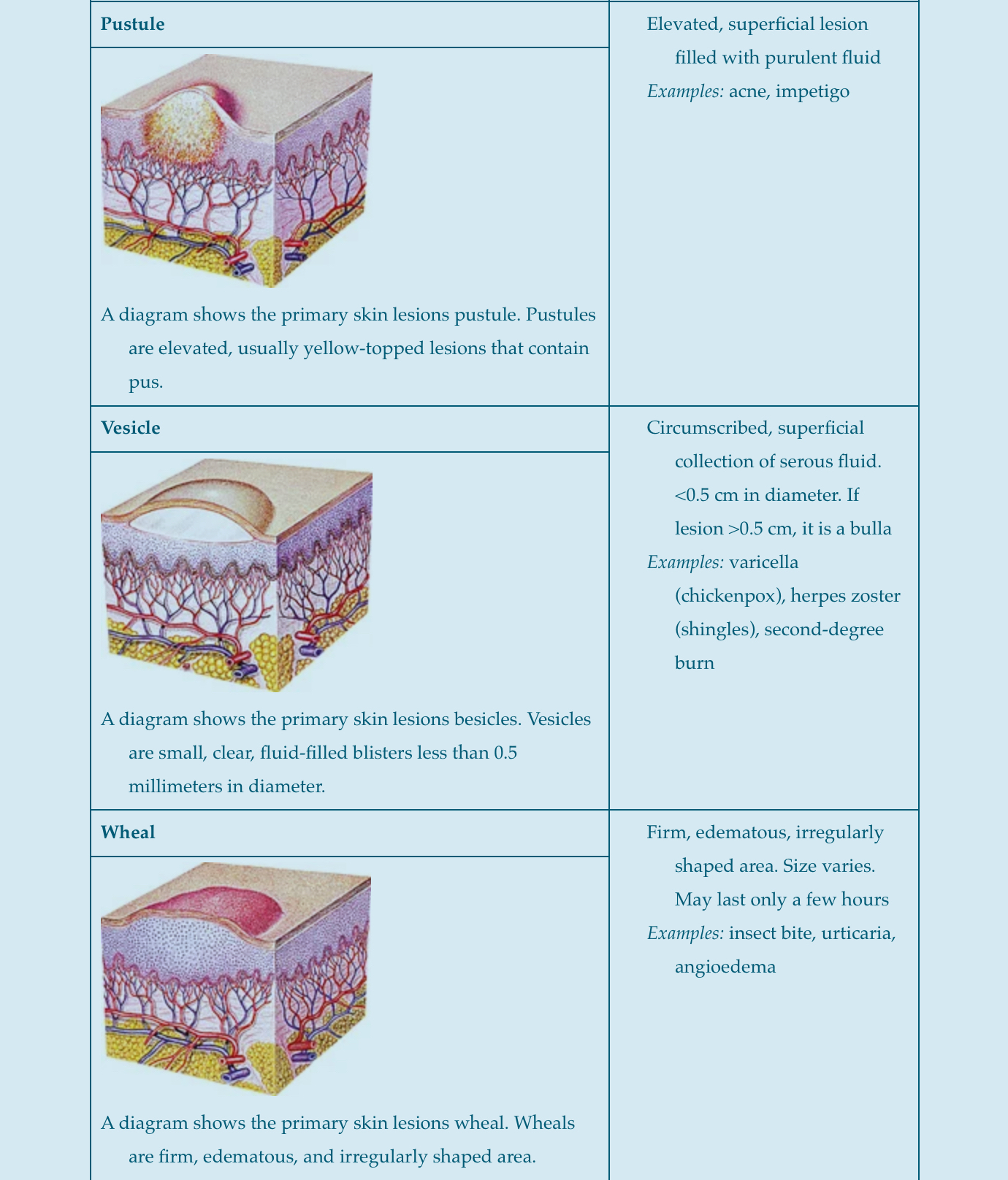
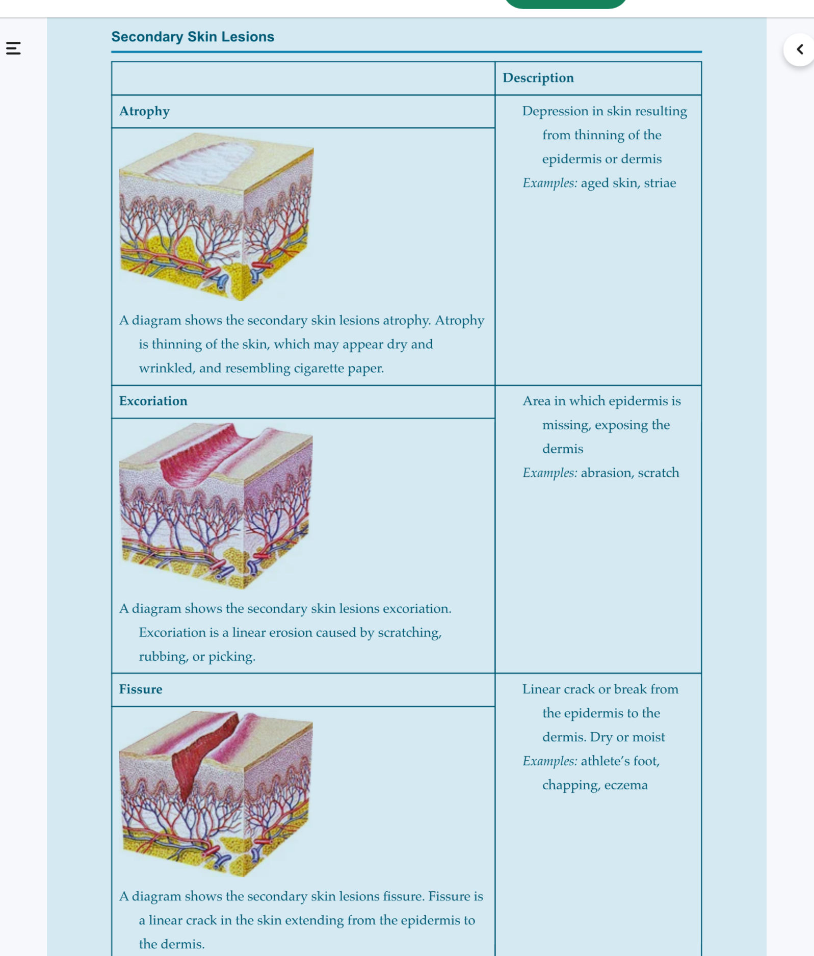
Primary Skin Lesions: Initial manifestations of skin disease.
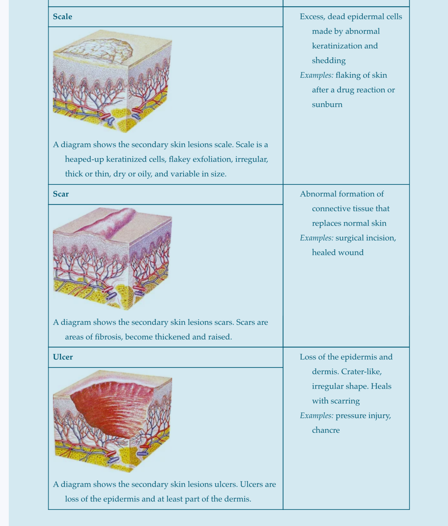
Secondary Skin Lesions: Results from primary lesions and factors like infection, inflammation, etc.
Lesion Distribution: Arrangement of lesions.
Configuration: Patterns (e.g., solitary, grouped).
Assessment in Dark Skin
Color assessment at areas with thinner epidermis.
Awareness of predisposition to keloids and vitiligo.
Diagnostic Evaluations
Techniques:
Dermatoscopy
Biopsy
Stains and Cultures
Immunofluorescence
Skin Cancer and Risk Factors
Skin Cancer Types
Nonmelanoma Skin Cancers:
Basal Cell Carcinoma: Most common, least deadly.
Definition: Basal cell carcinoma (BCC) is the most common type of skin cancer, originating in the basal cells of the epidermis.
Characteristics:
Appearance: BCC typically appears as a small, shiny bump or a red patch on the skin. It may also present as a sore that does not heal or a growth with a central depression.
Growth Rate: Slow-growing and rarely metastasizes (spreads to other parts of the body).
Risk Factors:
Prolonged exposure to ultraviolet (UV) radiation from the sun or tanning beds.
Fair skin, light hair, and blue or green eyes.
Previous history of skin cancer or pre-cancerous lesions.
Weak immune system or genetic predispositions (e.g., basal cell nevus syndrome).
Diagnosis:
Skin examination by a healthcare professional and confirmation through biopsy.
Treatment Options:
Surgical Excision: Removal of the tumor along with a margin of healthy tissue.
Cryotherapy: Freezing of the cancerous tissue.
Topical Chemotherapy: Application of cream containing anti-cancer medications.
Mohs Surgery: A specialized surgical technique that removes cancerous tissue layer by layer, ensuring complete removal while preserving healthy skin.
Prevention:
Regular skin checks and monitoring for any changes in moles or skin lesions.
Use of sunscreen with a high SPF, protective clothing, and avoiding excessive sun exposure, particularly during peak hours.
Squamous Cell Carcinoma: Aggressive, can metastasize.
Definition: Squamous cell carcinoma is a type of skin cancer that originates in the squamous cells, which are flat cells found in the epidermis.
Characteristics:
Appearance: SCC may appear as a firm red nodule, a flat lesion with a scaly crust, or a sore that bleeds and does not heal.
Growth Rate: Generally, SCC grows faster than basal cell carcinoma and has a higher likelihood of metastasis, particularly if not treated promptly.
Risk Factors:
Prolonged exposure to ultraviolet (UV) radiation, especially from the sun or tanning beds.
Fair skin, light hair, and light-colored eyes.
History of sunburns or chronic skin conditions like actinic keratosis.
Weakened immune system or previous skin cancers.
Diagnosis:
Diagnosis is typically confirmed through a skin examination and a biopsy, where a small sample of the lesion is analyzed for cancer cells.
Treatment Options:
Surgical Excision: The most common treatment, where the cancerous tissue is removed along with a margin of healthy skin.
Cryotherapy: Freezing the cancerous cells with liquid nitrogen.
Topical Chemotherapy: Application of creams containing chemotherapy agents, especially useful for superficial forms.
Radiation Therapy: Used for patients who are not surgical candidates or have recurrent SCC.
Prognosis:
Early detection and treatment generally lead to a favorable prognosis, but there is a risk of recurrence and metastasis if neglected.
Prevention:
Strategies include regular skin checks, using sunscreen with high SPF, wearing protective clothing, and avoiding tanning beds.
Melanoma- Leading cause of skin cancer deaths; can metastasize.
Definition: Melanoma is a serious type of skin cancer that develops from melanocytes, the cells that produce melanin, which gives skin its color.
Types of Melanoma:
Superficial Spreading Melanoma:
Most common type, often appearing as a brown or black flat lesion with irregular borders and multiple colors.
Typically arises on the trunk, legs, or arms.
Nodular Melanoma:
Second most common form.
Appears as a dark blue, black, or brown nodule that can be firm to the touch.
Grows vertically and can metastasize quickly.
Lentigo Maligna Melanoma:
Typically develops in sun-exposed areas like the face, neck, and arms.
Starts as a flat, tan or brown discoloration that may gradually become more irregular and darker.
Acral Lentiginous Melanoma:
Least common form, primarily found in people with darker skin.
Appears on the palms of the hands, soles of the feet, or underneath the nails.
Risk Factors:
Prolonged exposure to ultraviolet (UV) radiation.
Family history of melanoma.
Having a lot of moles or atypical moles (dysplastic nevi).
Fair skin that freckles easily.
Diagnosis:
Confirmed through skin examination and biopsy of the suspicious lesion.
ABCDE Rule for Detection:
Asymmetry: One half of the mole is different from the other.
Border: Irregular, scalloped, or poorly defined edges.
Color: Varied from one area to another; may include different shades of brown or black, or even white, red, or blue.
Diameter: Larger than 6mm (roughly the size of a pencil eraser).
Evolving: Changes in size, shape, or color.
Treatment Options:
Surgical Excision: Removal of the melanoma and some surrounding tissue.
Immunotherapy: Uses the body’s immune system to fight cancer.
Targeted Therapy: Focuses on specific genetic mutations in melanoma.
Chemotherapy: Typically used in advanced cases.
Prognosis:
Early detection is crucial; the prognosis is significantly better when diagnosed at an early stage.
Regular skin checks and monitoring for any changes in moles are essential for prevention.
Risk Factors for Melanoma
Red/blonde hair, light skin that freckles, chronic UV exposure.
Living near equator or in high altitude
History of skin cancer
Outdoor occupations or hobbies
Tanning
Sunscreen
Minimum of 15 SPF
Use every day and reapply during day
ABCDE Rule for Mole Assessment
Asymmetry
Border irregularity
Color variation
Diameter > 6 mm
Evolving characteristics over time.
Breslow Measurement
The Breslow measurement is a critical assessment used to determine the depth of invasion of melanoma, which is a key factor in staging the disease and predicting patient outcomes.
Key Points:
Definition: The Breslow thickness measures the vertical depth of the melanoma from the top of the granular layer of the epidermis to the deepest point of tumor invasion in the dermis or subcutaneous tissue.
Units: It is measured in millimeters (mm).
Significance:
Prognosis: Thicker tumors (greater Breslow depth) are associated with a higher risk of metastasis and poor prognosis.
Staging: Breslow thickness is often used in the AJCC (American Joint Committee on Cancer) staging system to classify melanoma and guide treatment decisions.
Interpretation:
Less than 1 mm: Thin melanoma, usually better prognosis.
1 mm to 4 mm: Intermediate thickness, moderate prognosis.
Greater than 4 mm: Thick melanoma, often associated with worse outcomes.
Atypical/Dysplastic Nevus
Definition: Atypical or dysplastic nevi are moles that differ from common moles in size, shape, and color. They often bear irregular features and can be a sign of increased risk for melanoma.
Characteristics:
Size: Typically larger than 6mm (the size of a pencil eraser).
Shape: Irregular, with uneven or scalloped borders.
Color: Variation in color that may include shades of brown, black, red, or blue.
Texture: Often flat or slightly raised with a rough surface.
Risk Factors:
Family history of melanoma or atypical nevi.
Presence of numerous moles (over 50).
Fair skin, light hair, and light-colored eyes, which are more susceptible to UV damage.
Management:
Regular skin examinations to monitor changes in size, shape, or color of existing moles.
Dermatological evaluation and possibly biopsy if changes are detected.
Preventive strategies include the use of sunscreen and protective clothing to reduce UV exposure.
Conclusion: Atypical/dysplastic nevi require close observation due to their association with melanoma risk, emphasizing the importance of regular skin checks and healthy sun habits.
Patient Education Topics
Bacterial Infections
Drainage is contagious; importance of hygiene and antibiotics.
Viral Infections
Education on herpes zoster and simplex; medications (acyclovir).
Fungal Infections
Common types: candidiasis, tinea infections.
Treatment includes hygiene and antifungal agents.
Infestations
Pediculosis (lice) and scabies management; non-contagiosity info.
Infections and Infestations of the Skin
Bacterial Infections
Bacterial infections of the skin are commonly caused by pathogens such as Staphylococcus aureus and Streptococcus pyogenes.
Common types:
Impetigo: Highly contagious, often seen in children; presents as red sores that can rupture and ooze.
Folliculitis: Inflammation of hair follicles, often caused by bacteria; can result in red bumps or pimples.
Cellulitis: A deeper skin infection that causes redness, swelling, and pain; can lead to systemic infection if untreated.
Treatment: Antibacterial medications (topical or systemic), wound care, and hygiene education to prevent spread.
Viral Infections
Viral infections are caused by various viruses that can affect the skin such as herpes simplex virus and varicella-zoster virus.
Common types:
Herpes Simplex: Causes cold sores (HSV-1) or genital herpes (HSV-2); characterized by painful blisters.
Herpes Zoster (Shingles): Reactivation of varicella-zoster virus; presents as a painful rash with blisters, typically on one side of the body.
Treatment: Antiviral medications (e.g., acyclovir) to decrease severity and duration, pain management, and patient education on contagion.
Fungal Infections
Fungal infections are caused by fungi and can affect the skin, nails, and mucous membranes.
Common types:
Tinea: Includes 'ringworm' infections, affecting various body parts (e.g., tinea corporis, tinea pedis (athlete's foot)).
Candidiasis: Caused by Candida species; often affects moist areas like the armpits and groin, resulting in red, itchy rashes.
Treatment: Antifungal medications (topical or systemic), management of moisture and hygiene to prevent recurrence.
Infestations
Infestations involve the presence of parasites on the skin.
Common types:
Pediculosis (Lice): Infestation of head, body, or pubic hair; manifests as itching and visible lice or eggs (nits).
Scabies: Caused by the Sarcoptes scabiei mite; results in intense itching, especially at night, and a rash from burrows.
Insect Bites: Bites from insects like mosquitoes, ticks, and fleas can cause localized redness, swelling, and itching. Serious reactions may occur, such as allergic responses or disease transmission (e.g., Lyme disease from ticks).
Management:
Pediculosis: Topical insecticides and education on hygiene and treatment of personal items.
Scabies: Topical scabicides, oral medications, and treatment of close contacts to prevent re-infestation.
Insect Bites: Clean the area, apply antihistamines for itching, and monitor for signs of infection or allergic reaction. Treat severe reactions as needed.
Allergic and Benign Skin Conditions
Contact Dermatitis
Definition: Contact dermatitis is an inflammation of the skin resulting from direct contact with an irritant or allergen.
Causes:
Irritant Contact Dermatitis: Caused by exposure to substances like soaps, detergents, or chemicals that damage the skin's barrier.
Allergic Contact Dermatitis: A delayed hypersensitivity reaction to allergens such as poison ivy, nickel, or certain fragrances.
Symptoms:
Redness and swelling
Itching and burning
Blisters or rash at the site of contact
Treatment:
Identify and avoid the triggering substance
Topical corticosteroids to reduce inflammation
Emollients to soothe the skin
Psoriasis
Definition: Psoriasis is a chronic autoimmune condition characterized by the rapid turnover of skin cells, leading to thick, red patches on the skin covered with silvery scales.
Symptoms:
Thick, red patches with silvery scales
Dry, cracked skin that may bleed
Itching and burning sensations
Causes:
Exact cause is unknown, but involves immune system dysfunction and genetic factors. Triggers may include stress, infections, and weather changes.
Treatment:
Topical treatments (corticosteroids, vitamin D analogs)
Phototherapy to reduce symptoms
Systemic medications (methotrexate, biologics) for moderate to severe cases
Allergic Skin Problems
Allergic skin problems can manifest as various conditions resulting from hypersensitivity reactions to environmental triggers. Common types include:
Atopic Dermatitis (Eczema)
Definition: A chronic inflammatory skin condition characterized by dry, itchy skin and rashes.
Symptoms: Red, inflamed skin, dryness, and intense itching, often appearing in patches.
Treatment: Moisturizers, topical corticosteroids, and antihistamines to relieve itching.
Urticaria (Hives)
Definition: Raised, itchy welts on the skin caused by an allergic reaction.
Symptoms: Red or skin-colored welts, often accompanied by swelling and itching.
Treatment: Antihistamines, corticosteroids for severe cases, and avoidance of known triggers.
Allergic Contact Dermatitis
Definition: Skin inflammation resulting from direct contact with an allergen.
Causes: Common allergens include poison ivy, nickel, and certain fragrances.
Symptoms: Redness, swelling, itching, and blistering at the site of contact.
Treatment: Avoidance of the allergen, topical corticosteroids, and soothing agents.
Steven Johnson Syndrome (SJS)
Definition: Steven Johnson Syndrome is a rare, serious condition that affects the skin and mucous membranes, often as a reaction to medications or infections. It is characterized by extensive skin detachment and blistering, resembling burns.
Causes:
Medications: Commonly triggered by sulfa medications, anticonvulsants, and non-steroidal anti-inflammatory drugs (NSAIDs).
Infections: Viruses such as herpes or Mycoplasma pneumoniae infections can also precipitate SJS.
Genetic Factors: Certain genetic predispositions may increase the risk of SJS.
Symptoms:
Flu-like symptoms (fever, sore throat, fatigue) preceding skin involvement.
Painful red or purplish rash that spreads, causing blisters and skin shedding.
Mucosal lesions affecting the eyes, mouth, and genital areas, leading to pain and swelling.
Diagnosis:
Diagnosis is typically based on clinical examination and patient history. Biopsy of the skin may also be performed to confirm.
Treatment:
Immediate discontinuation of the offending medication.
Supportive care in a hospital setting, similar to burn treatment, which may include fluid and electrolyte management.
Pain management and wound care to prevent infection and promote healing.
In severe cases, treatments such as corticosteroids, intravenous immunoglobulins (IVIG), or other immunosuppressive therapies may be considered.
Prognosis:
The prognosis can vary significantly depending on the severity of the condition and timely intervention. Early recognition and management are crucial for improving outcomes.
Nursing Management
General Principles
Wet Compresses and Bathing
Topical Medications Application
Pruritus Control: Hydration, cool environments, medications.
Post-procedure Care
Prevention of infections, control of pruritus.
Cosmetic Procedures and Management
Types: Laser surgery, liposuction, grafts.
Preoperative Management: Consent and patient teaching.
Postoperative Management: Pain control, monitoring for infections.